ARTHROPODS. In the contemporary world. Edited by. Alicja Buczek Czesław Błaszak Katarzyna Bartosik KOLIBER
|
|
|
- Teodor Łuczak
- 7 lat temu
- Przeglądów:
Transkrypt
1 ARTHROPODS In the contemporary world Edited by Alicja Buczek Czesław Błaszak Katarzyna Bartosik KOLIBER LUBLIN 2015
2
3 STAWONOGI We współczesnym świecie Pod Redakcją Alicji Buczek Katedra i Zakład Biologii i Parazytologii Uniwersytet Medyczny w Lublinie Czesława Błaszaka Zakład Morfologii Zwierząt Uniwersytet im. Adama Mickiewicza w Poznaniu KOLIBER LUBLIN
4 ISBN KOLIBER Oficyna Wydawnicza Fundacji na Rzecz Zwalczania Kleszczy i Profilaktyki w Chorobach Odkleszczowych fundacja@kleszcze.pl 4
5 Spis Treści I Stawonogi - Rozprzestrzenienie w różnych środowiskach Parasitic arthropods of mammals and their adaptations for living in the hosts in aquatic environment Joanna N. Izdebska, Leszek Rolbiecki, Paulina Kozina, Michał Skrzypczak 13 Threats to human health posed by ticks (Ixodida) in different climate zones Alicja Buczek, Katarzyna Bartosik, Zbigniew Zając, Weronika Buczek 27 The occurrence of Ixodes ricinus (Linnaeus, 1758) (Acari: Ixodidae) ticks in central, south-eastern, and eastern Poland in relation to habitats Zbigniew Zając, Alicja Buczek, Katarzyna Bartosik, Patrycja Tokarzewska, Paweł Szczepan Błaszkiewicz 37 Environmental background of tick occurrence with special emphasis on Ixodes ricinus the major vector of tick-borne pathogens in Poland Katarzyna Bartosik, Alicja M. Buczek, Weronika Buczek, Zbigniew Zając, Halina Cios, Alicja Buczek 47 Ticks from the genus Dermacentor Koch, 1844 (Acari: Amblyommidae) with the greatest epidemiological importance worldwide Buczek Alicja, Daniel Bzowski, Bartosik Katarzyna, Patrycja Tokarzewska, Zbigniew Zając 61 Endogenic factors influencing tick s (Acari: Ixodida) behaviour and life cycle Zbigniew Zając, Alicja Buczek, Katarzyna Bartosik 71 Dermacentor reticulatus (Fabr.) in Wroclaw area - planning field measurements and analyzing spatial distribution of ticks using GIS tools Dorota Kiewra, Mariusz Szymanowski, Elżbieta Lonc, Aleksandra Czułowska 79 TICKS (ACARI: IXODIDA) ATTACKING DOMESTIC DOGS IN THE MALOPOLSKA VOIVODESHIP, POLAND Paula Zajkowska 87 Invasive alien arthropod species and their adverse impact in Poland Małgorzata Kłyś, Magdalena Nowak-Chmura 101 Nieproszeni goście w naszych łóżkach Krzysztof Solarz 111 Problem obecności Arthropoda w przestrzeni placówki opieki społecznej Dorota Wodzisławska-Czapla, Grzegorz Hudzik, Krzysztof Solarz 135 POWIĄZANIA ROZTOCZY Z OWADAMI (ACARI INSECTA) UTRWALONE W BURSZTYNIE PRZED MILIONAMI LAT Wit Chmielewski 145 Mono-infestacja jeża wschodniego (Erinaceus roumanicus) pchłami (Archaeopsylla erinacei) na terenach rekreacyjnych Poznania* Sylwia Bialik, Jerzy Michalik 155 5
6 II Stawonogi i patogeny - wpływ na inne organizmy Candidatus Neoehrlichia mikurensis: nowe szerzące się infekcje i ich eko-epidemiologia Edward Siński 165 Bartonella spp. infections in bats and their hematophagous ectoparasitic Acari* Jerzy Michalik, Agnieszka Szubert-Kruszyńska, Justyna Liberska, Mirosława Dabert, J oanna Stańczak 173 Pathogens transmitted by ticks parasitizing humans Katarzyna Bartosik, Zbigniew Zając, Alicja Buczek, Patrycja Tokarzewska, Ewelina Wojczuk 183 Effect of Borrelia burgdorferi infection on the expression of genes related to oxidative stress in normal human dermal fibroblasts Małgorzata Kimsa, Magdalena Kimsa, Sławomir Dudek, Hubert Okła, Krzysztof Jasik, Grzegorz Hibner, Tomasz Janikowski, Aleksandra Skubis, Bartosz Sikora, Urszula Mazurek 197 Borrelia burgdorferi infection initiates changes in the expression of CD1 gene family in skin fibroblasts Magdalena Kimsa-Dudek, Sławomir Dudek, Hubert Okła, Małgorzata Kimsa, Katarzyna Pawłowska-Góral, Krzysztof Jasik, Urszula Mazurek 211 Expression pattern of genes associated with chromatin organization in human dermal fibroblasts infected with Borrelia burgdorferi sensu lato Grzegorz Hibner, Tomasz Janikowski, Olga Pawełczyk, Małgorzata Kimsa, Aleksandra Skubis, Bartosz Sikora, Sławomir Dudek, Małgorzata Ciałoń, Krzysztof Solarz, Krzysztof Jasik, Karol Juszczyk, Urszula Mazurek The effect of Borrelia burgdorferi sensu lato spirochetes on gene expression of stem signaling pathways responsible for wound healing in cultured normal human dermal fibroblasts Tomasz Janikowski, Olga Pawełczyk, Grzegorz Hibner,,Aleksandra Skubis, Bartosz Sikora,Małgorzata Porc, Sławomir Dudek, Krzysztof Jasik, Grażyna Janikowska, Agnieszka Lubczyńska, Martyna Bednarczyk, Urszula Mazurek 239 Interleukina 8 (CXCL8) i jej receptory w fibroblastach skóry zakażonych krętkami Borrelia burgdorferii Muc -Wierzgoń M., Rajs A., Nowakowska Zajdel E., Kokot T, Wierzgoń A, Gola J., Kruszniewska Rajs C., Dudek S., Jasik K., Kozieł P., Walkiewicz K.,Benjamin Grabarek, Mazurek U. 253 Analiza histologiczna wątroby szczura pod wpływem terapii przeciwpierwotniakowej azytromycyną, atowakwonem i prokwanilem Jan Słodki, Hubert Okła, Krzysztof P. Jasik, Aleksandra Słodki, Aniela Grajoszek, Beata Rozwadowska, Marta Albertyńska 263 Analiza histologiczna śledziony płodowej szczura zmienionej pod wpływem inwazji Babesia microti Aleksandra Słodki, Krzysztof P. Jasik, Jan Słodki, Hubert Okła, Aniela Grajoszek, Beata Rozwadowska, Marta Albertyńska 275 Trudności w diagnostyce boreliozy u dzieci Barbara Koleżyńska, Józef Kurek, Krzysztof Solarz, Marek Ślusarz 283 6
7 Ocena występowania zakażenia krętkami Borrelia u pracowników Nadleśnictwa Kobiór w latach Marta Albertyńska, Beata Rozwadowska, Grzegorz Hudzik, Hubert Okła, Aleksandra Słodki, Jan Słodki, Justyna Jakubas-Zawalska, Krzysztof P. Jasik, Andrzej S. Swinarew 293 Urban foci of Ixodes ticks borreliosis or Lyme disease on the example of Kyiv, Ukraine Igor Nebogatkin 301 The tick-borne diseases occurring among dogs and cats of Wysokie Mazowieckie county and Siemiatycze county Adam Kamil Trzeszczkowski, Bożena Kiziewicz 311 The role of developmental stages of Plodia interpunctella (Arthropoda, Insecta) as reservoirs/vectors of pathogenic fungi distributed in the human environment Lidia Chomicz, Bohdan Starościak, Monika Dybicz, Agnieszka Chruścikowska, Wanda Baltaza, Ilona Pilich, Konrad Perkowski, Julita Nowakowska, Paweł Zawadzki, Marcin Padzik, Piotr Wroczyński 327 Prognosis of the incidence of infestation by the head louse Pediculus humanus (Anoplura: Pedicullidae) in children in the Lublin Province as indicated by own long-term investigations Alicja Buczek, Katarzyna Bartosik, Zbigniew Zając, Patrycja Tokarzewska 335 Methods for control of malaria spread worldwide Bartosik Katarzyna, Buczek Alicja, Patrycja Lachowska-Kotowska, Paweł Szczepan Błaszkiewicz, Patrycja Tokarzewska, Zbigniew Zając 342 Pyralid moths as risk factors for spread of opportunistic/ pathogenic microbiota in urban and suburban dwellings Agnieszka Chruścikowska, Bohdan Starościak, Marek Asman, Wanda Baltaza, Monika Dybicz, Konrad Perkowski, Paweł Zawadzki, Piotr Piekarczyk, Beata Biernat, Lidia Chomi 351 Drobnoustroje występujące na powłokach ciała karaczanów prusaków (Blattella germanica L.) odłowionych w budynkach mieszkalnych niedocenione zagrożenie Ewa Mikulak, Agnieszka Królasik, Aleksandra Gliniewicz, Grażyna Młynarczyk, Kornelia Dobrzaniecka 365 III Stawonogi Sposoby zwalczania Susceptibility of Ixodes ricinus and Dermacentor reticulatus ticks to Beauveria bassiana and Metarhizium anisopliae fungi strains. Preliminary in vitro studies Anna Szczepańska, Elżbieta Lonc and Dorota Kiewra 379 Pythium carolinianum entomopathogenic fungus-like organisms isolated from the Supraśl River, Podlasie Province Bożena Kiziewicz, Anna Godlewska, Elżbieta Muszyńska, Paulina Pawłowska, Natalia Rogoz, Adam Kamil Trzeszczkowski 387 7
8 8
9 Wprowadzenie Pasożytnicze i alergogenne stawonogi ze względu na szeroki zakres szkodliwego oddziaływania na inne organizmy znajdujące się w tych samych ekosystemach stanowią jeden z najważniejszych czynników biologicznych w XXI wieku zagrażających zdrowiu człowieka i zwierząt. Duże zdolności adaptacyjne pasożytniczych i alergogennych owadów i pajęczaków sprawiły, że przystosowały się one do bytowania w różnych środowiskach. Stawonogi mogą przebywać nawet w ekstremalnych warunkach temperatury i wilgotności, jak na przykład w wysokich pasmach gór. Pasożytnicze i alergogenne stawonogi rozprzestrzeniają się w różny sposób, co jest sprzyjającym czynnikiem w zasiedlaniu nowych środowisk. Duży udział w pojawieniu się gatunków tych bezkręgowców na obszarach miejskich i podmiejskich ma człowiek, który poprzez swoje zachowanie i działania w znacznym stopniu przyczynia się do zwiększenia zasięgów ich występowania. Szerokie rozprzestrzenienie stawonogów w przyrodzie, przy jednoczesnym zmniejszeniu odporności ludzi i zwierząt skutkuje wzrostem częstości pojawiania się chorób alergicznych o tej etiologii i chorób transmisyjnych wywołanych przez przenoszone patogeny. Wśród pasożytniczych pajęczaków nadal duży problem medyczny i epidemiologiczny stanowią kleszcze i patogeny odkleszczowe. W świecie obserwuje się stały wzrost zachorowań ludzi i zwierząt na choroby bakteryjne, riketsjowe i pierwotniacze, m.in. na boreliozę, gorączki plamiste i babeszjozę. Szczególnie zainteresowanie wciąż wzbudza borelioza, której rozpoznanie i leczenie nadal stwarza wiele trudności. Coraz częściej na nowych obszarach u stawonogów odnotowuje się też nieznane dotychczas patogeny i przypadki chorób przez nie wywołanych. Powyższe względy sprawiają, że w wielu ośrodkach naukowych podejmowane są badania nad fauną, rozprzestrzenieniem i ekologią stawonogów alergogennych i pasożytniczych oraz nad skutkami ich szkodliwego oddziaływania na inne organizmy. W niniejszej monografii przedstawiamy prace nadesłane z kilku uniwersytetów i instytutów naukowych, w których prowadzone są badania nad stawonogami w nadziei, że zainspirują one biologów, lekarzy, diagnostyków i pracowników służ epidemiologicznych do kontynuowania doświadczeń i obserwacji terenowych. Alicja Buczek Czesław Błaszak Katarzyna Bartosik 9
10 10
11 I Stawonogi Rozprzestrzenienie w różnych środowiskach 11
12 12
13 Parasitic arthropods of mammals and their adaptations for living in the hosts in aquatic environment Joanna N. Izdebska, Leszek Rolbiecki, Paulina Kozina, Michał Skrzypczak Department of Invertebrate Zoology and Parasitology, University of Gdańsk, Wita Stwosza 59, Gdańsk , Poland, Abstract A typical parasitofauna of mammals, as the animals associated with various types of terrestrial ecosystems, is represented by different groups of terrestrial arthropods mites and insects. However, the mammals that secondarily adopted to aqueous life type, usually retain, at least partially, a set of parasitic arthropods typical for related to them terrestrial mammals. This is probably conditioned by a long-term evolution of the parasite-host system, where the relationship of parasite and mammal was so strong that it stayed with the host regardless of its place of living, gradually yielding new adaptations. A significant similarity of acaro- or entomofauna communities is usually characteristic for semiaquatic or aquatic mammals with a similar pedigree. In the case of parasites related to the hair, the presence of lice from Echonophthiridae family is characteristic for pinnipeds, while rich fauna of hair mites from Chirodiscidae or Listrophoridae families for holarcitc rodents (beavers, muskrats). However, the greatest potential to survive is noted here for parasites living in more stable conditions posed by skin or internal organs of the hosts, for example skin mites. The representatives of this group, Demodecidae mites, common in terrestrial mammals, were also reported in different mammals associated with aquatic environment, for example otters, beavers, seals and sea lions. Introduction Parasitofauna of the mammals, as animals associated with different types of terrestrial ecosystems, is typically composed of terrestrial arthropods - insects and mites. A number of taxa are host-specific parasites showing a long-term phylogenetic relationship with them, and therefore perfectly matched. But what if the hosts change the life environment in the course of evolution, secondarily returning to the aquatic mode, or adapting to semiaquatic lifestyle? It seems that such changes would result in a loss of parasitofauna, or acquisition of a new one. In fact, parasites from some groups mites or insects remained with the host acquiring an adaptation to the new conditions. However, for parasites, particularly stationary ones, the host constitutes the first order environment, but the external environment (second order environment) is also of a great importance. Meanwhile, living conditions in the aquatic environment significantly differ from those in the terrestrial environment, which concerns the availability of oxygen, medium density, or thermal properties. This particularly concerns ectoparasites, they 13
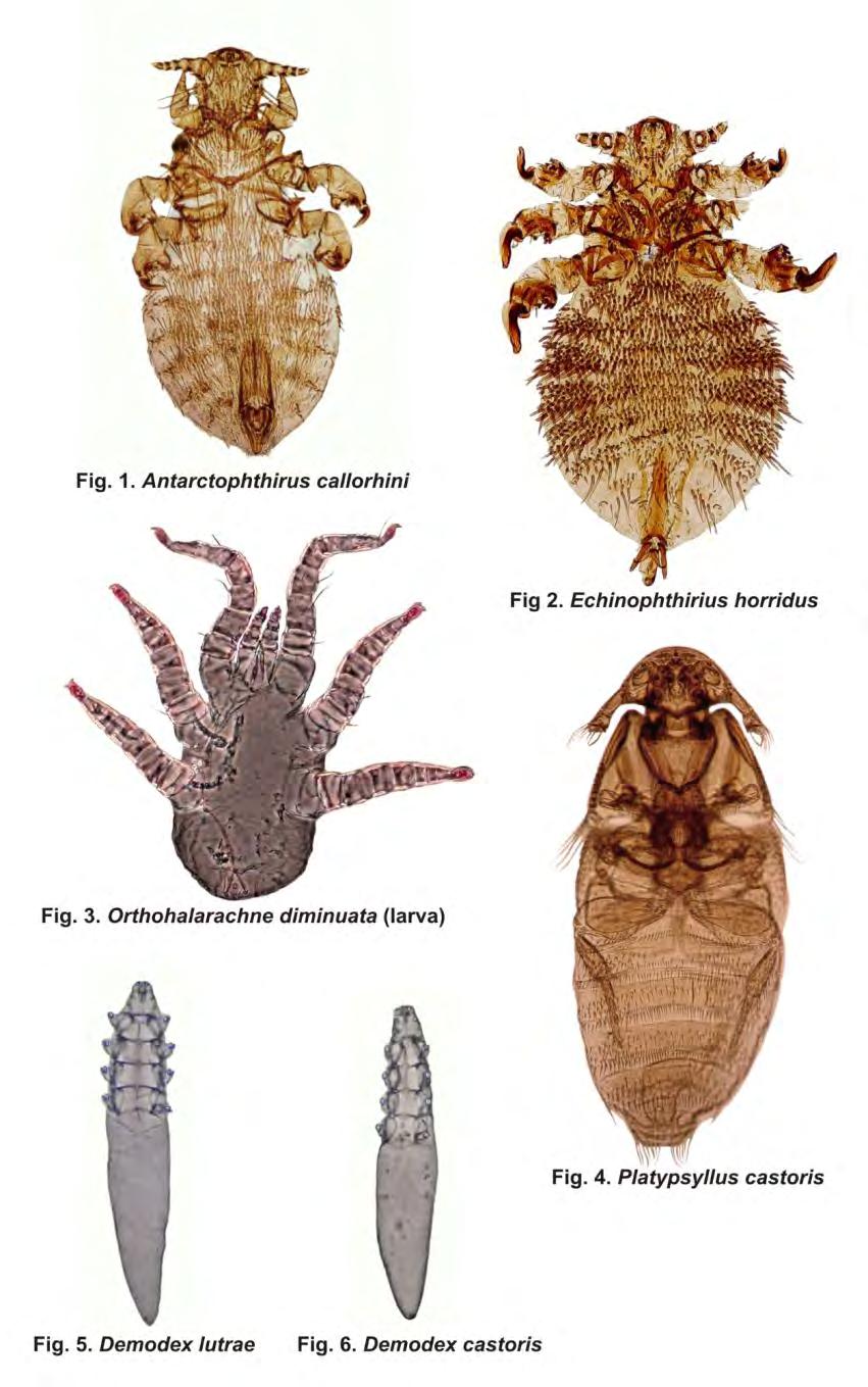
14 constantly stay in a direct contact with that environment. Various representatives of mites and insects exhibit here different adaptive strategies. They can obtain the required features allowing the functioning in aquatic conditions. They can also get adaptation to colonize other microhabitats within the host, moving to endoparasitism. Probably, a lot depends here on the life strategies of the host itself, which is important not only for the functioning of parasites in it, but also the mechanisms of their transmission to the subsequent hosts. And so, higher potential is undoubtedly created here by a semiaquatic lifestyle, which favors to gain parasites from both environments, but also increases the possibility of the survival of parasites from typically terrestrial groups. While a long-term contact with water typical, for example for marine mammals, is the limitation for insects and mites presence. Parasitic arthropods related to semiaquatic mammals Semiaquatic mammals are the animals partly connected to the aquatic environment, in which they usually prey. They stay on land during their resting or reproduction, where they are sometimes attacked by terrestrial parasites. However, this applies primarily to temporary parasites, for a short time related to the host, usually only in the phase of feeding. Stationary parasites had to develop the adaptations that allow them to function in both environments of the host, adjusting to this not only the phase of feeding, but also their entire life cycles. This is perfectly illustrated by parasitofauna communities of otters, beavers, muskrats, or coypus. For example, European otter Lutra lutra (Linnaeus, 1758) is a predatory mammal of the mustelids family. It is observed on the banks of rivers, ponds and lakes, and even salt waters is also sometimes noted on the shores of the seas (the Baltic Sea). It builds the burrows with an entrances under water surface, which is a feeding place for it (Mason and Macdonald 1986, Romanowski et al. 2011). Parasitofauna of the otter is an interesting combination of elements typical for the communities of aquatic vertebrates parasites, as well as characteristic for related to them terrestrial mammals, which specifically refers to arthropods (Rolbiecki and Izdebska 2014). Chewing louse Lutridia exilis (Nitzsch, 1861) (Jefferies et al. 1989, Haitlinger and Łupicki 2009), ticks Ixodes canisuga Johnston, 1848, I. hexagonus Leach, 1815, I. ricinus (Linnaeus, 1758) and mites Zachvatkinia lutrae Volgin, 1967 have been noted here so far (Haitlinger and Łupicki 2009, Christian 2012). And the fist in Lutrinae representative of Demodecidae Demodex lutrae Izdebska et Rolbiecki, 2014 (Fig. 5) has been described recently (Izdebska and Rolbiecki 2014). Only two species of this group D. melesinus Hirst, 1921 from badger Meles meles (Linnaeus, 1758) and D. erminae Hirst, 1919 from stoat Mustela erminea Linnaeus, 1758 have been described so far in other mustelids (Izdebska 2005), although they constitute parasitofauna common in various groups of mammals. However fur mites have not been noted in European otter, but this group representative has been described in North American river otter Lontra canadensis (Schreber, 1777). This is the species from Listrophoridae family, Lutracarus canadensis Fain et Yunker, 1980 (Fain and Yunker 1980), the group also noted in other mustelids. For example, Lynxacarus visoni Fain et Bochkov, 2002 has been described in American mink Neovison vison (Schreber, 1777) (Fain and Bochkov 2002), a mammal also related to aquatic environment. Other semiaquatic mammals are beavers. These are the rodents from Castorimorpha suborder, including, except the castorids (Castoridae), also American rodents kangaroo rats and mice Heteromyidae and pocket gophers Geomyidae. Castorids are currently represented only by Castor genus widely spread in Holarcitc, with two species Eurasian 14
15 beaver Castor fiber Linnaeus, 1758 and North American beaver Castor canadensis Kuhl, 1820 (Kuehn et al. 2000, Durka et al. 2005). Eurasian beaver is the largest rodent of Eurasia, which is in many regions extinct, or threatened with extinction and protected (Nolet and Rosell 1998, Bathbold et al. 2008). The works describing the rich acarofauna of beavers fur show that semiaquatic lifestyle here is not the limiting factor for parasitic arthropods. A typical parasitofauna is represented by fur mites of Schizocarpus genus (Listrophoridae) (Fain and Whitaker 1988, Dubinina et al. 1993), and as many as 44 species of them have been described (Bochkov 2012, Bochkov and Saveljev 2012, Bochkov et al. 2012). Perhaps the limited possibilities of these parasites transmission on other hosts contributed to speciation within the different microhabitats posed by a dense fur of the beavers. Schizocarpus spp. is also a predominant ectoparasite of North American beaver where 18 species have been described (Whitaker et al. 2009, Bochkov and Saveljev 2012). It is interesting, that these parasites are frequently and in large numbers present in the fur of beavers, even though the animals clean fur very intensively after leaving the water, and they use for this purpose a forked claw of second toe of the hind limb and teeth; in turn, to achieve water resistance, they impregnate it spreading the secretion of anal glands (Czech 2010). Except the rich fur acarofauna, also ticks I. apronophorus Schulze, 1924, I. hexagonus (Haitlinger 1991, Kadulski 1998) and parasitic beetle Platypsyllus castoris Ritsema, 1869 (Leiodidae) (Fig. 4) have been demonstrated in Eurasian beaver, which was in fact recorded in both species of Castor genus (Buchholz and Sikora 1984, Haitlinger 1991, Kadulski 1998, Peck 2006, Buchholtz et al. 2008, Moskovitz 2011, Arzanov et al. 2013, Pushkin 2014). The parasitic beetles pose some exception in this typically free living, the largest group of insects. Both imagines, and larval forms are noted in beaver. The copulation takes place on the host, and the fertilized female leaves the beaver fur and lay eggs in the lodges ground. The larvae are transferred to the skin of the host where they feed on the skin and the skin secretions, especially fatty substances. In case of a high abundance, these parasites can cause hard to heal damages in the beaver s skin. The larvae, after two moulting, leave the host and pupate in a chamber constructed in the mud or soil, within the lodges. The adults recolonize beaver fur, where they feed with exfoliating epidermis and secretions from damages caused by the larvae; they overwinter on the hosts. The specific adaptation are the legs of pairs II and III of these beetles, which have longitudinal grooves on the femurs where they can delve the tibias. This forms a prehensile organ allowing them to stay and move in the fur of the beaver (Wood 1964, Peck 2006). However P. castoris is a typical parasite of the beaver, it has also been noted in other mammal, i.e. Canadian otter L. canadensis (Schreber, 1777) (Belfiore 2006). It seems that skin mites should also be associated with the beaver, although from that ecological group, only Psororbia castori (Psorergatidae) has been noted so far in North American beaver (Kok et al. 1970, Giesen 1990). However, the first in castorids representative of Demodecidae, D. castoris (Fig. 6), has been fund recently, and it was demonstrated in head skin, in nose area of Eurasian beaver (Izdebska et al., in press). Undoubtedly, this is a region particularly predisposed to colonization by parasites, since it is especially protected when the beavers stay in water. During diving, ear and nasal canals are closed with skin folds, and the mouth is tightly closed due to the divided upper lip and exceptionally wide diastema. During swimming, nose, eyes and ears stay in one plane, which is held above water surface (Czech 2010). Another semiaquatic mammal is the muskrat Ondatra zibethicus (Linnaeus, 1776), a rodent from a large and diverse family of Cricetidae. It is an American species, introduced to 15
16 Europe in 1905 (Kowalski et al. 1981). Its dense fur is inhabited by numerous fur mites, including six species from Listrophoridae (Listrophorus americanus Radford, 1944, L. dozeri Radford, 1944, L. faini Dubinina, 1972, L. kingstownensis Fain et Hyland 1973, L. ondatrae Fain, Kok et Lukoschus, 1970, L. validus Banks, 1910) (Bochkov 2010, Whitaker 1982, Whitaker 1988, Prendergast and Jensen 2011), as well as from Myocoptidae (Myocoptes ondatrae Lukoschus et Rouwet, 1968) and Myobiidae (Radfordia zibethicalis (Radford, 1936)) (Whitacher 1982). Among other mites, Laelaps multispinosa (Banks, 1910) (Laelapidae) is regularly noted in muskrat (Prendergast and Jensen 2011). Similar ectoparasites composition is demonstrated in related to muskrat round-tailed muskrat Neofiber alleni True, 1884, in which three species from Listrophoridae two from Listrophorus and one from Prolistrophorus genus have been described (Bochkov 2010). In turn, the coypu Myocastor coypus (Molina, 1782) is also semiaquatic rodent with a dense fur, which is used (as the other mentioned rodents) as fur animal. However, it is a mammal of different origins, the South American representative of Myocastoridae family, introduced currently in various parts of the world. No specific Listrophotidae, or Chirodiscidae have been noted here, and its typical parasite is mite Myocastorobia myocastor (Fain, 1970) (Atopomelidae) (Whitacker 1982). In addition, the fur parasite is a specific of amblyceran chewing louse from Gyropidae family Pitrufquenia coypus Marelli, 1932 (Freeman 1946, Newson and Holmes 1968). Moreover, also ticks, e.g. I. ricinus, I. trianguliceps Birula, 1895, I. hexagonus, I. apronophorus (= I. arvicolae Warburton, 1926), Trombiculidae (Trombicula autumnalis (Shaw, 1790)), and even fleas (e.g. Ceratophyllus gallinae Schrank, 1809) (Newson and Holmes 1968), i.e. temporary parasites acquired in terrestrial region, have been found in the coypu in various regions of occurrence. Aquatic mammals arthropods Prolonged stay of the mammals in aquatic environment and related adaptations are undoubtedly the factors limiting the presence of these parasitic arthropods. Parasites of the insects and mites are, however, a typical parasitofauna of belonging to Carnivora pinnipeds, which, despite long periods of stay in water, also inhabit the terrestrial environment. In addition, they have a dense fur, constituting an adaptation to living in cold regions of the world, which allowed for the preservation of parasites associated with this habitat. And so, a typical parasitofauna of the pinnipeds are the sucking lice, which acquired an adaptation in the form of setae on the body modified in spines and scales, which facilitates the gas exchange in the aquatic environment (Castro et al. 2002, Mehlhorn et al. 2002, Izdebska and Rolbiecki 2010, Leonardi et al. 2012, Izdebska 2014). The group typical for pinnipeds are sucking lice from Echinophthiriidae, also observed in mustelids, with Echinophthirius horridus also noted in Poland (Durden and Musser 1994b, Kadulski 2001). However, various species of seals, sea lions and walruses are colonized by the representatives of Antarctophthirus, Lepidophthirus, or Proechinophthirus genera (Tab. 1, Figs. 1, 2) (Aznar et al. 2009, Thomson 1998, Leonardi and Palma 2013). The body of pinnipeds creates moreover a number of microhabitats to life for the skin and internal parasites. Two specific species of Demodecidae - D. phocidi Desch et al., 2003 from common seal Phoca vitulina Linnaeus, 1758, and D. zalophi Dailey et Nutting, 1980 from California sea lion Zalophus californianus (Lesson, 1828) have been described (Dailey and Nutting 1979, Desch et al. 2003). A typical acarofauna are also Halarachnidae (Mesostigmata) mites that adapted to the internal parasitism inhabiting the nasal ducts. The 16
17 representatives of Halarachne, or Orthohalarachne genera have been observed in pinnipeds (Tab. 2, Fig. 3) (Seawright 1964, Dunlap et al. 1976, Katz et al. 2012). In turn, parasitic Phthiraptera, or Acari were not found in aquatic mammals that do not have well-developed hairiness, like hippopotamus, sirens, or cetaceans. However, it seems possible to find in them skin or endoparasitic mites. A specific host group is represented here by marine mammals that spend their whole life in the aquatic environment - whales. However, no parasites from mites or insects have been found in them so far. In turn, typically aquatic arthropods may be noted in them - parasitic crustaceans, Amphipoda of Caprellidea suborder, Cyamidae family (so-called whale lice) and even copepods (Boxshall 2005, Lützen 2005). Summary The mammals which secondarily adopted to aquatic lifestyle typically retain at least in part a set of parasitic arthropods typical to related with them terrestrial mammals. This is probably conditioned by a long-term evolution of the parasite-host system, where the relationship of parasite and mammal was so strong that it stayed with the host regardless of its place of living, gradually yielding new adaptations. Thus, these are mainly species that, as closely related to the host, shared with it the common evolutionary pathway from typically terrestrial forms, through varying degrees of connection with the aquatic environment, to secondarily/typically aquatic. This is primarily related to the formation of various morphological, anatomical, and physiological modifications, concerning e.g. way of breathing, but also adaptation of reproduction and development to new living conditions. References Alonso-Farré J.M., Díaz D Silva J.I., Gestal C Naso-pharyngeal mites Halarachne halichoeri (Allman, 1847) in Grey seals stranded on the NW Spanish Atlantic Coast. Vet. Parasitol. 183: Aznar F.J., Leonardi M.S., Vera B.B., Vales D.G., Ameghino S., Raga J.A., Crespo E.A Population dynamics of Antarctophthirus microchir (Anoplura: Echinophthiriidae) in pups from South American sea lion, Otaria flavescens, in Northern Patagonia. Parasitology 136: Arzanov Yu.G., Valov G.V., Bakhtadze G.B Platypsyllus castoris Ritsema, 1869 (Coleoptera: Leiodidae) new species from Rostov Region (Russia). Cauc. Entomol. Bull. 9: Batbold J., Batsaikhan N., Shar S., Amori G., Hutterer R., Kryštufek B., Yigi N., Mitsain G., Palomo L.J Castor fiber. In: The IUCN red list of threatened species, ver Becker G.K., Robaldo R.B., Bianchini A., Colares E.P, Martinez P.E, Muelbert M.M., Brum J.G.W Lepidophithirus macrorhini (Anoplura: Echinophthiridae) in elephant seals (Mirounga leonina) from Elephant Island (South Shetlands - Antarctica). Arq. Inst. Biol. 67:
18 Belfiore N.M Observation of a beaver beetle (Platypsyllus castoris Ritsema) on a North American river otter (Lontra canadensis Schreber) (Carnivora: Mustelidae: Lutrinae) in Sacramento County, California (Coleoptera: Leiodidae: Platypsyllinae). Coleopt. Bull. 60: Bochkov A.V A review of mammal-associated Psoroptidia (Acariformes: Astigmata). Acarina 18: Bochkov A.V Schizocarpus saveljevi sp. nov. (Acariformes: Chirodiscidae) parasitizing the Eurasian beaver Castor fiber Linnaeus, 1758 (Rodentia: Castoridae) from Leningrad province. Russia Proc. Zool. Inst. RAS 316: Bochkov A.V., Labrzycka A., Skoracki M., Saveljev A.P Fur mites of the genus Schizocarpus Trouessart (Acari: Chirodiscidae) parasitizing the Eurasian beaver Castor fiber belorussicus Lavrov (Rodentia: Castoridae) in NE Poland (Suwałki). Zootaxa 3162: Bochkov A.V., Saveljev A.P Fur mites of the genus Schizocarpus Trouessart (Acari: Chirodiscidae) from the Eurasian beaver Castor fiber tuvinicus Lavrov (Rodentia: Castoridae) in the Azas River (Tuva Republic, Russia). Zootaxa 3410: Boxshall G Copepoda (copepods). In: Rhode K. (ed.), Marine parasitology. CABI Publishing, Wallingford, United Kingdom: Buchholz L., Sikora S Platypsyllus castoris Ritsema, 1869 (Coleoptera, Platypsyllidae) nowy dla fauny Polski przedstawiciel chrząszczy. Przegl. Zool. 28: Buchholz L., Czerwiński S., Komosiński K., Niewęgłowski H., Ruta R New records of the Platypsyllus castoris Ritsema, 1869 (Coleoptera: Leiodidae) from Poland. Wiad. Entomol. 27: Castro D.D.C., Romero M.D., Dreon M Ultrastructure of Proechinophthirus zumpti (Anoplura, Echinophthiriidae) by scanning electron microscopy. Mem. Inst. Oswaldo Cruz 97: Christian A Tick infestation (Ixodes) on the Eurasian Otter (Lutra lutra) a long-term study. Soil Org. 84: Czech A Bóbr budowniczy i inżynier. Fundacja Wspierania Inicjatyw ekologicznych, Kraków. Dailey M.D., Nutting W.B Demodex zalophi sp. nov. (Acari: Demodicidae) from Zalophus californianus, the California sea lion. Acarologia 21: Desch C.E., Dailey M.D., Tuomi P Description of a hair follicle mite (Acari: Demodecidae) parasitic in the earless seal familiy Phocidae (Mammalia: Carnivora) from the harbor seal Phoca vitulina Linnaeus, Int. J. Acarol. 29:
19 Domrow R Halarachne mirounge Ferris redescribed (Acarina: Laelaptidae). Pac. Insects 4: Dong W.G., Song S., Guo X.G., Jin D.C., Yang Q., Barker S.C., Shao R Fragmented mitochondrial genomes are present in both major clades of the blood-sucking lice (suborder Anoplura): evidence from two Hoplopleura rodent lice (family Hoplopleuridae). BMC Genomics 15: 751. Dubinina E.V., Bočkov A.V., Bodrovskaâ V.I Zamietki po sistematike kleŝej roda Schizocarpus (Acariformes: Chirodiscidae). Parazitologâ 27: Dunlap J.S., Piper R.C., Keyes M.C Lesions associated with Orthohalarachne attenuata (Halarachnidae) in the northern fur seal (Callorhinus ursinus). J. Wildl. Dis. 12: Durden L.A., Musser G.G. 1994a. The mammalian hosts of the sucking lice (Anoplura) of the world: a host-parasite list. Bull. Soc. Vector Ecol. 19: Durden L.A., Musser G.G. 1994b. The sucking lice (Insecta, Anoplura) of the world: a taxonomic checklist with records of mammalian hosts and geographical distributions. Bull. Am. Mus. Nat. Hist. 218: Durka W., Babik W., Ducroz J.F., Heidecke D., Rosell F., Samjaa R., Saveljev A.P., Stubbe A., Ulevičius A., Stubbe M Mitochondrial phylogeography of the Eurasian beaver Castor fiber L. Mol. Ecol. 14: Fain A., Bochkov A.V A new fur mite Lynxacarus (Lutracarus) visoni sp. nov. (Acari, Listrophoridae) from Mustela vison (Carnivore, Mustelidae) in North America. Acta Parasitol. 47: Fain A., Yunker C.E Lutracarus canadensis, n. g., n. sp. (Acari: Listrophoridae) from the river otter, Lutra canadensis. J. Med. Entomol. 17: Fain A., Whitaker J.O Mites of the genus Schizocarpus Trouessart, 1896 (Acari, Chirodiscidae) from Alaska and Indiana, USA. Acarologia 29: Fay F.H., Furman D.P Nasal mites (Acari: Halarachnidae) in the spotted seal, Phoca largha Pallas, and other pinnipeds of Alaskan waters. J. Wild. Dis. 18: Freeman R.B Pitrufquenia coypus Marelli (Mallophaga, Gyropidae), an ectoparasite on Myocastor coypus Mol. Entomol. Mon. Mag. 82: Furman D.P., Dailey M.D The genus Halarachne (Acari: Halarachnidae), with the description of a new species from the Hawaiian monk seal. J. Med. Entomol. 17: Giesen K.M.T A review of the parasitic mite family Psorergatidae (Cheyletoidea: Prostigmata: Acari) with hypotheses on the phylogenetic relationships of species and species groups. Zool. Verhandel. 259:
20 Haitlinger R Arthropods occurring on European beaver (Castor fiber L.) in Poland. Wiad. Parazytol. 37: Haitlinger R., Łupicki D First records of arthropods (Phthiraptera: Trichodectidae, Acari: Ixodidae) from Lutra lutra (Linnaeus, 1758) (Carnivora: Mustelidae) in Poland. Zesz. Nauk. UP Wroc., Biol. Hod. Zwierz. 59: Izdebska J.N Demodecid mites (Acari, Actinedida) in carnivorous mammals (Mammalia, Carnivora) in Poland. In: Buczek A., Błaszak C. (eds.), Arthropods. A variety of forms and interactions. Koliber, Lublin: Izdebska J.N Wszy? Poznaj i pokonaj problem. Wyd. Naukowe PWN SA, Warszawa. Izdebska J.N., Rolbiecki L Parasitic arthropods as the cause of parasitoses in aquatic animals. In: Buczek A., Błaszak C. (eds.), Arthropods. Ecological and pathological aspects of parasite-host relationships. Akapit, Lublin: Izdebska J.N., Rolbiecki L Demodex lutrae n. sp. (Acari) in European otter Lutra lutra (Carnivora: Mustelidae) with data from other demodecid mites in carnivores. J. Parasitol. 100: Jefferies D.J., Hanson H.M., Lyal C.H.C A further record of Lutridia exilis (Nitzsch) (Phthiraptera, Trichodectidae) in Britain, with notes on the presence and absence of lice on otters. Entomol. Mon. Mag. 125: Kadulski S Pasożyty zewnętrzne bobra Castor fiber L. z Popielna. Wiad. Parazytol. 44: Kadulski S Echinophthirius horridus (Olfers, 1816) (Anoplura) rzadki pasożyt fok. Wiad. Parazytol. 47: Katz H., Morgades D., Castro-Ramos M Pathological and parasitological findings in south American fur seal pups (Arctocephalus australis) in Uruguay. ISRN Zoology: 1-7. Kim K.C., Life stages and population of Proechinophthirus zumpti (Anoplura: Echinophthiriidae), from the cape fur of seal (Arctocephalus pusillus). J. Med. Entomol. 16: Kim K.C., Emerson K.C Latagophthirus rauschi, new genus and new species (Anoplura: Echinophthiriidae) from the river otter (Carnivora: Mustelidae). J. Med. Entomol. 11: Kim K.C., Haas V.L., Keyes M.C Populations, microhabitat preferences and effects of infestation of two species of Orthohalarachne (Halarachnidae: Acarina) in the northern fur seal. J. Wildl. Dis. 16:
21 Kok N.J.J., Lukoschus F.S., Clulow F.V Psorobia castoris spec. nov. (Acarina, Psorergatidae), a new itch mite from the beaver, Castor canadensis. Can. J. Zool. 48: Kowalski K., Pucek Z., Ruprecht A.L Rodents - Rodentia. In: Pucek Z. (ed.), Keys to vertebrates of Poland. Mammals. PWN, Warszawa: Kuehn R., Schwab G., Schroeder W., Rottmann O Differentiation of Castor fiber and Castor canadensis by noninvasive molecular methods. ZOO Biology 19: Leonardi M.S, Crespo E.A., Raga J.A., Fernández M Scanning electron microscopy of Antarctophthirus microchir (Phthiraptera: Anoplura: Echinophthiriidae): studying morphological adaptations to aquatic life. Micron 43: Leonardi M.S., Palma R.L Review of the systematics, biology and ecology of lice from pinnipeds and river otters (Insecta: Phthiraptera: Anoplura: Echinophthiriidae). Zootaxa 3630: Leonardi M.S., Poljak S., Carlini P., Galliari J., Bobinac M., Santos M., Márquez M.E., Negrete J Antarctophthirus carlinii (Anoplura: Echinophthiriidae), a new species from the Weddell seal Leptonychotes weddelli. Parasitol. Res. 113: Lützen J Amphipoda (amphipods). In: Rhode K. (ed.), Marine parasitology. CABI Publishing, Wallingford, United Kingdom: Martino P.E., Radman N., Parrado E., Bautista E., Cisterna C., Silvestrini M.P., Corba S Note on the occurrence of parasites of the wild nutria (Myocastor coypus, Molina, 1782). Helminthologia 49: Mason C.F., Macdonald S.M Otters: ecology and conservation. Cambridge University Press, New York. Mehlhorn B., Mehlhorn H., Plötz J Light and scanning electron microscopical study on Antarctophthirus ogmorhini lice from the Antarctic seal Leptonychotes weddellii. Parasitol. Res. 88: Scarabino F., Conde D. (eds.), Bases para la conservación y el manejo de la costa uruguaya. Vida Silvestre, Uruguay: Moskovitz D First record of the ectoparasitic beaver beetle, Platypsyllus castoris Ritsema (Coleoptera: Leiodidae: Platypsyllinae), in New Jersey, USA. Colleopt. Bull. 65: Murray M.D Ecology of the louse Lepidophthirus macrorhini Enderlein 1904 on the elephant seal Mirounga leonina (L.). Nature 182: Newson R.M., Holmes R.G Some ectoparasites of the coypu (Myocastor coypus) in Estern England. J. Anim. Ecol. 37:
22 Nolet B.A., Rosell F Comeback of the beaver Castor fiber. An overview of old and new conservation problems. Biol. Cons. 83: Peck S.B Distribution and biology of the ectoparasitic beaver beetle Platypsyllus castoris Ritsema in North America (Coleoptera: Leiodidae: Platypsyllinae). Insecta Mundi 20: Polechla P.J New host records of ticks (Acarina: Ixodidae) parasitizing the river otter (Lutra canadensis). IUCN Otter Spec. Group Bull. 13: Prendergast J.A., Jensen W.E Consequences of parasitic mite infestation on muskrat (Ondatra zibethicus). West. N. Am. Naturalist. 71: Pushkin S.V A new record of the parasitic beaver beetle (Platypsyllus castoris) (Coleoptera: Leiodidae) from Stavropol Territory (Russia). Entomol. Appl. Sci. Lett. 1: 1-3. Rolbiecki L., Izdebska J.N New data on the parasites of the Eurasian otter (Lutra lutra). Oceanol. Hydrobiol. St. 43: 1-6. Romanowski J., Orłowska L., Zając T The protection of the otter Lutra lutra in Poland. National Management Strategy for otter. SGGW, Warszawa. Seawright A.A Pulmonary acariasis in a tasmanian fur seal. J. Comp. Path. 74: Thompson P.M., Corpe H.M., Reid R.J Prevalence and intensity of the ectoparasite Echinophthirus horridus on harbour seals (Phoca vitulina): effects of host age and interannual variability in host food availability. Parasitology 117: Whitaker J.O Ectoparasites of Mammals of Indiana. Monograph 4. Indiana Academy of Science, Indianapolis, Indiana. Whitaker J.O Listrophorid mites and other ectoparasites of muskrats, Ondatra zibethicus, from the Chena River, Alaska. Murrelet 69: Whitaker J.O., Ruckdeschel C., Bochkov A.V Species of the genus Schizocarpus Trouessart, 1896 (Acari: Chirodiscidae) from Florida and Georgia beavers. Biol. Sci. 72: Wood D.M Studies on the beetles Leptinillus validus (Horn) and Platypsyllus castoris Ritsema (Coleoptera: Leptinidae) from beaver. Proc. Ent. Soc. 95:
23 Table 1. Checklist of sucking lice from the family Echinophthiriidae of aquatic mammals (based on Kim and Emerson 1974, Kim 1979, Durden and Musser 1994b, Becker et al. 2000, Leonardi and Palma 2013, Leonardi et al. 2014) Echinophthiriidae Antarctophthirus callorhini (Osborn, 1899) Antarctophthirus carlinii Leonardi, Poljak, Carlini, Galliari, Bobinac, Santos, Márquez et Negrete, 2014 Antarctophthirus lobodonis Enderlein, 1909 Antarctophthirus mawsoni Harrison, 1937 Antarctophthirus microchir (Trouessart et Neumann, 1888) Antarctophthirus ogmorhini Enderlein, 1906 Antarctophthirus trichechi (Bohemann, 1865) Echinophthirius horridus (von Olfers, 1816) Host Northern fur seal Callorhinus ursinus (Linnaeus, 1758) Weddell seal Leptonychotes weddelli (Lesson, 1826) Weddell seal Leptonychotes weddelli (Lesson, 1826) Crabeater seal Lobodon carcinophagus Hombron et Jacquinot, 1842 Ross seal Ommatophoca rossii Gray, 1844 South American fur seal Arctocephalus australis (Zimmermann, 1783) Steller sea lion Eumetopias jubatus (Schreber, 1776) Australian sea lion Neophoca cirenea (Peron, 1866) Southern sea lion Otaria bryonia (= O. flavescens) (de Blainville, 1820) New Zeland sea lion Phocarctos hookeri (Gray, 1844) California sea lion Zalophus californicus (Lesson, 1828) Leopard seal Hydrurga leptonyx (de Blainville, 1820) Weddell seal Leptonychotes weddelli (Lesson, 1826) Walrus Odobenus rosmarus (Linnaeus, 1758) Hooded seal Cystophora crystata (Erxleben, 1777) Bearded seal Erignathus barbatus (Erxleben, 1777) Harp seal Phoca groenlandica Erxleben, 1777 Ringed seal Phoca hispida Schreberk, 1775 Baikal seal Phoca sibirica Gmelin, 1788 Common seal Phoca vitulina Linnaeus, 1758 Grey seal Halichoerus grypus (Fabricius, 1791) 23
24 Latagophthirus rauschi Kim et Emerson, 1974 Lepidophthirus macrorhini Enderlein, 1904 Lepidophthirus piriformis Blagoveshtchensky, 1966 Proechinophthirus fluctus Ferris, 1916 Proechinophthirus zumpti Werneck, 1955 North American river otter Lontra canadensis Schreber, 1777 Southern elephant seal Mirounga leonina Linnaeus, 1758 Mediterranean monk seal Monachus monchus (Hermann, 1779) Steller sea lion Eumetopias jubatus (Schreber, 1776) Northern fur seal Callorhinus ursinus (Linnaeus, 1758) Cape fur seal Arctocephalus pusillus (Schreber, 1775) South American fur seal Arctocephalus australis (Zimmermann, 1783) Southern sea lion Otaria bryonia (= O. flavescens) (de Blainville, 1820) Table 2. Checklist of mites from the family Halarachnidae of aquatic mammals (based on Domrow 1962, Furman and Dailey 1980, Fay and Furman 1982, Alonso et al. 2012) Halarachnidae Host Steller sea lion Eumetopias jubatus (Schreber, 1776) Grey seal Halichoerus grypus (Fabricius, 1791) Halarachne halichoeri (Allman, 1847) Spotted seal Phoca largha Pallas, 1811 Common seal Phoca vitulina Linnaeus, 1758 Hawaiian monk seal Halarachne laysanae Furman et Dailey, 1980 Monachus schauinslandi Matschie, 1905 Northern elephant seal Halarachne miroungae Ferris, 1925 Mirounga angustirostris Gill, 1866 South American fur seal Arctocephalus australis (Zimmermann, 1783) Cape fur seal Orthohalarachne attenuata (Banks, 1910) Arctocephalus pusillus (Schreber, 1775) Northern fur seal Callorhinus ursinus (Linnaeus, 1758) South American fur seal Arctocephalus australis (Zimmermann, 1783) Orthohalarachne diminuata (Doetschman, Northern fur seal 1944) Callorhinus ursinus (Linnaeus, 1758) Steller sea lion Eumetopias jubatus (Schreber, 1776) 24
25 25
26 26
27 Threats to human health posed by ticks (Ixodida) in different climate zones Alicja Buczek, Katarzyna Bartosik, Zbigniew Zając, Weronika Buczek Chair and Department of Biology and Parasitology, Medical University, Radziwiłłowska 11 St., Lublin, Poland Department of Basic Nursing and Medical Teaching, Medical University, Staszica 3 St., Lublin, Poland Abstract The study presents tick species that can attack humans on different continents. It is based on own findings as well as results obtained by other Polish and foreign authors in their investigations of the fauna of human parasitic ticks conducted in , including observations of the immediate effects of tick activity on the host. Introduction The fauna of ticks (Ixodida) comprises species that are adapted to different habitats from different climate zones. Hence, these arthropods occur in the living habitats of all terrestrial vertebrates. Since the active tick stages (larvae, nymphs, and adult stages) are obligate blood-ingesting parasites and each stage parasitizes different animals, the host range for many species of these arthropods is extremely wide. Among tick species, there are some with high host specificity and others with low host specificity. Given their three-host or two-host developmental cycle and their parasitism of different hosts, ticks from the latter group play the greatest role in the spread of tickborne pathogens. Humans can also be a host to some species from both physiological groups. The present study, based on literature data from , is focused on tick species occurring on different continents and posing a serious threat to human health (Tab. 1). We have focused on continents mostly visited by European tourists. Ticks found on human in different parts of the globe The tick fauna is richer in tropical and subtropical zones than that occurring in the temperate climate zone. In a hot climate, the number of tick species infesting humans is usually higher. In South Asia, threat to humans is posed by representatives of six genera of hard ticks, i.e. Amblyomma, Dermacentor, Hyalomma, Rhipicephalus, and Haemaphysalis from the family Amblyommidae and Ixodes from the family Ixodidae and three genera of soft ticks, i.e. Argas, Carios, and Ornithodoros (Yu et al. 2015). For example, out of the 30,461 ticks collected in Sri Lanka (South Asia) in , 75 specimens were removed from human skin, 25,566 from domestic animals, and 1385 from 27
28 wild-living animals. Man was infested by 12 tick species. Dermacentor auratus (37.3%) and Amblyomma testudinarium (26.6%) were the most common ticks infesting humans, while other species were found to attach to human skin less frequently. These were represented by Hyalomma isaaci (9.3%), Rhipicephalus haemaphysaloides (5.3%), Rh. sanguineus (4%), Am. integrum (4%), Am. clypeolatum (4%), Rh. boophilus, and I. petauristae (2.6% each), as well as Hae. intermedia, Hae turturis, and Hae. cuspidata (1.3% each) (Liyanaarachchi et al. 2015). Many tick species have been found on humans in Turkey, the western part of which lies within the borders of Europe and the eastern part belongs to Asia. In the Tokat Province situated in the mid Black Sea region of Anatolia, samples of human-infesting ticks comprised 24 species from 5 genera, i.e. Hyalomma, Rhipicephalus, Haemaphysalis, Dermacentor, and Ixodes. The most numerous group was constituted by representatives of Hyalomma (Hy. aegyptium, Hy. anatolicum, Hy. dromedarii, Hy. detritum, Hy. marginatum, Hy. turanicum, and Hy. isaaci), Haemaphysalis (Hae. sulcata, Hae. concinna, Hae. inremis, Hae. erinacei, Hae. parva, and Hae. punctata), and Rhipicephalus (Rh. bursa, Rh. sanguineus, Rh. turanicus, Rh.(B.) annulatus, Rh. sp.) (Bursali et al. 2010). In the Aegean Region (western Turkey), species from the family Argasidae parasitizing birds and humans were found, i.e. Argas (A.) reflexus, Argas (P.) persicus (Keskin et al after Bursali et al. 2012), while Argas (C.) vespertilionis, a species ingesting bat and human blood was found in the Eastern Anatolia Region (eastern Turkey) (Bursali et al. 2012). In Africa, 20 tick species belonging to six genera were found, with the most common Amblyomma hebraeum, Haemaphysalis leachi, Hyalomma marginatum rufipes, Hyalomma turanicum, Rhipicephalus appendiculatus, Rhipicephalus gertrudae, Rhipicephalus simus, and Rhipicephalus maculatus (Horak et al. 2002). In turn, three of the 28 species reported in South America are regarded as frequent human parasites in certain regions of the continent. These include Amblyomma neumanni, a frequent parasite in Argentina, Amblyomma triste in Uruguay, and Amblyomma parvum in Argentina and Brazil (Guglielmone et al. 2006). The list of species capable of attachment to human skin occurring on this continent is obviously much longer. It mainly includes ticks from the genus Amblyomma, i.e. Am. brasiliense, Am. incisum, Am. tigrinum, Am. triste, Am. aureolatum, Am. cajennense, Am. Coelebs, Am. oblongoguttatum, Am. ovale, Am. parvum, and Am. scalpturatum. In North America, the greatest medical importance is attributed to the Ixodes scapularis, Ixodes pacificus, Amblyomma americanum, Amblyomma maculatum, Dermacentor variabilis, Dermacentor andersoni, and Rhipicephalus sanguineus species (Merten and Durden 2000, Childs and Paddock 2003, Stromdahl et al. 2011). Over the last 15 years, ticks from seven genera and three families have been found to infest humans in Europe. These included Argas and Carios from the family Argasidae, Ixodes from the family Ixodidae, and Dermacentor, Hyalomma, Haemaphysalis, and Rhipicephalus from the family Amblyommidae. Given their widespread occurrence, some of them play a substantial role in triggering local and systemic reactions in humans and, hence, pose a serious threat to public health. The most common tick species parasitizing humans in Europe is Ixodes ricinus, commonly occurring in forests and in wooded suburban and urban areas. This tick predominates in the collections from the western, central, and eastern parts of the continent. For instance, among the 10,050 ticks collected on different animals (cattle, sheep, goats, dogs, rodents), plants, and humans in Bosnia and Herzegovina in 2004, as many as 28
29 7,085 specimens, i.e. 70.5%, represented the I. ricinus species (Omeragic 2011). 23 ticks (0.2% of all specimens) were removed from human skin. The author identified 19 specimens parasitizing humans as I. ricinus and only 4 specimens as Rhipicephalus sanguineus. Similarly, in Romania, the collection of 308 ticks removed from patients skin over three months in 2010 comprised as many as 296 specimens identified as I. ricinus. A considerably lower number, i.e. 10 specimens, represented the Dermacentor marginatus species, and only 1 Haemaphysalis concinna specimen and 1 Haemaphysalis punctata tick were found (Briciu et al. 2011). In Poland, the I. ricinus species infests humans and animals most frequently; hence, it dominates in tick collections from these hosts (Michalik et al. 2003, 2005, Siński et al. 2006, Siuda et al. 2009, Bartosik and Buczek 2013). It is assumed that I. ricinus has a mosaic distribution across Poland. Noteworthy is the fact that in recent years, there have been reports of humans parasitized by ticks that have previously been found exclusively on animals, e.g. Ixodes vespertilionis (Piksa et al. 2013). Ticks have a direct, harmful effect on their hosts by triggering local and systemic reactions (Beaudouin et al. 1997, Moneret-Vautrin et al. 1998, Buczek 2000, Bartosik et al. 2011a). The parasitism effects are induced by the bioactive components of tick saliva, which are injected in the various stages of feeding (Buczek and Bartosik 2006). The range of skin lesions produced by tick parasitism can be varied. It depends on the species and the developmental stage of the feeding tick (Buczek et al. 2000, Szabó et al. 2006) and on human response to the tick saliva components, which is related to the physiological state and genetic traits of the host. The I. ricinus-induced local reactions observed in our investigations included erythema in 66.8% of the patients, pruritus in 50%, pain in 15.2%, formication in 11.4%, and burning in 9.7%. The systemic symptom reported most frequently by patients from the Lublin Province was headache (10.8%) and, less frequently, lymphadenitis (5.9%), fever (5.4%), and arthralgia (4.3%) (Bartosik et al. 2011). Tick paralysis can be an immediate effect of tick infestation on humans (Gordon and Giza 2004, Gürbüz et al. 2010, Doğan et al. 2012). Symptoms associated with introduction of the neurotoxin into the human organism begin with malaise, fatigue, severe headaches, tingling sensation in extremities (lips and face) and projectile vomiting (Mans et al, 2004). In case of tick paralysis maximal neurologic impairment occurs within hours to days after the onset of symptoms and may lead to death in case of respiratory muscles involvement (Kunze and Gothe 1971). Two tick species, Dermacentor andersoni and D. variabilis, are most often responsible for tick paralysis in humans (Mans et al. 2004). The occurrence of ticks capable of infesting humans prompts a necessity of undertaking integrated action by specialists in various disciplines in order to reduce tick populations and develop effective methods for anti-tick prophylaxis. 29
30 Table 1. Examples of ticks parasitizing humans based on documented research conducted on different continents in Continent Species References Ixodes ricinus Sanogo et al. 2003, Fernandez-Soto et al. 2006, Bartosik et al. 2011, Ixodes vespertilionis Piksa et al Ixodes persulcatus Eremeeva et al Argas reflexus Śpiewak et al. 2006, Buczek et al Argas vespertilionis Bursali et al Dermacentor marginatus Fernandez-Soto et al. 2003, 2006, Vatansever et al. 2008, Földvári et al. 2013, Europe Fernandez-Soto et al. 2006, Dermacentor reticulatus Bartosik et al. 2011, Földvári et al Hyalomma marginatum Fernández-Soto et al. 2003, Africa South America Haemaphysalis punctate Fernández-Soto et al. 2003, Orkun et al Rhipicephalus bursa Fernández-Soto et al. 2003, 2006, Orkun et al Rhipicephalus turanicus Fernández-Soto et al. 2003, Orkun et al Rhipicephalus sanguineus Omeragic 2011, Orkun et al Amblyomma hebraeum Horak et al Amblyomma marmoreum Horak et al Amblyomma variegatum Jensenius et al Rhipicephalus (Boophilus) decoloratus Portillo et al Haemaphysalis leachi De Matos et al Hyalomma marginatum rufipes Mediannikov et al Hyalomma truncatum Mediannikov et al Ixodes rubicundus Horak et al Rhipicephalus appendiculatus Horak et al Rhipicephalus gertrudae De Matos et al Rhipicephalus glabroscutatum Walker et al Rhipicephalus maculatus Horak et al Rhipicephalus simus Horak et al Ambyomma brasiliense Szabó et al Ambyomma incisum Szabó et al
31 Ambyomma tigrinum Venzal et al. 2003, 2006 Ambyomma triste Venzal et al. 2003, 2006 Amblyomma aureolatum Guglielmone et al Amblyomma cajennense Guglielmone et al Amblyomma coelebs Guglielmone et al Amblyomma oblongoguttatum Amblyomma ovale Guglielmone et al Guglielmone et al. 2006, Szabó et al Amblyomma parvum Guglielmone et al Amblyomma scalpturatum Guglielmone et al North America Asia Rhipicephalus sanguineus Venzal et al. 2003, 2006 Amblyomma americanum Childs and Paddock 2003 Dermacentor andersoni Merten and Durden 2000 Dermacentor variabilis Stromdahl et al. 2011, Sheele et al Ixodes scapularis Merten and Durden 2000, Sheele et al. 2013, 2014; Ixodes pacificus Hyalomma aegiptum Bursali et al Hyalomma anatolicum Hyalomma dromedarii Hyalomma excavatum Karaer et al. 2013, Orkun et al Hyalomma marginatum Gürbüz et al, 2010 Rhipicephalus bursa Rhipicephalus sanguneus Rhipicephalus turanicus Rhipicephalus (Boophilus) annulatus Haemaphysalis concinna Haemaphysalis inermis Haemaphysalis parva Orkun et al Haemaphysalis punctata Dermacentor daghestanicus Dermacentor niveus Dermacentor marginatus Dermacentor auratus Liyanaarachchi et al Amblyomma testudinarium Ixodes aguri Ixodes redikorzevi Ixodes ricinus Ixodes persulcatus Eremeeva et al
32 Ixodes nipponensis Ko et al References 1. Bartosik K., Buczek A Determination of the parameters of the parasitic stage in Ixodes ricinus females. Ann Agric Environ Med. 20: Bartosik K., Sitarz M., Szymańska J., Buczek A. 2011a. Tick bites on humans in the agricultural and recreational areas in south-eastern Poland. Ann Agric Environ Med. 18: Beaudouin E., Kanny G., Guerin B., Guerin L., Plenat F., Moneret-Vautrin D. A Unusual manifestations of hypersensitivity after a tick bite: report of two cases. Ann Allergy Asthma Immunol. 79: Briciu V. T., Titilincu A., Ţăţulescu D. F., Cârstina D., Lefkaditis M., & Mihalca A. D First survey on hard ticks (Ixodidae) collected from humans in Romania: possible risks for tick-borne diseases. Exp Appl Acarol. 54(2): Buczek A Interakcje między stawonogami, patogenami i żywicielami. W: Buczek A., Błaszak Cz. (red.). Stawonogi pasożytnicze i alergogenne. Wydawnictwo KGM, Lublin Buczek A., Bartosik K Tick-host interactions. Przegl Epidemiol 60(S1). V Ogólnokrajowa Konferencja Naukowo-Szkoleniowa nt, Neuroinfekcji. Białystok IV s Buczek A., Bartosik K., Szymańska J., Buczek S Obrzeżek gołębi Argas reflexus (Fabr.) (Ixodida: Argasidae) w południowo-zachodniej Polsce cechy biologiczne i objawy kliniczne. Zdr Publ. 121(4): Buczek A., Czerny K., Łańcut M., Buczek L., Kuśmierz A., Olszewski K Ultrastructural examination of rabbit skin afterfeeding of females Ixodes ricinus (L.) (Acari: Ixodida:Ixodidae). Acta Parasitol. 45: Bursali A., Keskin A., Tekin S A review of the ticks (Acari: Ixodida) of Turkey: species diversity, hosts and geographical distribution. Exp Appl Acarol. 57(1): Bursali A., Tekin S., Orhan M., Keskin A., Ozkan M Ixodid ticks (Acari: Ixodidae) infesting humans in Tokat Province of Turkey: species diversity and seasonal activity. J Vector Ecol. 35(1): Childs J. E., Paddock C. D The Ascendancy of Amblyomma americanum as a Vector of Pathogens Affecting Humans in The United States. Annu Rev Entomol. 48(1): De Matos C., Sitoe C., Neves L., Bryson N. R., Horak I. G Ixodid ticks on dogs belonging to people in rural communities and villages in Maputo Province, Mozambique. Onderstepoort J Vet Res. 75(2): Doğan M1, Devge C, Tanrıöver O, Pata YS, Sönmezoğlu M Facial nerve paralysis due to intra-aural Hyalomma tick infestation. Turkiye Parazitol Derg. 36(4): Eremeeva M. E., Oliveira A., Moriarity J., Robinson J. B., Tokarevich N. K., Antyukova L. P. & Dasch G. A Detection and identification of bacterial agents in Ixodes persulcatus Schulze ticks from the north western region of Russia. Vector Borne Zoonotic Dis. 7(3): Földvári G, Rigó K, Lakos A Transmission of Rickettsia slovaca and Rickettsia raoultii by male Dermacentor marginatus and Dermacentor reticulatus ticks to humans. Diagn Microbiol Infect Dis. 76(3):
33 16. Fernández-Soto P., Encinas Grandes A., Pérez Sánchez R Rickettsia aeschlimannii in Spain: molecular evidence in Hyalomma marginatum and five other tick species that feed on humans. Emerg Infect Dis. 9(7): Fernández-Soto P, Pérez-Sánchez R, Encinas-Grandes A, Alamo Sanz R Rickettsia slovaca in Dermacentor ticks found on humans in Spain. Eur J Clin Microbiol Infect Dis. 25(2): Gordon B.M., Giza C.C Tick paralysis presenting in an urban environment. Pediatr Neurol. 30(2): Guglielmone A. A., Beati L., Barros-Battesti D. M., Labruna M. B., Nava S., Venzal J. M., Estrada-Peña A Ticks (Ixodidae) on humans in South America. Exp Appl Acarol. 40(2): Gürbüz M.K., Erdoğan M., Doğan N., Birdane L., Cingi C., Cingi E Case report: isolated facial paralysis with a tick. Turkiye Parazitol Derg. 34(1): Horak I. G., Fourie L. J., Heyne H., Walker J. B., Needham G. R Ixodid ticks feeding on humans in South Africa: with notes on preferred hosts, geographic distribution, seasonal occurrence and transmission of pathogens. Exp Appl Acarol. 27(1-2): Horak I. G., Mckay I. J., Heyne H., Spickett A. M Hosts, seasonality and geographic distribution of the South African tortoise tick, Amblyomma marmoreum. Onderstepoort J Vet Res. 73(1): Jensenius M., Fournier P. E., Vene S., Hoel T., Hasle G., Henriksen A. Z. & Myrvang B African tick bite fever in travelers to rural sub-equatorial Africa. Clin Infect Dis. 36(11): Keskin A., Bursali A., Tekin S Sivas Bolgesinde Insanlar Uzerinde Parazitlenen Kenelerin (Acari, Ixodidae) Sistematik Yonden Incelenmesi. XX. National Congress of Biology. Denizli. 25. Ko J. H., Cho D. Y., Chung B. S., Kim S. I Two human cases of tick bite caused by Ixodes nipponensis. Korean J Parasitol. 40(4): Kunze K., Gothe R Tick paralysis in chickens caused by Argas (Persicargas) persicus- larvae. 3. Neurophysiological investigations. Z Parasitenkd. 36(3): Liyanaarachchi D. R., Rajakaruna R. S., Dikkumbura A. W., Rajapakse R. P. V. J Ticks infesting wild and domestic animals and humans of Sri Lanka with new host records. Acta Trop. 142: Mans B.J., Gothe R., Neitz A.W Biochemical perspectives on paralysis and other forms of toxicoses caused by ticks. Parasitology 129:S Mayne P., Song S., Shao R., Burke J., Wang Y., Roberts T Evidence for Ixodes holocyclus (Acarina: Ixodidae) as a vector for human lyme Borreliosis infection in Australia. J Insect Sci. 14: Mediannikov O., Fenollar F., Socolovschi C., Diatta G., Bassene H., Molez J. F. & Raoult D Coxiella burnetii in humans and ticks in rural Senegal. PLoS Negl Trop Dis. 4(4):e Merten H. A., Durden L. A A state-by-state survey of ticks recorded from humans in the United States. J Vector Ecol. 25(1): Michalik J., Hofman T., Buczek A., Skoracki M., Sikora B Borrelia burgdorferi s.l. in Ixodes ricinus (Acari:Ixodidae) ticks collected from vegetation and small rodents in recreational areas of the city of Poznań. J Med Entomol. 40:
34 33. Michalik J., Skotarczak B., Skoracki M., Wodecka B., Sikora B., Hofman T., Rymaszewska A., Sawczuk M Borrelia burgdorferi sensu stricto in yellownecked mice and feeding Ixodes ricinus ticks in a forest habitat of west central Poland. J Med Entomol. 42: Moneret-Vautrin D. A., Beaudouin E., Kanny G., Guerin L., Roche J. F Anaphylactic shock caused by ticks (Ixodes ricinus). J Allergy Clin Immunol. 101: Omeragic J Ixodid ticks in Bosnia and Herzegovina. Exp Appl Acarol. 53(3): Orkun Ö., Karaer Z., Çakmak A., Nalbantoğlu S Identification of tick-borne pathogens in ticks feeding on humans in Turkey. PLoS Negl Trop Dis. 8(8):e Piksa K., Nowak-Chmura M., Siuda K First case of human infestation by the tick Ixodes vespertilionis (Acari: Ixodidae). Int J Acarol. 39(1): Portillo A., Pérez-Martínez L., Santibáñez S., Blanco J. R., Ibarra V., Oteo J. A Detection of Rickettsia africae in Rhipicephalus (Boophilus) decoloratus ticks from the Republic of Botswana, South Africa. Am J Tropl Med Hyg. 77(2): Sanogo Y. O., Zeaiter Z., Caruso G., Merola F., Shpynov S., Brouqui P., Raoult D Bartonella henselae in Ixodes ricinus ticks (Acari: Ixodida) removed from humans, Belluno province, Italy. Emerg Infect Dis. 9(3): Sheele J.M., Byers P.A., Sonenshine D.E Initial assessment of the ability of ivermectin to kill Ixodes scapularis and Dermacentor variabilis ticks feeding on humans. Wilderness Environ Med. 24(1): Sheele J.M., Ford L.R., Tse A., Chidester B., Byers P.A., Sonenshine D.E The use of ivermectin to kill Ixodes scapularis ticks feeding on humans. Wilderness Environ Med. 25(1): Siński E., Bajer A., Welc R., Pawełczyk A., Ogrzewalska M., Behnke J. M Babesia microti: prevalence in wild rodents and Ixodes ricinus ticks from the Mazury Lakes District of North-Eastern Poland. Int J Med Microbiol. 296: Siuda K., Stanko M., Piksa K., Górz A Ticks (Acari: Ixodida) parasitizing bats in Poland and Slovakia. Wiad Parazytol. 55(1): Śpiewak R., Lundberg M., Johansson S. G. O., Buczek A Allergy to pigeon tick (Argas reflexus) in Upper Silesia, Poland. Ann Agric Environ Med. 13: Stromdahl E. Y., Jiang J., Vince M., Richards A. L Infrequency of Rickettsia rickettsii in Dermacentor variabilis removed from humans, with comments on the role of other human-biting ticks associated with spotted fever group rickettsiae in the United States. Vector Borne Zoonotic Dis. 11(7): Szabó M. P., Labruna M. B., Castagnolli K. C., Garcia M. V., Pinter A., et al Ticks (Acari: Ixodidae) parasitizing humans in an Atlantic rainforest reserve of Southeastern Brazil with notes on host suitability. Exp Appl Acarol. 39(3-4): Vatansever Z., Gargili A., Aysul N. S., Sengoz G., Estrada-Peña A Ticks biting humans in the urban area of Istanbul. Parasitology research. 102(3): Venzal J. M., Guglielmone A. A., Estrada Peña A., Cabrera P. A., Castro O Ticks (Ixodida: Ixodidae) parasitising humans in Uruguay. Ann Trop Med Pparasitol. 97(7): Venzal J. M., Pérez-Martínez L., Felix M. L., Portillo A., Blanco J. R., Oteo J. A Prevalence of Rickettsia felis in Ctenocephalides felis and Ctenocephalides canis from Uruguay. Ann N Y Acad Sci. 1078(1):
35 50. Walker J. B., Keirans J. E., Horak I. G The genus Rhipicephalus (Acari, Ixodidae): a guide to the brown ticks of the world. Cambridge University Press. 51. Yu Z., Wang H., Wang T., Sun W., Yang X., Liu J Tick-borne pathogens and the vector potential of ticks in China. Parasit Vectors. 8(1):24. 35
36 36
37 The occurrence of Ixodes ricinus (Linnaeus, 1758) (Acari: Ixodidae) ticks in central, south-eastern, and eastern Poland in relation to habitats Zbigniew Zając, Alicja Buczek, Katarzyna Bartosik, Patrycja Tokarzewska, Paweł Szczepan Błaszkiewicz Chair and Department of Biology and Parasitology, Medical University, Radziwiłłowska 11St., Lublin Abstract Ixodes ricinus ticks have the greatest medical and veterinary significance in Europe due to the possibility of transmission of many dangerous human and animal pathogens by this species. I. ricinus inhabits the whole continent. It is important to know what habitats are preferred by this tick species in the central, south-eastern, and eastern areas of Poland to reduce the risk of bites. Our studies were conducted in different ecological habitats on the territory of three provinces (Świętokrzyskie, Lubelskie, and Podkarpackie) in The greatest numbers of ticks were found in mixed forest with rich groundcover with domination of fir (Abies spp.), birch (Betula spp.), hornbeam (Carpinus spp.), spruce (Picea spp.), alder (Alnus spp.), beech (Fagus sylvatica), maple (Acer spp.), rowan (Sorbus aucuparia), aspen (Populus tremula), and ash (Fraximus excelsior). The comparison of field studies shows that the abundance of I. ricinus ticks was higher in study areas overgrown by mixed forests than in those dominated by coniferous forests. The higher abundance of I. ricinus in mixed forests is promoted by the prevailing abiotic conditions that are more favourable for different tick developmental stages and by the presence of a wider spectrum of hosts for the juvenile and adult stages. Given the considerable numbers of animals that are zoonotic reservoirs of tick-borne pathogens, the habitats are associated with the highest risk of tick attack and maintenance of transmission disease foci. Introduction The sheep tick Ixodes ricinus (Linnaeus, 1758) is the main European species of hard ticks. It is found across the European continent from the western coast of Portugal to the Ural Mountains in Russia. I. ricinus occurs in all countries of southern Europe, and its northern range is mainly limited by thermal conditions (Cinco et al. 1997, Gern et al. 1999, Lindgren et al. 2000, Alekseev et al. 2001, Jouda et al. 2004, Carvalho et al. 2008). Moreover, there are numerous reports of this tick species in neighbouring areas: north parts of Africa and the Middle East (Zhioua et al. 1999, Sarih et al. 2003, Güner et al. 2003). I. ricinus is a parasite with the greatest medical and veterinary importance in Europe due to the possibility of transmission of many dangerous human and animal pathogens by 37
38 this tick (Jongejan and Uilenberg 2004). It is important to know what habitats are preferred by this tick species to reduce the risk of bites, possible pathogen transmission, and negative systemic effects of feeding on the host (both humans and animals). This paper aims to show the occurrence of I. ricinus depending on the types of the ecological habitat in central, south-eastern, and eastern Poland. It is based on studies conducted by researchers from the Chair and Department of Biology and Parasitology of the Medical University of Lublin. Material and methods The investigations of the distribution of I. ricinus were conducted in in the area of three provinces, i.e. Świętokrzyskie Province (area of 11,672 square kilometres), Podkarpackie Province (area 17,844 square kilometres), and Lubelskie Province (area of 25,155 square kilometres) differing in terms of the climatic and biological characteristics. The selected investigated localities are shown in Table 1. Hungry I. ricinus nymphs and adults (males and females) were collected from vegetation with the flagging method in spring, i.e. in the period of the highest seasonal activity of this species in Europe. In order to compare the abundance of ticks in the different habitats, each collection of these arthropods was carried out for one hour at the same time of the day, i.e. between 10 a.m. and 11 a.m. During the investigation, special attention was placed on the vegetation structure. Plant communities were identified based on identification keys (Matuszkiewicz 2006). Results and discussion The distribution and activity of ticks is influenced by a variety of environmental factors presented in monographs of parasitic arthropods (Siuda 1993, Nowak-Chmura 2013) based on numerous papers from world literature focused on this issue, e.g. Lindgren et al. (2000), Gray (2002), Estrada-Pena et al. (2006), Jouda et al. (2004), Dobson (2011), Buczek and Bartosik (2011). The most notable of these factors include air temperature, humidity, the type of vegetation, and occurrence of hosts for different developmental stages of ticks. Abiotic factors vary in the different periods of seasonal activity and the years of the research. In turn, the vegetation structure and plant communities are more characteristic and stable factors in each area. Our investigations indicate that I. ricinus ticks are common across the area of central, south-eastern, and eastern Poland, but their abundance varies depending on the ecological type of the habitat. Similar findings were reported upon the analysis of research results conducted in other regions located in northern (e.g. Stańczak et al. 1999), western (e.g. Skotarczak et al. 2002, Michalik et al. 2005, Kiewra et al. 2009), central-eastern (e.g. Siński et al. 2006, Sytykiewicz et al. 2012), and southern Poland (e.g. Kiewra and Sobczyński 2006), in which I. ricinus specimens were collected. However, since different methods were applied and the investigations were carried out in different periods, the results of our research cannot be fully compared with findings of the abundance of the species in other regions of the country reported by other authors. In our studies the greatest numbers of ticks were found in mixed forests with a rich groundcover. In the locality of Kazimierz Dolny (Puławy district), during one hour, we collected on average 47 ticks (this includes on average 11.7 nymphs and a similar number of active females and males, on average 18.2 and 17.8, respectively) (Tab. 1). In this locality, mixed forest prevails with dominance of fir (Abies spp.), birch (Betula spp.), hornbeam 38
39 (Carpinus spp.), spruce (Picea spp.), alder (Alnus spp.), beech (Fagus sylvatica), maple (Acer spp.), rowan (Sorbus aucuparia), aspen (Populus tremula) and ash (Fraximus excelsior) (Bartosik et al. 2011a). In the locality of Wietrzno (Krosno district), the tick collection sites are covered by mixed forest too, with dominance of fir (Abies spp.) and rarely pine (Pinus sylvestris). In this site, the structure of the ticks population was predominated by juvenile stages (nymphs). During one hour of collection, on average as many as individuals were found (Tab. 1). The results obtained allow a conclusion that this habitat is preferred by I. ricinus ticks, particularly by juveniles (Bartosik et al. 2011a). It is also a preferred habitat for small rodents, i.e. zoonotic reservoirs of pathogens, which feed larvae and nymphs (Kurtenbach et al. 1995, Siński et al. 2006). Therefore, there is a particularly high risk of infection with tickborne pathogens in these areas. Considerably fewer I. ricinus ticks were collected in the locality of Kozłówka (Lubartów district). During one hour, we collected on average 1.5 females, and more males (on average 3.0) and juvenile stages nymphs (on average 5.2) (Tab. 1). The tree stand is a mixed forest with clear dominance of old pine (P. sylvestris) over deciduous trees such as oak (Quercus spp.), aspen, hornbeam, linden (Tilia cordata), and spruce (Bartosik et al. 2011a). In the south eastern part of Lublin Province (Solska Primeval Forest Józefów area and Roztoczański National Park Zwierzyniec area), in total adult I. ricinus individuals were collected in field studies conducted in In this part of Lublin province, active females (43-50), next males (36-37) and nymphs, which accounted for the smallest numbers, prevailed (Bartosik et al. 2011a). We identified on average adult ticks per one collection during one hour. About 75% of this area is covered by forest, where the dominant species are fir and Carpathian beech (Dentario glandulosae-fagetum) (Bartosik et al. 2011b). We collected greater numbers of active adult ticks in the northern and north-eastern part of Lublin Province. There are two largest forest complexes, i.e. Parczewskie Forest (Sosnowica area) and Kozłowieckie Forest (area of Majdan Kozłowiecki). Pine coniferous and mixed forests dominated in this area. The main species were alder, hornbean-oak, and riparian forest communities. The neighbouring areas comprise fishponds, lakes, meadows, and cultivated fields (Bartosik et al. 2011b). In these areas, we collected on average from 2.5 to 42 ticks per one hour (Tab. 1). In total, during the period of two-year field studies, more nymphs (60) were collected than females (25) and males (15) in Parczewskie Forests (Bartosik et al. 2011b). In Kozłowieckie Forests, we collected on average 42 individuals during one hour of tick collection. The comparison of field studies conducted in two different ecological habitats shows that the density of I. ricinus ticks was higher in Kozłowieckie and Parczewskie Forests (mixed forests) than in Solska Primeval Forest (coniferous forests). In this type of biotope, there is a higher risk of infection with tick-borne encephalitis virus and Borrelia burgdorferi (Daniel et al. 1998, Daniel et al. 1999, Wielinga et al. 2006, Bartosik et al. 2011b). In turn, studies conducted in Świętokrzyskie province in in different types of biotopes show I. ricinus ticks habitat preferences and places with the highest risk of tick bites. The biggest numbers of ticks were sampled in the locality of Skarżysko (on average 42 specimens per one hour), Końskie (38), Starachowice (39) and Kielce (29.4). These areas are overgrown by pine (58 to 87% in the structure of the tree cover), oak, alder, birch, and beech (Bartosik et al. 2011c). 39
40 Furthermore, in the localities of Kielce-skocznia, Borków, and Kielce-stadion we observed more active I. ricinus ticks during autumn than in spring time (Bartosik et al. 2011c). The greater activity of sheep ticks in autumn, especially nymph stages, is confirmed by literature data (Gray 2008). In eastern Poland, ticks occur in recreational areas (including national parks) but also very close to human habitats and in urban parks. Research conducted by scientific workers of the Chair and Department Biology and Parasitology shows that the ticks of this species are found in parks within the administrative borders of Lublin (Dąbrowa site). In this place, we collected 35 females and 36 nymphs per one hour (Tab. 1). This area is covered by deciduous and mixed forest belonging to the Tilio-Carpinetum community with rich undergrowth. A large area is also covered by coniferous trees with the dominance of pine and spruce. In close vicinity to buildings, we collected 6 females in urban areas and much more in rural sites (64 females) (Dębówka and Nowy Staw sites) (Tab. 1). The occurrence of I. ricinus ticks in urban and suburban recreational areas in other regions of Poland (e.g. Wegner i wsp. 1997, Michalik et al. 2003, Stańczak i wsp. 2004, Buczek et al. 2014) and Europe (e.g. Gray et al. 1999, Dobson et al. 2011, Pangrácová et al. 2013) poses a risk of maintenance of tick-borne disease foci in these areas. The results of our studies show that I. ricinus ticks from central-eastern Poland prefer wetland habitats and mixed forests with alder, hornbean-oak pine, aspen, hornbeam, linden, and pine. Many researchers (e.g. Daniel et al. 1976, Hubálek et al. 2006, Medlock et al. 2008, Schwarz et al. 2009, Tack et al. 2011) describe a similar type of habitats preferred by I. ricinus ticks. This means that, in this type of ecological habitats, people and animals are at a higher risk of being infected with tick borne pathogens. Until now, the following pathogens have been identified in ticks in central-eastern Poland: tick-borne encephalitis virus (from 0.11 to 4.30% of infected ticks) (Wójcik-Fatla et al. 2011, Biernat et al. 2014, Cuber et al. 2015), Anaplasma phagocytophilum (from 0.7 to 28.1% of infected ticks) (Chmielewska-Badora et al. 2007, Wójcik-Fatla et al. 2009, Tomasiewicz et al. 2004), Rickettsia helvetica (Stańczak 2006, Chmielewski et al. 2009), Bartonella spp. (from 0.9 to 3.4% of infected ticks) (Zając et al. 2009), Borrelia burgdorferi (from 2.0 to 22.6% of infected ticks) (Wójcik-Fatla et al. 2009, Sroka et al. 2009), Leptospira spp. (Wójcik-Fatla et al. 2012), a protozoan organism Babesia microti (from 0.4 to 50.87% of infected ticks) (Wójcik- Fatla et al. 2009, Wójcik-Fatla et al. 2006, Asman et al. 2015), and Toxoplasma gondii (Sroka et al. 2009, Asman et al. 2015). The common prevalence of I. ricinus in eastern, south-eastern, and central Poland as and identification of various pathogens in this species prompts a necessity of monitoring the area and creation of efficient diagnostic-therapeutic facilities in these parts of the country. 40
41 Table 1. The occurrence of Ixodes ricinus in central-eastern Poland collected during one-hour flagging. Number of ticks Place Season References Nymphs Females Males Total Wietrzno 87.7% 4.1% 8.2% 100% Bartosik K., Kazimierz Dolny Szymańska J., 25.0% 36.2% 38.8% 100% Buczek A. 2011a Kozłówka 53.9% 30.8% 15.3% 100% Dąbrowa 39.1% 22.8% 38.1% Buczek A., Bartosik Dębówka K., Lachowska- 00.0% 33.3% 66.7% 100% Kotowska P., Nowy Staw Kozłowska-Łój J., 00.0% 34.7% 65.3% 100% Zając Z Wierzchowiska 00.0% 33.3% 66.7% 100% Kielce stadion Bartosik K., 30.0% 40.0% 30.0% 100% Lachowska Borków Kotowska P., 50.0% 25.0% 25.0% 100% Szymańska J., Ameliówka Wójcik-Fatla A., 30.1% 34.6% 35.3% Pabis A., Buczek A Kielce skocznia 2011c 50.5% 13.8% 35.7% 100% Kielce-stadion Bartosik K., 36.6% 36.6% 26.7% 100% Lachowska Borków 7.00 Kotowska P., 19.0% 47,6% 33.4% Szymańska J., Ameliówka Wójcik-Fatla A., 00.00% 68.1% 32.9% Pabis A., Buczek A Kielce-skocznia c 48.9% 28.7% 22.4% a Average number of ticks collected with one flag during one hour of collection spring spring spring autumn References 1. Alekseev A. N., Dubinina H. V., Van De Pol I., Schouls L. M Identification of Ehrlichia spp. and Borrelia burgdorferi in Ixodes Ticks in the Baltic Regions of Russia. J Clic Microbiol. 39(6): Asman M, Solarz K, Cuber P, Gąsior T, Szilman P, Szilman E, Tondaś E, Matzullok A, Kusion N, Florek K Detection of protozoans Babesia microti and Toxoplasma gondii and their co-existence in ticks (Acari: Ixodida) collected in Tarnogórski district (Upper Silesia, Poland). Ann Agric Environ Med. 22(1): Bartosik K., Lachowska-Kotowska P., Szymańska J., Wojcik-Fatla A., Pabis A., Buczek A. 2011c. Environmental conditioning of incidence of tick-borne encephalitis in the South Eastern Poland in Ann Agric Environ Med. 18(1): Bartosik K., Sitarz M., Szymańska J., Buczek A. 2011b. Tick bites on humans in the agricultural and recreational areas in south-eastern Poland. Ann Agric Environ Med. 18(1):
42 5. Bartosik K., Szymańska J., Buczek A. 2011a. Risk to human health posed by Ixodes ricinus ticks in relations to diagnostic and therapeutic possibilities in south-eastern Poland. Zdr Publ. 121(4): Biernat B, Cieniuch S, Stańczak J Detection of TBEV RNA in Ixodes ricinus ticks in north-eastern Poland. Ann Agric Environ Med. 21(4): Buczek A, Ciura D, Bartosik K, Zając Z, Kulisz J Threat of attacks of Ixodes ricinus ticks (Ixodida: Ixodidae) and Lyme borreliosis within urban heat islands in southwestern Poland. Parasit Vectors. 11;7: Buczek A., Bartosik K., Lachowska-Kotowska P., Kozłowska-Łój J., Zając Z Occurrence and abundance of Ixodes ricinus (Linnaeus, 1758) in relation to prevalence of tick-borne diseases transmitted by these arthropods in eastern Poland. In: Arthropods. The medical and economic importance. Buczek A., Błaszak Cz.(Eds.). Akapit. Lublin p Carvalho I. L. D., Milhano N., Santos A. S., Almeida V., Barros S. C., Sousa R. D., Núncio M. S Detection of Borrelia lusitaniae, Rickettsia sp. IRS3, Rickettsia monacensis, and Anaplasma phagocytophilum in Ixodes ricinus collected in Madeira Island, Portugal. Vector Borne Zoonotic Dis. 8(4): Chmielewska-Badora J., Zwoliński J., Cisak E., Wójcik-Fatla A., Buczek A., Dutkiewicz, J Prevalence of Anaplasma phagocytophilum in Ixodes ricinus ticks determined by polymerase chain reaction with two pairs of.?? Ann Agric Environ Med. 14: Chmielewski T., Podsiadly E., Karbowiak G., Tylewska-Wierzbanowska S Rickettsia spp. in ticks, Poland. Emerg Infect Dis. 15(3): Cinco M., Padovan D., Murgia R., Maroli M., Frusteri L., Heldtander M., Engvall E. O Coexistence of Ehrlichia phagocytophila and Borrelia burgdorferi sensu lato in Ixodes ricinus ticks from Italy as determined by 16S rrna gene sequencing. J Clic Microbil. 35(12): Cuber P, Andreassen Å, Vainio K, Asman M, Dudman S, Szilman P, Szilman E, Ottesen P, Ånestad G, Cieśla-Nobis S, Solarz K Risk of exposure to ticks (Ixodidae) and the prevalence of tick-borne encephalitis virus (TBEV) in ticks in Southern Poland. Ticks Tick Borne Dis. 6(3): Daniel M., Černy V., Dusbabek F., Honzakova E., Olejnicek J Influence of microclimate on the life cycle of the common tick Ixodes ricinus (L.) in an open area in comparison with forest habitats. Folia Parasitol. 24(2): Daniel M., Danielová V., Kříž B., Jirsa A., Nožička J Shift of the tick Ixodes ricinus and tick-borne encephalitis to higher altitudes in Central Europe. Eur J Clin Microbiol Infect Dis 22(5): Daniel M., Kolár J., Zeman P., Pavelka K., Sádlo J Predictive map of Ixodes ricinus high-incidence habitats and a tick-borne encephalitis risk assessment using satellite data. Exp App Acarol. 22(7): Daniel M., Kolar J., Zeman P., Pavelka K., Sadlo J Tick-borne encephalitis and Lyme borreliosis: comparison of habitat risk assessments using satellite data (an experience from the Central Bohemian region of the Czech Republic). Cent Eur J Public Health. 7(1): Dobson A. D., Taylor J. L., Randolph S. E Tick (Ixodes ricinus) abundance and seasonality at recreational sites in the UK: hazards in relation to fine-scale habitat types revealed by complementary sampling methods. Tick Tick Borne Dis. 2(2),
43 19. Estrada-Peña A., Venzal J.M., Sánchez Acedo C The tick Ixodes ricinus: distribution and climate preferences in the western Palaearctic. Med Vet Entomol. 20(2): Gern L., Hu C. M., Kocianova E., Vyrostekova V., Rehacek J Genetic diversity of Borrelia burgdorferi sensu lato isolates obtained from Ixodes ricinus ticks collected in Slovakia. Europ J Epidemiol. 5(7): Gray J. S Ixodes ricinus seasonal activity: implications of global warming indicated by revisiting tick and weather data. Int J Med Microbiol. 298: Gray J. S., Kirstein F., Robertson J. N., Stein J., Kahl O Borrelia burgdorferi sensu lato in Ixodes ricinus ticks and rodents in a recreational park in south-western Ireland. Exp App Acarol. 23(9), Gray JS Biology of Ixodes species ticks in relation to tick-borne zoonoses. Wien Klin Wochenschr. 114(13-14): Güner E. S., Hashimoto N., Takada N., Kaneda K., Imai Y., Masuzawa T First isolation and characterization of Borrelia burgdorferi sensu lato strains from Ixodes ricinus ticks in Turkey. J Med Microbiol. 52(9): Hubálek Z, Halouzka J, Juricová Z, Sikutová S, Rudolf I Effect of forest clearing on the abundance of Ixodes ricinus ticks and the prevalence of Borrelia burgdorferi s.l. Med Vet Entomol. 20(2): Jongejan F., Uilenberg G The global importance of ticks. Parasitology. 129(S1): S3-S Jouda F., Perret J. L., Gern L Density of questing Ixodes ricinus nymphs and adults infected by Borrelia burgdorferi sensu lato in Switzerland: spatio-temporal pattern at a regional scale. Vector Borne Zoonotic Dis. 4(1): Kiewra D., Lonc E., Rydzanicz K Mapping of parasitological environmental data: the tick Ixodes ricinus--a case of study. Wiad Parazytol. 55(4): (in Polish). 29. Kiewra D., Sobczyñski M Biometrical analysis of the common tick, Ixodes ricinus, in the Sleza Massif (Lower Silesia, Poland). J Vector Ecol. 31(2): Kurtenbach K., Kampen H., Dizij A., Arndt S., Seitz H. M., Schaible U. E., Simon M. M Infestation of Rodents with larval Ixodes ricinus (Acari; Ixodidae) is an Important Factor in the Transmission Cycle of Borrelia burgdorferi sl in German Woodlands. J Med Entomol. 32(6): Lindgren E., Tälleklint L., Polfeldt T Impact of climatic change on the northern latitude limit and population density of the disease-transmitting European tick Ixodes ricinus. Environ Health Perspect. 108(2): Matuszkiewicz W Przewodnik do oznaczania zbiorowisk roślinnych. PWN. Warszawa. 33. Medlock J. M., Pietzsch M. E., Rice N. V. P., Jones L., Kerrod E., Avenell D., Los S., Ratcliffe N., Leach S., Butt T Investigation of ecological and determinants for the presences of questing Ixodes ricinus (Acari: ixodidae) on Gower, South Wales. J Med Entomol 45(2): Michalik J, Hofman T, Buczek A, Skoracki M, Sikora B Borrelia burgdorferi s.l. in Ixodes ricinus (Acari:Ixodidae) ticks collected from vegetation and small rodents in recreational areas of the city of Poznań. J MedEntomol. 40: Michalik J., Skotarczak B., Skoracki M., Wodecka B., Sikora B., Hofman T., Sawczuk M Borrelia burgdorferi sensu stricto in yellow-necked mice and feeding Ixodes ricinus ticks in a forest habitat of west central Poland. J Med Entomol. 42(5):
44 36. Nowak M Ticks of Central Europe (Ixodida). Wydawnictwo Naukowe Uniwersytetu Pedagogicznego, Kraków pp Pangrácová L., Derdáková M., Pekárik L., Hviščová I., Víchová B., Stanko M., Hlavatá H., Peťko B Ixodes ricinus abundance and its infection with the tick-borne pathogens in urban and suburban areas of Eastern Slovakia. Parasit Vectors 16;6(1): Sarih M. H., Jouda F., Gern L., Postic D First isolation of Borrelia burgdorferi sensu lato from Ixodes ricinus ticks in Morocco. Vector Borne Zoonotic Dis. 3(3): Schwarz A., Maier W. A., Kistemann T., Kampen H Analysis of the distribution of the tick Ixodes ricinus L.(Acari: Ixodidae) in a nature reserve of western Germany using Geographic Information Systems. Int J Hyg Environ Health. 212(1): Siński E., Bajer A., Welc R., Pawełczyk A., Ogrzewalska M., Behnke J. M Babesia microti: prevalence in wild rodents and Ixodes ricinus ticks from the Mazury Lakes District of North-Eastern Poland. Int J Med Microbiol. 296: Siuda K Ticks of Poland (Acari: Ixodida). Część II. Systematics and distribution. Monografie Parazytologiczne Nr 12, Wyd. PTP, Warszawa. 42. Skotarczak B., Wodecka B., Cichocka A Coexistence DNA of Borrelia burgdorferi sensu lato and Babesia microti in Ixodes ricinus ticks from north-western Poland. Ann Agric Environ Med. 9(1): Sroka J., Szymanska J., Wójcik-Fatla A The occurrence of Toxoplasma gondii and Borrelia burgdorferi sensu lato in Ixodes ricinus ticks from east Poland with the use of PCR. Ann Agric Environ Med. 16(2): Stańczak J, Gabre RM, Kruminis-Łozowska W, Racewicz M, Kubica-Biernat B Ixodes ricinus as a vector of Borrelia burgdorferi sensu lato, Anaplasma phagocytophilum and Babesia microti in urban and suburban forests. Ann Agric Environ Med. 11: Stańczak J The occurrence of Spotted Fever Group (SFG) Rickettsiae in Ixodes ricinus ticks (Acari: Ixodidae) in northern Poland. Ann N Y Acad Sci. 1078: Stańczak J., Racewicz M., Kubica-Biernat B., Kruminis-Lozowska W., Dabrowski J., Adamczyk A., Markowska M Prevalence of Borrelia burgdorferi sensu lato in Ixodes ricinus ticks (Acari, Ixodidae) in different Polish woodlands. Ann Agric Environ Med. 6(2): Sytykiewicz H., Karbowiak G., Werszko J., Czerniewicz P., Sprawka I., Mitrus J Molecular screening for Bartonella henselae and Borrelia burgdorferi sensu lato coexistence within Ixodes ricinus populations in central and eastern parts of Poland. Ann Agric Environ Med. 19(3): Tack W. Madder M., De Frenne P., Vanhellemont M., Gruwez R., Verheyen K The effects of sampling method and vegetation type on the estimated abundance of Ixodes ricinus ticks in forests. Exp Applied Acarology. 54(3): Tomasiewicz K., Modrzewska R., Buczek A., Stańczak J., Maciukajć J The risk of exposure to Anaplasma phagocytophilum infection in Mid-Eastern Poland. Ann Agric Environ Med. 11(48): Wegner Z., Racewicz M., Kubica-Biernat B., Kruminis-Łozowska W., Stańczak J Występowanie kleszczy Ixodes ricinus (Acari, Ixodidae) na zalesionych obszarach Trójmiasta i ich zakażenie krętkami Borrelia burgdorferi. Przegl Epid. 51:
45 51. Wielinga P. R., Gaasenbeek C., Fonville M., de Boer A., de Vries A., Dimmers W., van der Giessen J. W Longitudinal analysis of tick densities and Borrelia, Anaplasma, and Ehrlichia infections of Ixodes ricinus ticks in different habitat areas in The Netherlands. Exp App Acarol. 72(12): Wójcik-Fatla A., Cisak E., Chmielewska-Badora J., Zwoliński J., Buczek A., Dutkiewicz J Prevalence of Babesia microti in Ixodes ricinus ticks from Lublin region (eastern Poland). Ann Agric Environ Med. 13(2): Wójcik-Fatla A., Cisak E., Zając V., Zwoliński J., Dutkiewicz, J Prevalence of tickborne encephalitis virus in Ixodes ricinus and Dermacentor reticulatus ticks collected from the Lublin region (eastern Poland). Tick Tick Borne Dis. 2(1): Wójcik-Fatla A., Szymanska J., Wdowiak L., Buczek A., Dutkiewicz J Coincidence of three pathogens Borrelia burgdorferi sensu lato, Anaplasma phagocytophilum and Babesia microti in Ixodes ricinus ticks in the Lublin macroregion. Ann Agric Environ Med. 16(1): Wójcik-Fatla A., Zając V., Cisak E., Sroka J., Sawczyn A., Dutkiewicz J Leptospirosis as a tick-borne disease? Detection of Leptospira spp. in Ixodes ricinus ticks in eastern Poland. Ann Agric Environ Med. (19) Wójcik-Fatla, A., Szymańska J., Wdowiak L., Buczek A., Dutkiewicz J Coincidence of three pathogens Borrelia burgdorferi sensu lato, Anaplasma phagocytophilum and Babesia microti in Ixodes ricinus ticks in the Lublin macroregion. Ann Agric Environ Med. 16(1): Zając V., Wójcik-Fatla A., Szymańska J Zakażenie kleszczy Ixodes ricinus Bartonella spp. w makroregionie lubelskim. Zdr Pub. 119(4): Zhioua E., Bouattour A., Hu C. M., Gharbi M., Aeschliman A., Ginsberg H. S., Gern L Infection of Ixodes ricinus (Acari: Ixodidae) by Borrelia burgdorferi sensu lato in North Africa. J Med Entomol. 36(2):
46 46
47 Environmental background of tick occurrence with special emphasis on Ixodes ricinus the major vector of tick-borne pathogens in Poland Katarzyna Bartosik, Alicja M. Buczek, Weronika Buczek, Zbigniew Zając, Halina Cios, Alicja Buczek Chair and Department of Biology and Parasitology, Medical University, Radziwiłłowska 11St., Lublin Abstract Among the representatives of hard tick genera, ticks from the genus Ixodes represent the most dangerous biological agents threatening human and animal health in the northern hemisphere. The world fauna comprises 243 species of this genus (Guglielmone et al. 2010). Some of them are most common in their geographical range and play the greatest role in transmission of pathogenic agents. This group includes the Palearctic species Ixodes ricinus (Linnaeus, 1758). The high competence of this tick in pathogen transmission and sustenance is determined by its biological and physiological traits, which facilitate pathogen proliferation in its body and spread within the populations of these arthropods and their hosts. As shown by numerous studies (e.g. Cinco et al. 1998, Christova et al. 2001, Barral et al. 2002, Guner et al. 2003, Bertolotti et al. 2006, Fingerle et al. 2008, Estrada-Peña et al. 2013a,b), I. ricinus serves an important function in sustenance of tick-borne disease foci in different regions of Eurasia. Moreover, the ticks exert a direct, harmful effect on their hosts by inducing local and systemic reactions (Beaudouin et al. 1997, Moneret-Vautrin et al. 1998, Buczek 2000, Bartosik et al. 2011). Bioactive components of tick saliva injected in the various stages of feeding are responsible for the adverse effects of tick parasitism (Buczek and Bartosik 2006). The prevalence of I. ricinus ticks in various habitats, particularly their presence close to the human environment posing a high risk of attack during leisure and occupational activities, raises biologists, epidemiologists, and doctors interest in these arthropods. In this context, it is extremely important that investigations should be conducted to determine the distribution of I. ricinus in different habitats and to assess the significance of environmental factors in the development of their populations, as this will provide a realistic assessment of the threat posed to human and animal health. This type of research is being increasingly taken up in various foreign and Polish research centres. 47
48 Distribution of Ixodes ricinus ticks I. ricinus occurs widely almost across the entire area of Europe and large parts of Asia. It is absent only in the north-eastern part of the tundra and on the steppes in the southeastern European part of the Eurasian continent. Besides the strict occurrence range, there are I. ricinus foci in north-western Africa and South East Asia (Filippowa 1977, Kolonin 2009). The species can also be found in localities in western Siberia (Korenberg et al. 1971). In Poland, I. ricinus has been reported from various localities. Most data on its occurrence have been provided in studies on the prevalence of tick-borne pathogens in I. ricinus, in which the localities of collection of specimens used for molecular analyses were indicated (e.g. Wegner et al. 1997, Pawełczyk and Siński 2004, Stańczak 2006, Cieniuch et al. 2009, Cisak et al. 2012, Asman et al. 2013, Wodecka et al. 2014, Dunaj et al. 2014, Kiewra et al. 2014), or in reports of accidental finding of these ticks by various authors. Much less information is based on faunistic and zoophenological investigations of ticks (e.g. Siuda 1993, Nowak-Chmura 2013). As indicated by previous investigations, I. ricinus frequently infests humans and animals; hence, it is numerously represented in collections of ticks removed from these hosts (Kadulski et al. 2006, Izdebska 2001, Karbowiak 2009, Michalik et al. 2003, 2005, Siński et al. 2006, Siuda et al. 2009, Bartosik and Buczek 2013). The view of a mosaic distribution of I. ricinus across Poland is widely accepted. The species is common in urban and forest areas. Yet, comparison of its prevalence between different regions and habitats is impossible due to the different methodologies of field research applied by the authors of other publications and different periods in which their investigations were carried out. Environmental conditions promoting tick spread There are many papers in the world literature concerning the abiotic and biotic factors that exert an impact on the spread of various tick species. They are summarised in the publications of many authors, e.g. Siuda et al. (1991), Siuda 1993, Gray (1998), Randolph (2004), Gern et al. (2008), Mannelli et al. (2012), Estrada-Peña et al. (2013a,b), Nowak Chmura (2013), Buczek et al. (2014), Kulisz et al. (2014), and Zając et al. (2014). Abiotic factors that have been analysed by researchers most frequently include temperature and humidity. The major biological factors investigated include mainly the type of vegetation and the presence of hosts. The investigations showed differences in the preferences of different tick species with regard to environmental factors that determine their geographical range and localities within the occurrence range. Persistence of populations of these arthropods in a given area is possible only when environmental conditions allow completion of their developmental cycle. The non-parasitic phase, during which hungry ticks questing for hosts and engorged specimens detached from hosts stay in the environment, is a particularly sensitive period in the life cycle. In this period, ticks are the most vulnerable to water loss through the surface of their body, which leads to disruption of homeostasis in the organism and, when not compensated, to tick death. Tick species differ in their resistance to drying. Water loss rapidly increases in critical environmental conditions. For instance, at a temperature of 25 C and 0% humidity, Hyalomma asiaticum females living in natural desert conditions lose 0.6% of their body weight and survive over 30 days (Balashov 1960). In the same temperature and humidity conditions, Dermacentor andersoni females inhabiting meadow ecosystems may lose 1-2% of body weight and survive for 27 days. In turn, females of Ixodes ricinus, a more hydrophilic species than the ticks mentioned above, reduce their body weight by as much as 50% due to 48
49 water imbalance in their organisms at a temperature of 25 C and 0% humidity, and survive only from 1 to 2 days (Lees 1946). Juvenile tick stages are even more sensitive to water deficiency (Gray 1984, 1991, Gern et al. 2008).This is related to the lower degree of chitinization of the body covers in larvae and nymphs as well as their lower body weight. Water loss can be compensated by active absorption thereof from the atmosphere. After detachment of the cuticle from integument hypodermal cells, moulting specimens lose the ability to absorb water. As shown in the investigations conducted by Kahl and Knülle (1988), Ixodes ricinus and Ixodes dammini regain the ability within one hour after moulting. Engorged Ixodes ricinus females can absorb water from the environment during oviposition (Karl et al after Needham and Teel 1991). According to Health (1979), eggs of Haemaphysalis longicornis, Ixodes holocyclus, and Rhipicephalus sanguineus do not absorb water from the environment. Ixodes ricinus ticks residing under soil clods or a layer of plants or stones survive even at an air temperature lower than 0 C (Dautel and Knülle 1997). Therefore, even after severe winters, tick abundance is usually unaffected. It was found in laboratory conditions that Ixodes scapularis females did not survive at a temperature of C for longer than 4 hours (Ogden et al. 2004). In turn, other tick species, e.g. Dermacentor marginatus, do not die even at 17 C and -23 C (Dautel and Knülle 1996). It is believed that climate change and in particular global warming contribute to expansion of the tick occurrence range and abundance (Lindgren et al. 2000). However, analysis of the results of the long-term field research concerning tick prevalence in the Lublin region and of climate data recorded in this region for over 50 years conducted by the research team from the Chair and Department of Biology and Parasitology, Medical University in Lublin, shows no simple relationship between the presence of ticks and climatic parameters. A similar view was presented by Randolph (2013). An important factor influencing tick behaviour is the length of the light and dark photoperiods (Belozerov 1982). In accordance with the preferences of species with regard to this environmental factor, short-day ticks (e.g. Ixodes rubicundus, Rhipicephalus turanicus) and long-day species (e.g. Ixodes persulcatus, Dermacentor marginatus) have been distinguished. The length of the light phase stimulates the transition of one developmental stage into another (Fourie et al. 2001, Belozerov et al. 2002). In many papers, authors (e.g. Randolph 2004, Estrada-Peña et al. 2005, Boyard et al. 2011, Mihalca and Sándor 2013, Obsomer et al. 2013, Mysterud et al. 2014) emphasise a correlation between tick abundance and the distribution range and the presence of hosts. A spectacular example supporting the considerable role of hosts in tick spread into new areas in Poland is the appearance of new foci of Dermacentor reticulatus occurrence on the routes of moose and other Cervidae (Karbowiak 2014). In Upper Silesia, Andrearczyk-Woźniakowska (2007) observed numerous Ixodes ricinus larvae at animal feeders and absence thereof in immediate surroundings. Therefore, the interpretation of all results of investigations of tick ecology should be based on many interacting abiotic and biotic factors. The role of Ixodes ricinus in pathogen transmission To date, the ability of I. ricinus to transmit viruses, rickettsiae and other bacteria, protozoa, and fungi has been confirmed. 49
50 In Poland, spirochetes from the complex Borrelia burgdorferi sensu lato are most frequently detected in this species. Among the 19 species belonging to the complex, three genospecies are dominant, i.e. Borrelia garinii, Borrelia afzelii, and Borrelia burgdorferi s.s., which are attributed the greatest role in causing Lyme disease. They differ in the tissue tropism, which results in different clinical symptoms in patients affected by borreliosis (Mannelli et al. 2012). The clinical picture of Lyme disease is also influenced by patient s immune status. Due to the different host preferences of the Borrelia burgdorferi s. l. genospecies, their distribution depends on the animal species composition in the ecosystem (Kurtenbach et al. 1998, Pichon et al. 1999, Pawełczyk and Siński 2001, Hanincova et al. 2003, Paulauskas et al. 2008). Borreliosis is a multiform disease manifesting mainly with dermatologic, arthral-osseous, neurologic, and cardiologic symptoms (Stanek and Strle 2003, Strle and Stanek 2009, Zajkowska et al. 2006). Therefore, recognition thereof still poses many diagnostic and therapeutic problems. As reported in recent investigations conducted in scientific centres dealing with this research problem, the proportion of ticks infected by Borrelia burgdorferi s.l. spirochetes and their genospecies varies in Poland. For instance, B. burgdorferi s.s. is most frequently detected in Ixodes ricinus (96% of infected ticks) in the north-western part of our country (Wodecka and Skotarczak 2000) and B. afzelii (68.5%) in the south-western part (Kiewra et al. 2014). Another pathogen detected in many geographical regions of Poland is Anaplasma phagocytophilum, an aetiological factor of anaplasmosis. It is estimated that in Poland this bacterium infects on average from a few to several dozen percent of Ixodes ricinus ticks; however, as in the case of other tick-borne pathogens, its prevalence varies even within nearby localities. In the north-west (Skotarczak et al. 2006, Grzeszczuk and Stańczak 2006), south-west (Asman et al. 2013, 2014, Kiewra et al. 2014), and east (Wójcik-Fatla et al. 2009, Sytykiewicz et al. 2012a), i.e. the most frequently investigated regions, Anaplasma phagocytophilum was diagnosed in 4.1%-23.7%, 6.8%-76.7%, and 4.94%-8.5% ticks, respectively. The manifestations of symptomatic anaplasmosis comprise high body temperature (over 38 C), headaches, myalgia (without arthralgia) and tenalgia, neurologic symptoms (convulsive disorders), and upper right abdominal pain. Laboratory analyses show low levels of white blood cells and elevated levels of liver enzymes (Dumler 2012). Bartonella spp. bacteria were detected in 3.7%-4.8 % of Ixodes ricinus ticks in central and eastern Poland (Podsiadły et al. 2009, Sytykiewicz et al. 2012b) and in 5.2% in other parts of the country (Skotarczak and Adamska 2005). Infection with these bacteria cause bartonellosis disease in humans, the CNS symptoms of which include bradykinesia, muscular contraction and tremor, insomnia, seizures, anxiety, mood swings, and aggressive outbursts. Patients may have a headache as well as pain localised on the sides of the thorax and on soles. They may present irritable bowel symptoms, soft subcutaneous nodules, and tachycardia. In some cases, lymphadenopathy is diagnosed (Breitschwerdt 2014). In Poland, Babesia microti, B. venatorum, B. divergens, and Babesia canis canis have so far been detected in Ixodes ricinus (e.g. Cieniuch et al. 2009, Welc-Falęciak et al. 2012, Asman et al. 2013, 2015, Stańczak et al. 2015, Wójcik-Fatla et al. 2015). These protozoa cause babesiosis, which is generally asymptomatic in humans; however, in the case of immunodeficiency or after splenectomy, symptoms that may appear include fever, sweating, shortness of breath and dyspnoea, severe headaches and vertigo, Bell s 50
51 palsy, nausea, blurred vision, and redness of the face. The symptoms worsen every 4-6 days. Patients are diagnosed with haemolytic anaemia and haemoglobinuria (Vannier and Krause 2012, Usmani-Brown et al. 2013). In this group of patients, babesiosis can lead to death. Babesiosis in dogs is typically severe and often ends dramatically with animal death (Gójska-Zygner and Zygner 2014). The greatest medical importance among the wide range of viruses transmitted by I. ricinus in Poland is attributed to the tick-borne encephalitis virus (TBEV). Tick-borne encephalitis is biphasic; the first phase is characterised by flu-like symptoms, whereas the second phase includes impaired consciousness, memory loss, palsy often combined with loss of consciousness and psychiatric symptoms (Gut and Prokopowicz 2002, Hermanowska- Szpakowicz et al. 1997, Kondrusik et al. 2004), and ocular symptoms (Ostrowska et al. 2006) or permanent hearing impairment (Rzewnicki et al. 1998). Despite these serious effects of TBEV infection, little research has been carried out so far on the prevalence of the virus in ticks (Hudson et al. 2001, Cisak et al. 2002). In the case of suspicion of tick-borne encephalitis in the patient, serological tests are recommended to detect the presence of antibodies against the virus. In recent years, rickettsiae from the spotted fever group (SFG) have been detected in I. ricinus (Stańczak 2006, Stańczak et al. 2009, Chmielewski et al. 2009). Rickettsiosis is associated with the following symptoms: fever, skin lesions, headaches, malaise, muscle pain, confusion, gastrointestinal symptoms (nausea, lack of appetite), and lymphadenopathy (Parola et al. 2005). Ticks usually infect the host during blood ingestion. Already after damaging human or animal skin with their mouthparts, ticks inject bioactive substances, which facilitate pathogen penetration thanks to their multiple anticoagulant (eicosanoids, apyrases, plasma coagulation inhibitors), anti-inflammatory (kininase, prostaglandin PGE2 histamine binding protein), proinflammatory (phospholipase A2, metabolites of arachidonic acid metabolism, histamine releasing factor, macrophage migration inhibitory factor), and immunomodulatory (apyrases, eicosanoids, antibody binding proteins) effects (Buczek 2000). Alekseev et al. (1996) suggest that pathogens may be present in a cement casing formed of tick saliva components during attachment to host s skin. Hence, the necessity of removal of the entire tick body without leaving the hypostome in the skin. The host can also be infected with a pathogen during incorrect removal of a feeding tick from the skin leading to damage to the tick s body. Transmission of the tick-borne encephalitis virus is accelerated by the SAT factor present in tick saliva (Jones et al. 1992). Hence, humans are most frequently infected with the virus during tick feeding. However, there are reports of human infection with the virus through consumption of milk (and/or dairy products) produced by infected animals (Vereta et al. 1991, Heinz and Kunz 2004, Balogh et al. 2010). Molecular biology techniques have confirmed the presence of the virus in cow, goat, and sheep milk (Cisak et al. 2010). Less frequently, the host can be infected with pathogens through regurgitation, i.e. expulsion of the content of tick s gut due to formation of a block in the digestive tract, and spilling it onto mechanically damaged skin (Halpern 1988). These multiple mechanisms of pathogen transmission contribute to the persistence of tick-borne disease foci in a given area. Biological and physiological traits of Ixodes ricinus promoting pathogen transmission I. ricinus is one of the most important groups of vectors of pathogenic microorganisms. 51
52 An important role in the spread of pathogens in the environment by these arthropods is ascribed to the tick three-host developmental cycle, during which each active stage can infect a host while ingesting its blood. Since the juvenile stages prefer feeding on small mammals, reptiles, and birds, and mature stages choose medium-sized and large mammals (over 1 kg), the group of animals infected with tick-borne pathogens can quickly increase and comprise host species from different taxonomic groups of the animal world. Ticks can be transferred to other regions or even other continents by migratory animals, mainly birds and mammals. Pathogens can proliferate and persist in different tick organs (Buczek 2000). Their localisation is associated with the length of tick feeding necessary to infect the host with pathogens. Likewise other representatives of the families Ixodidae and Amblyommidae, I. ricinus ticks feed for a long time: females from 7 to 10 days, nymphs from 3 to 6 days, and larvae from 2 to 6 days at a temperature of ca C (Buczek 2010). They ingest large amounts of blood, which improves the chance of introducing an effective dose of the pathogen to infect the host. In adverse environmental conditions (e.g. during autumn or winter months), blood meal ingestion may be slower but ticks, which are well attached to the skin with the hypostome, remain on the host until they can finish feeding. Hence, ticks were found on hosts skin in wintertime (Karbowiak 2009). Fully engorged I. ricinus females increase their volume, and their body weight increases from 0,262 to 0,397 g (Bartosik 2006). As demonstrated by the research conducted in the Lublin centre, the body weight of I. ricinus females in feeding phase I is 0,0003-0,0043 g (mean 0,0019 g) and increases in phases II and III to 0,0017-0,3075 g (mean 0,0263 g) and 0,0904-0,3122 g (mean 0,1913 g), respectively (Bartosik and Buczek 2013). The species is characterised by a large number of laid eggs; therefore, in favourable conditions, the abundance of its populations can increase. Fully engorged females can lay from 303 to 2092 eggs (Buczek et al. 2002). The occurrence of I. ricinus in ecologically diverse habitats confirms the ability of the species to adapt to changing environmental conditions. The tick has been reported from areas with Mediterranean and subtropical climates (Algeria and Tunisia in the northern part of Africa) as well as areas with temperate marine, cool, and subpolar climates (the southern part of the Scandinavian Peninsula and Karelia, western Siberia). The wide distribution of I. ricinus is an important factor determining the expansion of areas regarded as endemic foci of tick-borne diseases. References 1. Alekseev AN, Burenkova LA, Vasilieva IS, Dubinina HV, Chunikhin SP Preliminary studies on virus and spirochete accumulation in the cement plug of ixodid ticks. Exp Appl Acarol. 20: Andrearczyk-Woźniakowska A Liczebność i aktywność Ixodes ricinus (Linnaeus, 1758) (Acari: Ixodida: Ixodidae) w środowiskach o różnym stopniu antropopresji. Praca doktorska wykonana w Katedrze i Zakładzie Biologii i Parazytologii Uniwersytetu Medycznego w Lublinie. 3. Asman M, Nowak M, Cuber P, Strzelczyk J, Szilman E, Szilman P, Trapp G, Siuda K, Solarz K, Wiczkowski A The risk of exposure to Anaplasma phagocytophilum, Borrelia burgdorferi sensu lato, Babesia sp. and co-infections in Ixodes ricinus ticks on the territory of Niepołomice forest (southern Poland). Ann Parasitol. 59:
53 4. Asman M, Pindel Ł, Solarz K Ryzyko narażenia zawodowego na krętki Borrelia burgdorferi sensu lato, riketsje Anaplasma phagocytophilum i pierwotniaki Babesia microti na terenie Żywieckiego Parku Krajobrazowego. W: Buczek A, Błaszak C (red.) Stawonogi, zagrożenie zdrowia człowieka i zwierząt. Koliber, Lublin: Asman M, Solarz K, Cuber P, Gąsior T, Szilman P, Szilman E, Tondaś E, Matzullok A, Kusion N, Florek K Detection of protozoans Babesia microti and Toxoplasma gondii and their co-existence in ticks (Acari: Ixodida) collected in Tarnogórski district (Upper Silesia, Poland). Ann Agric Environ Med. 22(1): Balashov YS Water balance and the behawior of Hyalommaasiaticum in desert areas. Med Parazitol. Moscow 29: Balogh Z, Ferenczi E, Szeles K, Stefanoff P, Gut W, Szomor KN Tick-borne encephalitis outbreak in Hungary due to consumption of raw goat milk.j Virol Methods. 163: Barral M, García-Pérez AL, Juste RA, Hurtado A, Escudero R, Sellek RE, Anda P Distribution of Borrelia burgdorferi sensu lato in Ixodes ricinus (Acari: Ixodidae) ticks from the Basque Country, Spain. J Med Entomol. 39: Bartosik K, Buczek A Determination of the parameters of the parasitic stage in Ixodes ricinus females. Ann Agric Environ Med. 20: Bartosik K, Buczek A Determination of the parameters of the parasitic stage in Ixodes ricinus females. Ann Agric Environ Med. 20(3): Bartosik K, Sitarz M, Szymańska J, Buczek A Tickbites on humans in the agricultural and recreationalareas in south-eastern Poland. Ann Agric Environ Med 18: Bartosik K Interactions of ticks belonging to Prostriata and Metastriata (Acari: Ixodida: Ixodidae) in parasitic and nonparasitic phase of life cycle. Praca doktorska wykonana w Katedrze i Zakładzie Biologii i Parazytologii Uniwersytetu Medycznego w Lublinie pod kierunkiem Prof. dr hab. Alicji Buczek. 13. Beaudouin E, Kanny G, Guerin B, Guerin L, Plenat F, Moneret-Vautrin DA Unusualmanifestations of hypersensitivityafter a tick bite: report of two cases. Ann AllergyAsthmaImmunol. 79: Belozerov VN, Fourie LJ, Kok DJ Photoperiodic control of developmental diapause in nymphs of prostriateixodid ticks (Acari: Ixodidae). ExpApplAcarol. 28: Belozerov VN Diapause and biological rythms in ticks. In: Obenchain, F.C. Gaulin, R. (Eds) Physiology of ticks. Pergamon Press, New York: Bertolotti L, Tomassone L, Tramuta C, Grego E, Amore G, Ambrogi C, Nebbia P, Mannelli A Borrelialusitaniae and spottedfelergrouprickettsiae in Ixodes ricinus (Acari :Ixodidae) in Tuscany, central Italy. J Med Entomol. 43: Boyard C, Barnouin J, Bord S, Gasqui P, Vourc'h G Reproducibility of local environmental factors for the abundance of questing Ixodes ricinus nymphs on pastures.tickstick Borne Dis. 2: Breitschwerdt EB Bartonellosis: one health perspectives for an emerging infectious disease. ILAR J. 55: Buczek A, Bartosik K Interakcja w układzie kleszcz-żywiciel. PrzeglEpidemiol 60(S1). V Ogólnokrajowa Konferencja Naukowo-Szkoleniowa nt, Neuroinfekcji. Białystok, IV s
54 20. Buczek A, Ciura D, Bartosik K, Zaj C Z, Kulisz J Threat of attacks of Ixodes ricinus ticks (Ixodida:Ixodidae) and Lyme borreliosis within urban heat islands in southwestern Poland. Parasit Vectors. 7: Buczek A, Trzeciak T, Kubicka K, Bartosik K, Andrearczyk A Przebieg dojrzewania i składania jaj u kleszczy (Acari: Ixodida) o różnym typie pasożytnictwa. W: Buczek A, Błaszak C (red.) Stawonogi w medycynie. Wydawnictwo Drukarnia Liber, Lublin: Buczek A Interakcje między stawonogami, patogenami i żywicielami. W: Buczek A. Błaszak Cz. (red.). Stawonogi pasożytnicze i alergogenne. Wydawnictwo KGM, Lublin: Buczek A Choroby pasożytnicze, epidemiologia, diagnostyka, objawy. Wydanie czwarte. Koliber, Lublin: Chmielewski T, Podsiadly E, Karbowiak G, Tylewska-Wierzbanowska S Rickettsia spp. in ticks, Poland. Emerg Infect Dis. 15: Christova I, Schouls L, van de Pol I, Park J, Panayotov S, Lefterova V, Kantardjiev T, Dumler JS High prevalence of granulocyticehrlichiae and Borrelia burgdorferi sensu lato in Ixodes ricinus ticks from Bulgaria. J Clin Microbiol. 39: Cieniuch S, Stańczak J, Ruczaj A The first detection of Babesia EU1 and Babesia canis canis in Ixodes ricinus ticks (Acari, Ixodidae) collected in urban and rural areas in northern Poland. Pol J Microbiol. 58: Cinco M, Padovan D, Murgia R, Poldini L, Frusteri L, van de Pol I, Verbeek-De Kruif N, Rijpkema S, Maroli M Rate of infection of Ixodes ricinusticks with Borreliaburgdorferisensustricto, Borreliagarinii, Borreliaafzelii and Group VS116 in an endemic focus of Lyme Disease in Italy. Eur. J. Clin. Microbiol. Infect. Dis. 17: Cisak E, Chmielewska-Badora J, Rajtar B, Zwoliński J, Jabłonski L, Dutkiewicz J Study on the occurrence of Borrelia burgdorferi sensu lato and tick-borne encephalitis virus (TBEV) in ticks collected in Lublin region (eastern Poland). Ann Agric Environ Med. 9: Cisak E, Wójcik-Fatla A, Zając V, Sroka J, Buczek A, Dutkiewicz J Prevalence of tick-borne encephalitis virus (TBEV) in samples of raw milk taken randomly from cows, goats and sheep in eastern Poland. Ann Agric Environ Med. 17(2): Cisak E, Wójcik-Fatla A, Zając V, Sroka J, Dutkiewicz J Risk of Lyme disease at various sites and workplaces of forestry workers in eastern Poland. Ann Agric Environ Med. 19: Dautel H, Knülle W The supercooling ability of ticks (Acari: ixodoidea). J Comp Physiol B. 166: Dautel H, Knülle W Cold hardiness, supercooling ability and causes of lowtemperature mortality in the soft tick, Argas reflexus, and the hard tick, Ixodes ricinus (Acari: Ixodoidea) from Central Europe. J Insect Physiol. 43: Dumler JS The biological basis of severe outcomes in Anaplasma phagocytophilum infection. FEMS Immunol Med Microbiol. 64: Dunaj J, Zajkowska JM, Kondrusik M, Gern L, Rais O, Moniuszko A, Pancewicz S, Świerzbińska R Borreliaburgdorferigenospeciesdetection by RLB hybridization in Ixodes ricinusticks from differentsites of North-Eastern Poland. Ann Agric Environ Med. 21: Estrada- Peña A, Farkas R, Jaenson TGT, Koenen F, Madder M, Pascucci I, Salman M, Tarrrès-Call J, Jongejan F. 2013b. Association of environment al traits with the 54
55 geographicranges of ticks (Acari: Ixodidae) of medical and veterinaryimportance in the western Palearctic. A digital data set. Exp. Appl. Acarol. 59: Estrada-Peña A, Estrada-Sánchez A, Estrada-Sánchez D, de la Fuente J. 2013a. Assessing the effects of variables and background selection on the capture of the tick climate niche.int J Health Geogr. 12: Estrada-Peña A, Osácar JJ, Pichon B, Gray JS Hosts and pathogen detection for immature stages of Ixodes ricinus (Acari: Ixodidae) in North-Central Spain. ExpApplAcarol. 37: Filippowa NA Iksodowyjekleszczypodsiem. Ixodinae. - Fauna SSSR. Paukoobraznyje, 4 (4) Nauka, Leningrad. 39. Fingerle V, Schulte-Spechtel UC, Ruzic-Sabljic E, Leonhard S, Hofmann H, Weber K, Pfister K, Strle F, Wilske B Epidemiologicalaspects and molecularcharacterization of Borreliaburgdorferis.l. from southern Germany with specialrespect to the newspeciesborreliaspielmanii sp. nov.int J Med. Microbiol. 298: Fourie LJ, Belozerov VN, Needham GR Ixodes rubicundus nymphs are short-day diapause-induced ticks with thermolabile sensitivity and desiccation resistance. Med Vet Entomol. 15: Gern L, Morán-Cadenas F, Burri C Influence of some climatic factors on Ixodes ricinus ticks studied along altitudinal gradients in two geographic regions in Switzerland. Int J Med Microbiol. 298: Gójska-Zygner O, Zygner W Hyperaldosteronism and its association with hypotension and azotaemia in canine babesiosis. Vet Q. 18: Gray J Ecology of ticks transmitting Lyme borreliosis. Exp Appl Acarol 22: Gray JS Studies on the dynamics of active populations of the sheep tick, Ixodes ricinus L. in Co, Wicklow, Ireland. Acarologia 25: Gray JS The development and seasonal activity of the tick Ixodes ricinus: a vector of Lyme borreliosis. Rev Med Vet Entomol. 79: Grzeszczuk A, Stanczak J High prevalence of Anaplasma phagocytophilum infection in ticks removed from human skin in north-eastern Poland. Ann Agric Environ Med. 13: Guglielmone AA, Robbins RG, Apanaskevich DA, Petney TN, Estrada-Peňa A, Horák IG, Shao R, Barker SC The Argasidae, Ixodidae and Nuttalliellidae (Acari: Ixodida) of the world: a list of validspeciesnames. Zootaxa 2528: Guner E S, Hashimoto N, Takada N, Kaneda K, Imai Y, MasuzawaT First isolation and characterization of Borreliaburgdorferisensulato strains from Ixodes ricinusticks in Turkey. Internat.J Med Microbiol. 52: Gut W, Prokopowicz D Half century of the TBE in Poland. PrzeglEpidemiol. 56: Halpern JS Ticks removal. J Emerg Nurs. 14: Hanincova K, Schafer SM, Etti S, Sewell HS, Taragelova V, Ziak D, Labuda M, Kurtenbach K Association of Borrelia afzelii with rodents in Europe. Parasitology 126: Health ACG The temperature and humidity preferences of Haemaphysalis longicornis, Ixodes holocyclus and Rhipicephalus sanguineus (Ixodidae): Studies on egg. Int J Parasitol. 9:
56 53. Heinz FX, Kunz C Tick-borne encephalitis and the impact of vaccination.arch Virol. (18): Hermanowska-Szpakowicz T, Pancewicz S, Szulc A, Popławska R Mental disorders in patients after tick-borne encephalitis (TBE). Psychiatr Pol. 31: Hudson PJ, Rizzoli A, Rosà R, Chemini C, Jones LD, Gould EA Tick-borne encephalitis virus in northern Italy: molecular analysis, relationships with density and seasonal dynamics of Ixodes ricinus. Med Vet Entomol. 15: Jones LD, Kaufman WR, Nuttall PA Modification of the skin feeding site by tick saliva mediates virus transmission. Experientia 48: Kahl O, Knülle W Water vapour uptake from subsaturated atmospheres by engorged immature ixodid ticks. Exp Appl Acarol. 4: Karbowiak G Kleszcz łąkowy Dermacentor reticulatus występowanie, biologia i rola jako wektora chorób odkleszczowych. Rozprawa habilitacyjna. Instytut Parazytologii im W. Stefańskiego, Polska Akademia Nauk, Warszawa. 59. Karbowiak G The occurrence of the Dermacentor reticulatus tick--its expansion to new areas and possible causes. Ann Parasitol. 60: Kiewra D, Stańczak J, Richter M Ixodes ricinusticks (Acari, Ixodidae) as a vector of Borrelia burgdorferi sensu lato and Borrelia miyamotoi in Lower Silesia, Poland preliminary study. Ticks Tick Borne Dis. 5: Kolonin GV Fauna of Ixodidticks of the world (Acari, Ixodidae). Moscow ( 62. Kondrusik M, Biedzińska T, Pancewicz S, Zajkowska J, Grygorczuk S, Swierzbińska R, Saniutycz-Kuroczycki S, Hermanowska-Szpakowicz T Tick-borne encephalitis (TBE) cases in Białostocki and Podlaski regions in years Przegl Epidemiol. 58: Korenberg EI, Dzyuba MI, Zhukov VI ArealkleshchaIxodes ricinus v SSSR.Zool. Zhl. 50: Kulisz J, Zając Z, Bartosik K, Cios H, Buczek A Environmental determinants of ticks occurrence. In: Arthropods: threat to human and animal health, Buczek A, Błaszak C (Eds), Koliber, Lublin, s Kurtenbach K, Peacey M, Rijpkema SGT, Hoodless AN, Nuttall PA, Randolph SE Differential transmission of the genospecies of Borrelia burgdorferi sensu lato by game birds and small rodents in England. Appl Env Microbiol. 64: Lees AD The water balance in Ixodes ricinus and water balance of ticks. Parasitology 37: Lindgren E, Tälleklint L, Polfeldt T Impact of climatic change on the northern latitude limit and population density of the disease-transmitting European tick Ixodes ricinus. Environ Health Perspect. 108: Mannelli A, Bertolotti L, Gern L, Gray J Ecology of Borrelia burgdorferi sensu lato in Europe: transmission dynamics in multi-host systems, influence of molecular processes and effects of cli mate change. FEMS Microbiol Rev. 36: Michalik J, Hofman T, Buczek A, Skoracki M, Sikora B Borrelia burgdorferi s.l. In Ixodes ricinus (Acari: Ixodidae) ticks collected from vegetation and small rodents in recreational areas of the city of Poznań. J Med. Entomol. 40: Michalik J, Skotarczak B, Skoracki M, Wodecka B, Sikora B, Hofman T, Rymaszewska A, Sawczuk M Borrelia burgdorferi sensu stricto in yellow-necked mice and 56
57 feeding Ixodes ricinus ticks in a forest habitat of west central Poland. J Med Entomol. 42: Mihalca AD, Sándor AD The role of rodents in the ecology of Ixodes ricinus and associated pathogens in Central and Eastern Europe. Front Cell Infect Microbiol. 3: Moneret-Vautrin DA, Beaudouin E, Kanny G, Guerin L, Roche JF Anaphylacticshockcaused by ticks (Ixodes ricinus). J. Allergy Clin Immunol. 101: Mysterud A, Hatlegjerde I, Sørensen O Attachment site selection of life stages of Ixodes ricinus ticks on a main large host in Europe, the red deer (Cervus elaphus). Parasit Vectors. 7: Needham GR, Teel PD Off host physiological ecology of ixodid ticks. Ann Rev Entomol. 36: Nowak-Chmura M Tick fauna (Ixodida) of Central Europe. Wydawnictwo Naukowe Uniwersytetu Pedagogicznego, Kraków. 76. Obsomer V, Wirtgen M, Linden A, Claerebout E, Heyman P, Heylen D, Madder M, Maris J, Lebrun M, Tack W, Lempereur L, Hance T, Van Impe G Spatial disaggregation of tick occurrence and ecology at a local scale as a preliminary step for spatial surveillance of tick-borne diseases: general framework and health implications in Belgium. Parasit Vectors.6: Ogden NH, Lindsay LR, Charron D, Beauchamp G, Maarouf A, O Callaghan CJ, Waltner-Toews D, Barker IK Investigation of the relationships between temperature and development rates of the tick Ixodes scapularis (Acari: Ixodiae) in the laboratory and field. J Med Entomol. 41: Ostrowska JD, Zajkowska J, Krupa W, Pancewicz S, Kondrusik M, Grygorczuk S Severe course of tick-borne encephalitis (Encephalomeningomyelitis): a case report. Med Pr. 57: Parola P, Paddock CD, Raoult D Tick-borne rickettsioses around the world: emerging diseases challenging old concepts. ClinMicrobiol Rev : Paulauskas A, Arnbrasiene D, Radzijevskaja J, Rosef O, Turcinaviciene J Diversity in prevalence and genospecies of Borrelia burgdorferi sensu lato in Ixodes ricinus ticks and rodents in Lithuania and Norway. International J Med Microbiol. 298: Pawełczyk A, Siński E Współwystępowanie Borrelia garinii i B. afzelii wśród populacji gryzoni leśnych. Wiad Parazytol. 47: Pawełczyk A, Siński E Prevalence of Ixodes ricinus infection with Borrelia burgdorferi s.l.: seasonal and annual variations. Wiad Parazytol. 50: Pichon B, Moussona L, Figureaub C, Rodhaina F, Perez-Eid C Density of deer in relation to the prevalence of Borrelia burgdorferi s.l. in Ixodes ricinus nymphs in Rambouillet forest, France. Exp Appl Acarol 23: Podsiadly E, Karbowiak G, Tylewska-Wierzbanowska S Presence of Bartonella spp. in Ixodidae ticks. Clin Microbiol Infect. 15(S2): Randolph SE Evidence that climate change has caused 'emergence' of tickborne diseases in Europe? Int J Med Microbiol. 293(S) 37: Randolph SE Is expert opinion enough? A critical assessment of the evidence for potential impacts of climate change on tick-borne diseases. Anim Health Res Rev. 14:
58 87. Rzewnicki I, Snarska-Furła I, Pancewicz SA, Lachowicz M, Zajkowska JM, Hermanowska-Szpakowicz T Otoneurologic state estimation 2 years after tickborne encephalitis Otolaryngol Pol. 52: Siński E, Pawełczyk A, Bajer A, Behnke J Abundance of wild rodents, ticks and environmental risk of Lyme borreliosis: a longitudinal study in an area of Mazury Lakes district of Poland. Ann Agric Environ Med. 13: Siuda K, Buczek A, Solarz K, Deryło A, Sadowski T, Kwiatkowski S, Horak B, Procyk A Wstępne badania nad występowaniem Ixodes ricinus (Acari : Ixodida : Ixodidae) na obszarach Jury Krakowsko-Częstochowskiej w różnym stopniu zmienionych antropopresją. Wiad Parazytol. 37: Siuda K, Stanko M, Piksa K, Górz A Ticks (Acari: Ixodida) parasitizing bats in Poland and Slovakia. Wiad Parazytol. 55: Siuda K Ticks of Poland (Acari: Ixodida). Part II Systematics and distributions. PTP, Warszawa 1993: p Skotarczak B, Adamska M Capreolus capreolus and Ixodes ricinus as a reservoir of Bartonella in north-western Poland. Wiad Parazytol. 51: Skotarczak B, Rymaszewska A, Wodecka B, Sawczuk M, Adamska M, Maciejewska A PCR detection of granulocytic Anaplasma and Babesia in Ixodes ricinus ticks and birds in west-central Poland. Ann Agric Environ Med. 13: Stańczak J, Cieniuch S, Lass A, Biernat B, Racewicz M Detection and quantification of Anaplasma phagocytophilum and Babesia spp. in Ixodes ricinus ticks from urban and rural environment, northern Poland, by real-time polymerase chain reaction. Exp Appl Acarol. 66(1): Stańczak J, Racewicz M, Michalik J, Cieniuch S, Sikora B, Skoracki M Prevalence of infection with Rickettsia helvetica in feeding ticks and their hosts in western Poland. Clin Microbiol Infect. 15(S2): Stańczak J The occurrence of Spotted Fever Group (SFG) Rickettsiae in Ixodes ricinus ticks (Acari: Ixodidae) in northern Poland. Ann N Y Acad Sci. 1078: Stanek G, Strle F Lyme borreliosis. Lancet 9396: Strle F, Stanek G Clinical manifestations and diagnosis of Lyme borreliosis. Curr Probl Dermatol. 37: Strle F, Stanek G Clinical manifestations and diagnosis of Lyme borreliosis. Curr Probl Dermatol. 37: Sytykiewicz H, Karbowiak G, Hapunik J, Szpechciński A, Supergan-Marwicz M, Goławska S, Sprawka I, Czerniewicz P. 2012a. Molecular evidence of Anaplasma phagocytophilum and Babesia microti co-infections in Ixodes ricinus ticks in centraleastern region of Poland. Ann Agric Environ Med. 19: Sytykiewicz H, Karbowiak G, Werszko J, Czerniewicz P, Sprawka I, Mitrus J. 2012b. Molecular screening for Bartonella henselae and Borrelia burgdorferi sensu lato co-existence within Ixodes ricinus populations in central and eastern parts of Poland. Ann Agric Environ Med. 19: Usmani-Brown S, Halperin JJ, Krause PJ Neurological manifestations of human babesiosis. Handb Clin Neurol. 114: Vannier E, Krause PJ Human babesiosis. N Engl J Med. 366: Vereta LA, Skorobrekha VZ, Nikolaeva SP, Aleksandrov VI, Tolstonogova VI, Zakharycheva TA, Red'ko AP, Lev MI, Savel'eva NA The transmission of the tick-borne encephalitis virus via cow's milk. MedParazitol (Mosk). 3:
59 105. Wegner Z, Racewicz M. Kubica-Biernat B. Kruminis-Łozowska W, Stańczak J Występowanie kleszczy Ixodes ricinus (Acari, Ixodidae) na zalesionych obszarach Trójmiasta i ich zakażenie krętkami Borrelia burgdorferi. Przegl Epid. 51: Welc-Falęciak R., Bajer A., Paziewska-Harris A., Baumann-Popczyk A., Siński E Diversity of Babesia in Ixodes ricinus ticks in Poland. Adv Med Sci. 57(2): Wodecka B, Rymaszewska A, Skotarczak B Host and pathogen DNA identification in blood meals of nymphal Ixodes ricinus ticks from forest parks and rural forests of Poland. Exp Appl Acarol. 62: Wodecka B, Skotarczak B Genetic diversity of Borrelia burgdorferi sensu lato in Ixodes ricinus ticks collected in north-west Poland. Wiad Parazytol. 46: Wójcik-Fatla A, Szymańska J, Wdowiak L, Buczek A, Dutkiewicz J Coincidence of three pathogens (Borrelia burgdorferi sensu lato, Anaplasma phagocytophilum and Babesia microti) in Ixodes ricinus ticks in the Lublin macroregion. Ann Agric Environ Med. 16: Zając Z, Kulisz J, Bartosik K, Kulina D, Buczek A Dependences of distribution and abundance of ticks (Acari: Ixodida) on biotic factors. In: Arthropods: threat to human and animal health, Buczek A, Błaszak C (Eds), Koliber, Lublin s Zajkowska JM, Hermanowska-Szpakowicz T, Grygorczuk S, Kondrusik M, Pancewicz SA, Czeczuga A, Ciemerych M Neuroborelioza. Pol Przegl Neurol. 2:
60 60
61 Ticks from the genus Dermacentor Koch, 1844 (Acari: Amblyommidae) with the greatest epidemiological importance worldwide Alicja Buczek, Daniel Bzowski, Katarzyna Bartosik, Patrycja Tokarzewska, Zbigniew Zając Chair and Department of Biology and Parasitology, Medical University, Radziwiłłowska 11 St., Lublin, Poland Abstract Frequent human movement associated with political and economic emigration and the growing interest in tourism observed worldwide in the recent years increase the risk of the spread of tick-borne diseases in different climatic zones. A clear example is the prevalence of human diseases caused by pathogens transmitted by ticks from one continent to another, e.g. rickettsioses (McQuiston 2004). Among hard ticks, representatives of the genus Dermacentor Koch, 1844 play an important role in epidemiology. Currently, 34 species from the genus Dermacentor are known in the world fauna (Guglielmone et al. 2010, Apanaskevich 2013). Most of them (19 species) have been noted in the Eurasian continent and in North America and Latin America (13 species). In turn, considerably fewer Dermacentor tick species inhabit other continents, i.e. in South America (two species) and Africa (two species). The distribution range of several species from this genus covers areas located on more than one continent. Introduction Ticks from the genus Dermacentor can mainly be found in open areas, i.e. steppes, forest-steppes, and savannahs as well as grasslands, scrubs, and sparsely forested areas. In forests, ticks usually occupy sunlit biotopes, e.g. forest clearings and forest margins. In Eurasia, two species, i.e. Dermacentor marginatus (Schulzer, 1776) and Dermacentor reticulatus (Fabricius, 1794), have the greatest medical and veterinary importance. The occurrence range of the species Dermacentor marginatus extends in southern Europe from France, south-western Germany, Austria, southern Slovakia, and Ukraine to southern Russia and in Asia from Turkey, northern Syria and Iran, Georgia and other countries located at this latitude, and as far as China. Habitats of this species can also be found in Morocco, Algeria, and Tunisia in North Africa (Kolonin, 2009, Estrada-Peña et al. 1999). The main hosts for adult D. marginatus stages include domestic and wild animals (e.g. sheep, goats, and boars), while juvenile ticks feed on small mammals (Nosek 1972). Ticks transmit pathogenic agents of e.g. rickettsiosis (Parola et al. 2009), tularaemia, tick-borne encephalitis (TBE) (Kozuch and Nosek 1971), Crimean-Congo haemorrhagic fever (Tishkova 2011), and piroplasmosis (Walter 1982). The harmful effects of ticks activity on humans 61
62 include tick-borne lymphadenopathy (TIBOLA) and Dermacentor-borne necrosis and lymphadenopathy (DEBONEL) (Oteo et al. 2004, Chmielewski et al. 2011). The Dermacentor reticulatus (meadow tick) occurs in a belt stretching from western and north-western Europe (Martinod and Gilot 1991, Tijsse-Klasen et al. 2013) through central Europe (Liebisch and Walter 1986, Bauch 1990, Dautel 2006) and its eastern part (Szymański 1986, Razumova 1998) reaching the Yenisey River basin in Siberia (Kolonin 2009). Localities of this species have also been reported from the Crimea, the Caucasus, and East Transcaucasia as well as desert areas of Central Asia (Kulik and Vinokurova 1983, Filippova 1997). In Poland, D. reticulatus is common in the eastern and north-eastern provinces. However, in recent years, its occurrence range has extended substantially, which is evidenced by the newly described localities of the species in the central and western parts of the country (Kadulski and Izdebska 2009, Karbowiak and Kiewra 2010, Nowak 2011, Kiewra and Czulowska 2013). In the Lublin Province, Dermacentor reticulatus ticks occur in three large clusters located within the Hrubieszów focus (near Strzelce), Pobuże focus (Kotlina Pobuża Basin), and Polesie focus (the area of Lasy Parczewskie Forests, Lasy Kozłowieckie Forests, and Pagóry Chełmskie Hills (Dróżdż and Szymański 1965, Dutkiewicz and Siuda 1969, Szymański 1974, 1977, 1986, Siuda 1993, Dróżdż and Bogdaszewska 1997, Bartosik et al. 2011, Biaduń et al. 2007, Buczek et al. 2014, Zając 2015). The tick localities known in the area since 1969 and described by different authors are presented by Bartosik et al. (Buczek and Bartosik 2011) in a paper on the tick fauna in the Lublin Province. Over three years after publication of the report, the employees of the Department of Biology and Parasitology, Medical University in Lublin, found new D. reticulatus occurrence sites. It can be assumed that the species is widespread in the northern and eastern part of the Lublin Province. As demonstrated by our observations, meadow ticks in the Lublin Province usually inhabit areas close to natural water bodies, e.g. rivers, lakes, and streams, and sites located near irrigation channels, on meadows, forest clearings, as well as areas adjacent to forest complexes (Buczek and Bartosik 2011, Zając 2015). Wild-living and domestic animals are host to the tick species. The spectrum of the juvenile D. reticulatus hosts comprises representatives of the families Murinae, Microtinae, and Soricidae (Olsuf ev 1949, Černy 1972, Labuda et al. 1989, Vysotskaja et al. 1984, Szymański 1987, Karbowiak 2000), with birds as possible hosts for tick nymphs (Nosek 1972). Medical importance of Dermacentor spp. Literature documents the role of D. reticulatus in transmission of tick-borne pathogens, primarily babesiosis-causing protozoa from the genus Babesia (Wójcik-Fatla et al. 2012), bacteria Francisella tularensis (Hubalek et al. 1998), Anaplasma phagocytophilum (Paulauskas et al. 2012), and Coxiella burnetii (Rehacek et al. 1991), as well as spotted fever group rickettsiae (SFGR) (Stańczak et al. 2006, Wójcik - Fatla et al. 2013, Tijsse-Klasen et al. 2011), tick-borne encephalitis virus (TBEV) (Wójcik-Fatla et al. 2011), Russian spring-summer encephalitis virus (RSSEV), and Omsk haemorrhagic fever (OHF) (Kulik and Vinokurova 1983a, Filippova 1997, Teng and Jiang 1991). 62
63 In North and Central America, the most important epidemiological role is attributed to three species, i.e. Dermacentor andersoni (Stiles, 1908), Dermacentor variabilis (Say, 1821), and Dermacentor occidentalis (Marx, 1982). The D. andersoni tick occurs in south-western Canada (British Columbia, Alberta, and Saskatchewan) and in 14 states of the USA, mainly in three states of the north-western part of the country, i.e. Montana, Idaho, and Oregon (James et al. 2006), and rarely on the Pacific coast. The hosts for the adult stages of these ticks usually include deer and farm animals, porcupines and rabbits (Kolonin, 2009), as well as humans (Burgdorfer, 1969). Juvenile stages (larvae and nymphs) feed on small mammals - rodents and rabbits (Burgdorfer et al. 1966). The D. andersoni tick causes tick paralysis in humans and animals and the risk posed by the species exists throughout the period of its seasonal activity, i.e. between spring and autumn, and in particular during the peak activity of adults and nymphs, which takes place from March to June (Dvorkin et al. 1999). This tick species has the greatest epidemiological importance in North America since it transmits pathogens responsible for such diseases as Rocky Mountain spotted fever (RMSF) (rickettsiae Rickettsia rickettsi), Colorado tick fever (orbivirus), Powassan disease (Powassan virus), Lyme disease (Borrelia burgdorferi), anaplasmosis (Anaplasma phagocytophilum), and tularaemia (Francisella tularensis) (Estrada-Pena et al. 1999, Gordon et al. 1983, Matilla et al. 2007, Yunker et al. 1986, James et al. 2006). Dermacentor variabilis occurs in the southern (Saskatchewan, Manitoba, and Ontario Provinces) and eastern (Nova Scotia Province) part of Canada and in the USA on the east coast, in the belt from Montana to Texas in the west, and in the northern part of Mexico (Chan and Kaufman 2008). Adult stages usually infest dogs and wild predators (raccoons, foxes, skunks, coyotes) and porcupines (Kolonin, 2009, McNemee, 2003), but also humans (Parsons and Rossignol 1989). Juvenile stages parasitize rodents and lagomorphs (Lagomorpha) (Carroll and Nichols 1986, McNemee et al. 2003, Wobeser et al. 2009). D. variabilis toxins can cause tick paralysis in animals and humans (Gregson 1973, Bartosik 2011). This tick species is a vector of e.g. rickettsiae R. ricketsii and bacteria Francisella tularensis (Yunker et al. 1986, Guglielmone et al. 2003a, Stromdahl 2011, Reese et al. 2011). Dermacentor occidentalis inhabits areas in Oregon and California in the USA and northern Mexico (Baja California and Sinaloa). In the mature stage, the species parasitizes domestic animals, primarily horses and cattle, and the mule deer (Odocoileus hemionus), i.e. a representative of wild-living animals. The larval and nymph stages infest rodents and lagomorphs (Lagomorpha) (Lane et al. 2005). Adult stages often infest humans between autumn and spring and immature stages - between April and October, most frequently in July and August (Sonenshine 1991). Tick paralysis may be an immediate effect of D. occidentalis parasitism in domestic animals and deer (Botzler 1980). D. occidentalis transmits e.g. pathogens responsible for development of rickettsioses, tularaemia, and anaplasmosis (Brown et al. 2005, Lane et al. 2010, Wikswo et al. 2008, Yunker et al. 1986). Periods of the greatest threat of Dermacentor tick infestations Due to climatic differences, both the length of the life cycle and the periods of activity of different tick species may differ within their distribution range. For instance, the life cycle of D. variabilis in the northern areas of its occurrence lasts for 2 years; in turn, it 63
64 lasts only 1 year in the central and southern regions of its geographical range. In the northern part of the USA (Massachusetts and Nova Scotia), adults of the species are active from April to August with peak activity in May and June (Campbell 1979, McEnroe 1979a, Burg 2001) - Bluegrass region of Kentucky the northern part of the state in the middle east of the USA). In turn, in the central part (Virginia), adult ticks are active from April to September or October with peak activity in May and June (Sonenshine and Sout 1971, Carroll and Nichols 1986). Adult D. variabilis stages from an Ohio population (in the northeastern part of the USA) are active from April to September with two activity peaks: the one in April-May is higher than that in August-September (Conlon and Rockett 1982). Similar differences in the activity periods have been noted for D. reticulatus. In the warmer climate of Western Europe, (Jura- France), the activity of the species begins in February (Martinod and Gilot 1991), whereas in Poland (Eastern Europe) it starts in March- April and persists until late November or early December (Bartosik et al. 2011). The authors emphasise that even in wintertime, when the species usually undergoes diapause, the ticks of this species may exhibit questing behaviour in favourable climatic conditions (Buczek et al. 2014). Most ticks from the genus Dermacentor exhibit a three-host life cycle, while only a few species e.g. D. albipictus (Packard, 1869), D. nitens (Neumann, 1897), and probably D. dissimilis (Cooley, 1947) are one-host species. The three-host life cycle, which involves ingesting blood from a different host by each active stage, promotes interspecific transmission of tick-borne pathogens. A change of the host enlarges the circle of infected animals and contributes to pathogen spread in populations of ticks infesting wild-living and domestic animals. The great importance of Dermacentor ticks prompts a necessity to undertake integrated action aimed at better understanding their vector competence and biology as well as factors contributing to the spread and activity of these ticks. References 1. Buczek A., Bartosik K., Zając Z Changes in the activity of adult stages of Dermacentor reticulatus (Ixodida: Amblyommidae) induced by weather factors in eastern Poland. Parasit Vectors May 28: 7: Apanaskevich D.A., Bermúdez S.E Description of a new Dermacentor (Acari: Ixodidae) species, a parasite of wild mammals in Central America". Journal of Medical Entomology, 50, Bartosik K, Wiśniowski L, Buczek A Abundance and seasonal activity of adult Dermacentor reticulatus (Acari: Amblyommidae) in eastern Poland in relation to meteorological conditions and the photoperiod. Ann Agric Environ Med. 18(2): Bartosik K Tick paralysis. Zdr. Publ. 121(3): Bartosik K., Wiśniowski Ł., Buczek A. 2011; Abundance and seasonal activity of adult Dermacentor reticulatus (Acari: Amblyommidae) in eastern Poland in relation to meteorological conditions and the photoperiod. Annales of Agricultural and Environmental Medicine 18: Bauch R Ixodes ricinus, Haemaphysalis concinna und Dermacentor reticulatus (Ixodida, Ixodidae) im DDR-Bezirk Leipzig. Angewandte Parasitologie 31: Biaduń W., Chybowski J., Najda N A new records of Dermacentor reticulatus (Fabricius, 1794) in Lublin region. Wiad Parazytol 53:
65 8. Botzler, R.G., J. Albrecht, and T. Schaefer Tick paralysis in a western harvest mouse. J. Wildl. Dis. 16: Brown R.N., Lane R., Dennis D.T Geographic distributions of tick-borne diseases and their vectors. In Tick-Borne Diseases of Humans. J. L. Goodman, D. T. Dennis, D. E. Sonenshine (Eds), Washington DC, ASM Press. pp Buczek A., Bartosik K Ticks (Ixodida: Ixodidae, Amblyommidae) in southeastern Poland and their medical and epidemiological importance. Zdr. Publ. 121(4): Burg JG Seasonal Activity and spatial distribution of host-seeking adults of the tick Dermacentor variabilis. Medical and Veterinary Entomology 15: Burgdorfer W Ecology of Tick Vectors of American Spotted Fever, Bull. Org. mond Sante, Bull Wld Hlth Org, 40, Burgdorfer W., Friedhoff K.T., Lancaster Jr J.L Natural History of Tick-borne Spotted Fever in the USA, Bull. Org. mond Sante, Bull Wld Hlth Org, 35, Campbell A and MacKay PR. (1979). Distribution of the American dog tick. Dermacentor variabilis (Say), and its small-mammal hosts in relation to vegetation types in a study area in Nova Scotia. Can. J. Zool. 57: Carrol, J., J. Nichols Parasitization of meadow voles, Microtus pennsylvanicus (Ord), by American dog ticks, Dermacentor variabilis (Say), and adult tick movement during high host density. J. Entomol. Sci. 21: Carroll JF, Nichols JD Parasitization of meadow voles, Microtus pennsylvanicus (Ord), by American dog ticks, Dermacentor variabilis (Say), and adult tick movement during high host density. Journal of Entomological Science 21: Černý V The tick fauna of Czechoslovakia. Folia Parasitol. Praha 19: Chan W.H. and Kaufman P.E American dog tick: Dermacentor variabilis (Say) (Arachnida: Ixodida: Ixodidae). Featured Creatures: University of Florida Institute of Food and Agricultural Sciences; Dept. of Entomology and Nematology. 19. Chmielewski T., Rudzka D., Fiecek B., Maczka I., Tylewska-Wierzbanowska S Case of TIBOLA/DEBONEL (tick - borne lymphadenopathy/dermacentor spp. - borne necrosis - erythema - lymphadenopathy) in Poland. Przegl. Epidemiol. 65(4): (in Polish). 20. Conlon J.M., Rockett C.L Ecological investigations of the American dog tick, Dermacentor variabilis (Say), in northwest Ohio (Acari: Ixodidae). International Journal of Acarology 8: Dautel H, Dippel C, Oehme R, Hartelt K, Schettler E Evidence for an increased geographical distribution of Dermacentor reticulatus in Germany and detection of Rickettsia sp. RpA4. Int. J. Med. Microbiol. 296(Suppl 40): Dróżdż J. Bogdaszewska Z Ognisko Dermacentor reticulatus podtrzymywane przez jeleni i daniele w hodowli fermowej (Kosewo, Polska). Wiadomości Parazytologiczne, 43: Dróżdż J., Szymański S Dalsze dane o występowaniu i ekologii Dermacentor pictus Herm. w rejonie Rajgrodzko-Kobielańskim. Wiad. Parazytol. 11: Dutkiewicz J, Siuda K New focus of Dermacentor pictus ticks in the south of Lublin region. Med. Weter. 25: (in Polish). 25. Dvorkin M.S., Shoemaker P.C., Anderson Jr D.E Tick Paralysis: 33 Human Cases in Washington State, , Clin Infect Dis, 29:
66 26. Estrada-Pena A., Jongejan F Ticks feeding on humans: a review of records on human-biting Ixodoidea with special reference to pathogen transmission, Exp. Appl. Acarol., 23: Filippova N.A Ixodid Ticks of Subfamily Amblyomminae. Fauna of Russia and neighbouring countries, Arachnoidea 4(5), St. Petersburg: Nauka (in Russian) 28. Gordon J.R., McLaughlin B.G., Nitiuthai S Tularaemia Transmitted by Ticks (Dermacentor andersoni) in Saskatchewan, Can. J. Comp. Med. 47: Gregson JD Tick paralysis: an appraisal of natural and experimental data. Monograph No. 9. Canada Department of Agriculture, Ottawa 30. Guglielmone A.A., Estrada-Peña A., Keirans J.E., Robbins R.G. 2003a. Ticks (Acari: Ixodida) of the Neotropical Zoogeographic Region Guglielmone A.A., Robbins R.G., Apanaskevich D.A., Petney T.N., Estrada-Peña, Horak I.G., Shao R., Barker S. C The Argasidae, Ixodidae and Nuttalliellidae (Acari: Ixodida) of the world: a list of valid species names. Zootaxa 2528: Hubalek Z., Sixl W., Halouzka J Francisella tularensis in Dermacentor reticulatus ticks from the Czech Republic and Austria. Wien. Klin. Wochenschr. 110: James A.M., Freier J.E., Keirans J.E., Durden L.A., Mertins J.W., Schlater J.L Distribution, seasonality, and hosts of the rocky mountain wood tick in the United States. J. Med. Entom., v. 43, N 1, p Kadulski S., Izdebska J.N New data on distribution of Dermacentor reticulatus (Fabr.) (Acari, Ixodidae) in Poland. In: Stawonogi. Inwazje i ich ograniczanie. A. Buczek, Cz. Błaszak(Eds). Akapit, Lublin: Karbowiak G The role of Apodemus flavicollis and Clethrionomys glareolus as hosts of Ixodes ricinus and Dermacentor reticulatus in northern Poland. In: Kazimírová M., Labuda M., Nuttall P.A., Proceedings of the 3rd International Conference -Ticks and Tick - borne pathogens: Into the 21st century-. High Tatra Mountains, Slovakia, 30 August - 3 September Institute of Zoology SAS, Bratislava, Karbowiak G., Kiewra D New locations of Dermacentor reticulatus ticks in western Poland: the first evidence of the merge in D. reticulatus occurrence areas? Wiad. Parazytol. 56: Kiewra D., Czulowska A Evidence for an increased distribution range of Dermacentor reticulatus in south-west Poland. Exp. Appl. Acarol. 59: Kolonin G.V Fauna od Ixodid ticks of the world Kozuch O., Nosek J Transmission of tick-borne encephalitis (TBE) virus by Dermacentor marginatus and D.reticulatus ticks. Acta Wrol. 15(4): Kulik I.L., Vinokurova N.C Range of the tick Dermacentor silvarum in the USSR. Med. Parasit. Parasit. Dis. 3:23 28 (In Russian). 41. Kulik I.L., Vinokurova N.C. 1983a. Range of the tick Dermacentor pictus in the USSR (Ixodidae). Parasitology, v. 17, N 3, p (In Russian). 42. Labuda M., Lysý J., Krippel E Kliešte Ixodes ricinus, Haemaphysalis concinna a Dermacentor reticulatus (Acarina, Ixodidae) na drobných zemných cicavcoch vybraných lokalít západného Slovenska. Biológia 44: Lane R.S., Mun J., Parker J.M., White M Columbian black-tailed deer (Odocoileus hemionus columbianus) as hosts for Borrelia spp. in northern California. J. Wildl. Dis. 41(1):
67 44. Lane R.S., Mun J., Peribáñez M.A., Fedorova N Differences in prevalence of Borrelia burgdorferi and Anaplasma spp. infection among host-seeking Dermacentor occidentalis, Ixodes pacificus and Ornithodoros coriaceus ticks in northwestern California. Ticks Tick-Borne Dis. 1: Liebisch, A., Walter, G., Ticks of domestic and wild animals in Germany: on the occurrence and biology of the hedgehog tick (Ixodes hexagonus) and the fox tick (Ixodes canisuga). Dtsch Tierärztl Wochenschr. 93: Martinod S., Gilot B Epidemiology of canine babesiosis in relation to the activity of Dermacentor reticulatus in southern Jura (France). Exp. Appl. Acarol. 11: Mattila J.T., Munderloh U.G., Kurtti T.J Phagocytosis of the Lyme disease spirochete, Borrelia burgdorferi, by cells from the ticks, Ixodes scapularis and Dermacentor andersoni, infected with an endosymbiont, Rickettsia peacockii, J. Insect Sci. 7: McEnroe W.D. 1979a. Dermacentor variabilis (Say) in eastern Massachusetts. Recent Advances in Acarology. Rodriguez JG (editor). Volume 2: Mcnemee R.B., Sames W.J., Maloney Jr F.A Occurrence of Dermacentor variabilis (Acari:Ixodidae) around a porcupine (Rodentia: Erthethizontidae) Carcass at Camp Ripley, Minnesota. J. Med. Entomol. 40(1): McQuiston JH Imported spotted fever rickettsioses in United States travelers returning from Africa: a summary of cases confirmed by laboratory testing at the Centers for Disease Control and Prevention. Am J Trop Med Hyg 70: Nosek J The ecology and public health importance of Dermacentor marginatus and D. reticulatus ticks in Central Europe. Folia Parasitol. 19: Nowak M Discovery of Dermacentor reticulatus (Acari: Amblyommidae) populations in the Lubuskie Province (Western Poland). Exp. Appl. Acarol. 54: Olsuf ev N.G O naruzhnykh parazitakh seroy polevki Microtus arvalis Pall. i nekotorykh drugikh dikikh mlekopitayushschikh yuzhnoy chasti Moskovskoy oblasti. In: Voprosy kraevoy, obshschey i eksperimentaľnoy parazitologii. Pavlovski E.N. (Ed.). Izdateľstvo A 54. Oteo J.A., Ibarra V., Blanco J.R., Martínez de Artola V., Márquez F.J., Portillo A., Raoult D., Anda P Dermacentor-borne necrosis erythema and lymphadenopathy: clinical and epidemiological features of a new tick-borne disease. Clin Microbiol Infect. 10(4): Parola, P., Rovery, C., Rolain, J. M., Brouqui, P., Davoust, B., & Raoult, D Rickettsia slovaca and R. raoultii in Tick-borne Rickettsioses. Emerg. Infect. Dis, 15(7): Parsons G.L., Rossignol P.A Identifying adult hard ticks commonly found on humans in Oregon. EM Paulauskas, A., Radzijevskaja, J., and Rosef, O Molecular detection and characterization of Anaplasma phagocytophilum strains. Comp. Immunol. Microbiol. Infect. Dis. 35, Razumova I.V The activity of Dermacentor reticulatus Fabr. (Ixodidae) ticks in nature. Med. Parasitol. 4: Reese S.M., Petersen J.M., Sheldon S.W., Dolan M.C., Dietrich G., Piesman J., Eisen R.J Transmission efficiency of Francisella tularensis by adult american 67
68 60. Rehacek J., Urvolgyi J, Kocianova E., Sekeyova Z., Vavrekova M., Kovacova E., Extensive examination of different ticks species for infestation with Coxiella burnetii in Slovakia. Eur J Epidemiol 1991; 7: Siuda K Kleszcze Polski (Acari: Ixodida). Część II. Systematyka i rozmieszczenie. Monografie Parazytologiczne Nr 12, Wyd. PTP, Warszawa. 62. Sonenshine D.E, Stout I.J Ticks infesting medium-sized wild mammals in two forest localities in Virginia. Journal of Medical Entomology 8: Sonenshine D.E Biology of Ticks, Vol. 1. Oxford University Press, New York. 64. Stańczak J. Detection of spotted fever group (SFG) rickettsiae in Dermacentor reticulatus (Acari: Ixodidae) in Poland. Int J Med Microbiol. 2006; 296(Suppl. 40): Stromdahl E.Y., Jiang J., Vince M., Richards A.L Infrequency of Rickettsia rickettsii in Dermacentor variabilis removed from humans, with comments on the role of other human-biting ticks associated with spotted fever group rickettsiae in the United States. Vector Borne Zoonotic Dis. 11: Szymański S Seasonal activity of Dermacentor reticulatus (Fabricius, 1794) (Acarina: Ixodidae) in Poland. Acta Parasitol. Pol. 31: Szymański S., Distribution of the tick Dermacentor reticulatus (Fabricius, 1794) (Ixodidae) in Poland. Acta Parasitol. Pol. 31: Szymanski, S., New foci of Dermacentor reticulatus in Poland. Wiad. Parazytol. 23, (in Polish). 69. Szymanski, S., 1974: The seasonal dynamics of the numbers of larvae in Dermacentor reticulatus Fabricius, 1794 of the environs of Czerwone Bagno Red marsh. Wiadomosci parazytologiczne: 0 (5) Teng K.F., Jiang Z.J Acari: Ixodidae. Economic Insect Fauna of China. Science Press, Beijing, Fasc. 39, 359 p. 71. Tijsse-Klasen E, Jameson LJ, Fonville M, Leach S, Sprong H, Medlock JM. First detection of spotted fever group rickettsiae in Ixodes ricinus and Dermacentor reticulatus ticks in the UK. Epidemiol Infect. 2011; 139(4): Tijsse-Klasen E., Hansford K. M., Jahfari S., Phipps P., Sprong H., Medlock J. M Spotted fever group rickettsiae in Dermacentor reticulatus and Haemaphysalis punctata ticks in the UK. Parasit.Vectors 6: Tishkova F.H., Belobrova E.A., Valikhodzhaeva M., Atkinson B., Hewson R., Mullojonova M Crimean-Congo hemorrhagic fever in Tajikistan. Vector Borne Zoonotic Dis. 12: Vysotskaya S.O Comparative analysis of the components of invertebrate microbiota nests of mice Apodemus agrarius Pall. in the Transcarpathian region and Belarus / Vysotskaya S.O, Arzamasov I.T., Chikilevskaya I.V. etc. / / Parasitological collected articles.leningrad: Nauka,. V. 32. P (In Russian). 75. Walter G Transmission of Babesia microti by nymphs of Dermacentor marginatus, D. reticulatus, Haemaphysalis punctata, Rhipicephalus sanguineus and Ixodes hexagonus. Z. Parasitenkd. 66(3): Wikswo M.E., Hu R., Dasch G.A., Krueger L., Arugay A., Jones K., Hess B., Bennett S., Kramer V., Eremeeva M.E Detection and identification of spotted fever group rickettsiae in Dermacentor species from southern California. J Med Entomol. 45(3):
69 77. Wobeser, G., Campbell, G. D., Dallaire, A., McBurney, S Tularemia, plague, yersiniosis, and Tyzzer s disease in wild rodents and lagomorphs in Canada: A review. The Canadian Veterinary Journal, 50(12): Wójcik-Fatla A., Cisak E., Zajac V., Sroka J., Sawczyn A., Dutkiewicz J. Study on tickborne rickettsiae in Eastern Poland. I. Prevalence in Dermacentor reticulatus (Acari: Amblyommidae). Ann Agric Environ Med. 2013;20: Wójcik-Fatla A., Cisak E., Zając V., Zwoliński J., Dutkiewicz J Prevalence of tickborne encephalitis virus in Ixodes ricinus and Dermacentor reticulatus ticks collected from the Lublin region (eastern Poland). Ticks Tick Borne Dis.; 2: Wojcik-Fatla, A., Bartosik, K., Buczek, A., Dutkiewicz, J., Babesia microti in adult Dermacentor reticulatus ticks from eastern Poland. Vector Borne Zoonotic Dis 12, Yunker, C.E., J.E. Keirans, C.M. Cliffort, E.R. Easton Dermacentor ticks (Acari: Ixodoidae: Ixodidae) of the New World. A scanning electron microscope atlas. Proc. Entomol. Soc. Wash. 88: Zając Z Aktywność Dermacentor reticulatus (Fabricius, 1794) (Ixodida: Amblyommidae) na chronionych obszarach wschodniej Polski. Praca doktorska wykonana w Katedrze i Zakładzie Biologii i Parazytologii I Wydziału Lekarskiego z Oddziałem Stomatologicznym Uniwersytetu Medycznego w Lublinie. 69
70 70
71 Endogenic factors influencing tick s (Acari: Ixodida) behaviour and life cycle Zbigniew Zając, Alicja Buczek, Katarzyna Bartosik Chair and Department of Biology and Parasitology. Medical University of Lublin. Radziwiłowska 11 St., Lublin Ticks belong to ectoparasites of most important medical and veterinary significance in the world (Jongejan and Uilenberg 2004, Magnarelli 2009). These obligatory blood-feeding arthropods can transmit numerous pathogens including tick-borne viruses, bacteria, rickettsia, and protozoan parasites (Burgdorfer et al. 1982, Adelson et al. 2004, Guedes et al. 2005, Suss et al. 2006). Moreover, tick bites cause negative effects such as skin rushes, allergic reaction, tick paralysis, anaphylactic shock, and even death (Hattwick et al. 1973, Gauci et al. 1989, Elston 2010). It is becoming more important to understand and know the factors regulating their life cycles and behaviour that can be used in the future in the control of ticks. Chemical factors are among the most important, however, still little known ones. Like other animals, ticks (Ixodida) can secrete specific chemicals called volatile substances (semiochemicals) outside their body (Sonenshine 2004, Nchu et al. 2009). Ticks can receive chemical stimuli using very sensitive receptors in the body, mainly grouped in Haller s organ. These substances induce a specific response from other individuals of the same (pheromones) or another (allomones, kariomones, and synomones) species (Sonenshine 2004). Four different types of pheromones have been detected in ticks. Arrestment (assembly) pheromones (AP) reduce the distance between individuals of the same species. Attraction aggregation attachment pheromones (AAA) are produced by feeding ticks. This type of semiochemicals can attract another individual and help attach to the skin of the host and start feeding. Sex pheromones (SP) are involved in the process of mating and fusion of gametes. Primer pheromones (PP) mediate physiological processes. Pheromones are often secreted only in a certain stage of life and only for a certain stage of development. For example, a feeding female can produce sex pheromones, which are attractive only to fed preying males capable of copulation and fertilization (Buczek 1990, Sonenshine 2004). The tick s behaviour is mostly controlled by secreted pheromones. Arrestment (assembly) pheromones are common for both ixodid and argasid ticks but they are better known in soft ticks (Leahy et al. 1973, Graf 1978). Pheromones with a similar effect have also been found in some soft ticks e.g. Argas cooley, A. persicus, and Ornithodoros concanensis (Leahy 1973, George 1981). The argasid assembly pheromones can be produced not only by females but also by males. Leahy (1979) reported that A. persicus pheromone-regulated activity was common not only in the adult stage but was also present in the larva and nymph stage. 71
72 Leahy et al. (1975) reported that adult stages of Ornithodoros moubata clustered more on the surface with excreta from other ticks. Moreover, O. moubata female ticks respond faster to O. moubata male excreta than to female excreta. This tick s behaviour was observed by Dusbabek et al. (1991). Ticks grouped in cluster on vegetation were observed in Ixodes ricinus (Graf 1974). Research conducted by Allan and Soneshine (2002) showed that Ixodes scapularis ticks arrested in response to guanine, xanthine, inosine, hypoxanthine, and haematin. Yoder et al. (2013) reported that Rhipicephalus sanguineus faeces, which contain guanine, can acts as assembly pheromones. Moreover, these researchers confirm that adult females of R. sanguineus in the non-parasitic stage are more prone to assemble under dry conditions that match the arid microhabitats preferred by their potential hosts (at 30-50% relative humidity). In several hard tick species from the genera Dermacentor, Ixodes, Amblyomma, and Rhipicephalus, female aggregation-attachment pheromones have been found, which act primarily during feeding (Sonenshine et al. 1976, Rechav et al. 1977, Treverrow et al. 1977, Sonenshine et al. 2003). Rechav et al. (1997) found a male-produced attachment pheromone in Amblyomma cajennense, which is one of the most important ticks feeding on livestock and cattle. In laboratory conditions, A. hebraeum males start producing attachment pheromones after at least 5 days of continuous feeding (Rechav et al. 1977). Usually, the aggregation-attachment pheromones exhibit species-specificity, but the results of studies conducted by Price et al. (1994) show that these chemicals can also affect individuals of other species. The aggregation pheromones produced by certain species of Ixodes ticks cause aggregation of these arthropods on vegetation and soil in areas of potential host burrows, thus increasing the probability of contact of the individuals of the opposite sex and increasing the probability of copulation (Sonenshine 1985, Allan and Sonenshine 2002). The aggregation-attachment pheromones include o-nitrophenol (clustering ticks), methyl salicylate, and pelargonic acid (mating directly to females and males) (Schöni et al. 1984, Norval et al. 1991). Moreover, studies conducted by Norval et al. (1989) confirmed a similar effect of carbon dioxide on tick clustering. The attraction aggregation attachment (AAA) pheromone occurs only in some species of the genus Amblyomma (i.e. Amblyomma hebraeum, A. variegatum, A. lepidum, A. gemma, and A. marmoreum) where adults parasitize large cattle (Sonenshine 2004). The chemical structure of AAA consists of o- nitrophenol (2-nitropenol), methyl salicylate, and nonanoic acid (Schoni et al. 1984). Sex pheromones (an attractant sex pheromone, a mounting sex pheromone, and a genital sex pheromone) are chemical mixtures secreted by one individual that is attractive to individuals of the opposite sex. Mostly, they are secreted by feeding females causing an increase in the locomotor activity of other individuals (Allan et al. 1988). In ticks, these pheromones play an important role in the courtship behaviour. In the metastriates (i.e. Dermacentor genus), sexual activity occurs during blood feeding, as opposed to prostriates (Ixodes genus) where the process is initiated on a host by unfed adults. Thus, sex pheromones indirectly affect the oogenesis and spermatogesis (Sonenshine 1991, 2008, Berger et al. 1971, 1972). In terms of the chemical structure, sex pheromones are mainly esters of fatty acids and cholesterol (Subbhy et al. 1994). The most important are cholesteryl oleate, 2,6-dichlorophenol (plays a role as an attractant sex pheromone and as a mounting sex pheromone) as well as 20-hydroxyecdysone (a genital sex pheromone). 2,6- dichlorophenol initiates the mate-finding process in most hard tick species (Sonenshine et al. 1982, Hamilton et al. 1989, Taylor et al. 1991, Soneshine 2004). 72
73 Sex pheromones occur in argasid ticks as well and play an important role in regulation of their mating behaviour. This was confirmed by the results of studies conducted by Schlein and Gunders (1981) and Mohamed et al. (1990). Allomones produced and released by ticks could increase their potential chances for survival. For example, hydrocarbons emitted by these arthropods can deter predatory ants (Sonenshine 2008). Moreover, Dermacentor variabilis and Amblyomma americanum can secrete waxy material to protect their body from predation (i.e. fire ants Solenopsis invicta) (Yoder et al. 2003). Another group of chemical compounds that affect the behaviour of ticks are kairomones - substances secreted by vertebrates, where ticks may be recipients and can help to find a potential host easier and begin feeding. Their impact on the behaviour of ticks increases in combination with other stimuli, such as CO 2 (in animal breath), NH 3, and lactic acid (on host skin). Moreover, compounds deposited on vegetation by passing vertebrates may have an impact of tick behaviour (Carroll 2003, Sonenshine 2008). Ammonia is also known to attract R. sanguineus (Haggart and Davis 1981). Carrol et al. (1995) confirmed in their studies that glandular secretions and urine of white-tailed deer (Odocoileus virginianus) influences the activity of Ixodes scapularis. Hormones secreted by ticks affect the number of life processes and their development behaviour, regulate the maturation process, oogenesis, oviposition, and embryonic development, and stimulate the process of moulting (Ress 2004, 2008). As in insects and crustaceans, the moulting process in ticks is regulated by steroid hormones from the ecdycosteroid group. The main chemicals in this group are ecdysone and 20-hydroxyecdysone. During biochemical reactions, ecdysone is converted to α-ecdysone, and then to β-ecdysone, which in turn stimulates secretion of epidermal cells to facilitate removal of the moult during the metamorphosis of one stage into the next (Diehl et al. 1986, Sonenshine 1991 Grieneisen 1994 Lomas and Rees, 1998). The impact of 20-hydroxyecdysone on moulting in ticks was confirmed in laboratory studies conducted by Mango and Obenchain (1982, cited by Solomon 1982). They demonstrated that while the Ornithodoros porcinus nymph feeds blood with the hormone, the moulting process occurs rapidly, as opposed to the experiment conducted by Campbell and Oliver (1986) on the nymph stages of O.parkeri in which there was no such effect. Ecdycosteroid hormones are probably also involved in the development of male reproductive cells (Oliver and Dotson 1993). Spermatogenesis occurs during nymph metamorphosis in the male and in most species of ticks spermatogenesis stops at the stage of spermatogonia. The further sperm maturation process is possible only after blood meal feeding (Sonenshine 1991). Injection of 20-hydroxyecdysone into the hungry male tick body stimulates the reproductive cells (Olivier 1986). Ecdycosteroids also play a role in production of sex hormones in ticks. Secretion of 2,6-dihydroxyphenol, which is an attractant, takes place immediately after the moulting of the nymph to the adult stage. This coincides in time with a high concentration of 20- hydroxyecdysone in the body (Dees et al. 1984, 1985, Jaffe et al., 1986). In females, ecdycosteroid hormones, juvenile hormones, and neuropeptides regulate vitellogenesis. Moreover, in hard ticks, vitellogenesis can be stimulated by injection of a high dose of 20-hydroxyecdysone (Olivier and Dotsn 1993, Lomas et al. 1997). The structure and function of juvenile hormones (JH) in insects is very well known. In these arthropods, JH are responsible for regulation of metamorphosis and moulting in immature stages, while in adults they control reproduction. In ticks, juvenile hormones 73
74 probably have the same or similar chemical structure and function. In fed O. moubata females, JH I analogues and JH III analogues induce vitellogenesis and oviposition, whereas in unfed females of this species their action has not been detected (Connat et al. 1983, Taylor et al. 1991, Goodman and Granger 2004). References: 1. Allan S. A., Sonenshine D. E Evidence of an assembly pheromone in the blacklegged deer tick, Ixodes scapularis. J Chem Ecol. 28(1): Allan S. A., Phillips J. S., Taylor D., Sonenshine D. E Genital sex pheromones of ixodid ticks: evidence for the role of fatty acids from the anterior reproductive tract in mating of Dermacentor variabilis and Dermacentor andersoni. J Ins Physiol. 34: Berger R. S , 6-Dichilorophenol, sex pheromone of the lone star tick. Science. 177(4050): Berger R. S., Dukes J. G., Chow Y. S Demonstration of a sex pheromone in three species of hard ticks. J Med. Entomol. 8(1): Buczek A Feromony kleszczy (Ixodida). Przeg Zool. 34: In Polish. 6. Burgdorfer W., Barbour A. G., Hayes S. F., Benach J. L., Grunwaldt E., Davis J. P Lyme disease-a tick-borne spirochetosis?. Science. 216(4552): Campbell J. D., Oliver J. H. Jr Membrane feeding and developmental effects of ingested β-ecdysone on Ornithodoros parkeri (Acari: Argasidae). [in:] D. A. Griffiths, C. E. Bowman (eds.). Acarology VI, vol. 1. pp Chichester, UK: Ellis Horwood. S: Carroll J. F How specific are host-produced kairomones to host-seeking ixodid ticks?. In: Ticks and Tick-Borne Pathogens (pp ). Springer Netherlands. 9. Carroll J. F., Klun J. A., Schmidtmann E. T Evidence for kairomonal influence on selection of host-ambushing sites by adult Ixodes scapularis (Acari: Ixodidae). J Med Entomol. 32: Connat J. L., Ducommun J., Diehl P. A Juvenile hormone-like substances can induce vitellogenesis in the tick Ornithodoros moubata (Acarina: Argasidae). Int J Inver Rep. 6: Dees W. H., Sonenshine D. E., Breidling E Ecdysteroids in Hyalomma dromedarii and Dermacentor variabilis and their effects on sex pheromone activity. [in:] D. A. Griffiths, C. E. Bowman (red.). Acarology VI, vol. 1. Chichester, UK: Ellis Horwood. S: Dees W. H., Sonenshine D. E., Briedling E Ecdysteroids in the camel tick, Hyalomma dromedarii (Acari:Ixodidae) and comparison with sex pheromone activity. J Med Entomol. 22: Diehl P. A., Connat J. L., Dotson E. M Chemistry, function, and metabolism of tick ecdysteroids. [in:] J. R. Sauer, J. H. Hair (red.). Morphology, Physiology and Behavioural Biology of Ticks. Chichester, UK: Ellis Horwood. 14. Dusb abek F., Simek P., Jegorov A., Troska J Identification of xanthine and hypoxanthine as components of assembly pheromone in excreta of argasid ticks. Exp App Acarol. 11: Elston D. M Tick bites and skin rashes. Current opinion in infectious diseases. 23(21):
75 16. Gauci M., Loh R. K. S., Stone B. F., Thong Y. H Allergic reactions to the Australian paralysis tick, Ixodes holocyclus: diagnostic evaluation by skin test and radioimmunoassay. Clinic Exp Allergology. 19: George J. E The influence of aggregation pheromones on the behavior of Argas cooleyi and Ornithodoros concanensis (Acari: Ixodoidea: Argasidae). J Med Entomol. 18(2): Goodman W., Granger N The juvenile hormone. [in:] Gilbert L. I., K. Iatrou, S. Gill (red.). Comprehensive Insect Science, vol. 3, Endocrinology. Amsterdam: Elsevier. S: Graf J. F Ecologie et ethologie d Ixodes ricinus L. en Suisse (Ixodoidea: Ixodidae). III. Copulation, nutrition et ponte. Acarologia. 16, Graf J. F Ecologie et ethologie d lxodes ricinus L. en Suisse (Ixodoidea: Ixodidae). III. Bulletin de la Soci et e Entomologique Suisse 51: Grieneisen M. L Recent advances in our knowledge of ecdysteroid biosynthesis in insects and crustaceans. Insect Biochemi Molecular Biology. 24: Guedes E., Leite R. C., Prata M. C., Pacheco R. C., Walker D. H., Labruna M. B Detection of Rickettsia rickettsii in the tick Amblyomma cajennense in a new Brazilian spotted fever-endemic area in the state of Minas Gerais. Memórias do Instituto Oswaldo Cruz. 100(8): Hamilton J. G. C., Sonenshine D. E., Lusby W. R Cholesteryl oleate: mounting sex pheromone of the hard tick Dermacentor variabilis (Say)(Acari: Ixodidae). J Ins Physiol 35: Jongejan F., Uilenberg G The global importance of ticks. Parasitology. 129(S1), S3-S Leahy M. G Assembly pheromone (s) in the soft tick Argas persicus (Oken). Nature, 246: Leahy M. G Pheromones of argasid ticks. Recent Advan Acarol. 2: Leahy M. G., Karuhize G., Mango C. C. Galun, R An assembly pheromone and its perception in the tick, Ornithodoros moubata (Murray) (Acari: Argasidae). J Med. Entomol. 12: Leahy M. G., Vandehay R. Galun R Assembly pheromones in the soft tick, Argas persicus (Olsen). Nature. 246: Lomas L. O., Rees H. H Endocrine regulation of development and reproduction in acarines. [in:] G. M. Coast, S. G. Webster (red.). In Recent Advances in Arthropod Endocrinology. Cambridge, UK: Cambridge University Press. 30. Lomas L. O., Turner P. C., Rees H. H A novel neuropeptide endocrine interaction controlling ecdysteroid production in ixodid ticks. Proc Biol Soci. 1381: Magnarelli L. A Global importance of ticks and associated infectious disease agents. Clinc Microbiol News. 31: Mohamed F. S. A., Khalil G. M., Marzouk A. S. Roshdy M. A Sex pheromone recognition of mating behavior in the tick Ornithodoros (Ornithodoros) savignyi (Audouin) (Acari: Argasidae). J Med. Entomol. 27: Nchu F., Maniania N. K., Touré A., Hassanali A., Eloff J. N The use of a semiochemical bait to enhance exposure of Amblyomma variegatum (Acari: Ixodidae) to Metarhizium anisopliae (Ascomycota: Hypocreales).Vet parasitol. 160(3):
76 34. Norval R. A. I., Butler J. F., Yunker C. E Use of carbon dioxide and natural or synthetic aggregation-attachment pheromone of the bont tick, Amblyomma hebraeum, to attract and trap unfed adults in the field. Exp App Acarol. 7(3), Norval R. A. I., Peter T., Yunker C. E., Sonenshine D. E., Burridge M. J Responses of the ticks Amblyomma hebraeum and A. variegatum to known or potential components of the aggregation-attachment pheromone. I. Long-range attraction. Exp Appl Acarol. 13(1): Oliver J. H. Jr Relationship among feeding, gametogenesis, mating and syngamy in ticks. [in:] D. Borovsky, A. Spielman (ed.). Host Regulated Development Mechanisms in Vector Arthropods. Vero Beach, FL: University of Florida Press. s: Oliver J. H. Jr, Dotson E. M Hormonal control of molting and reproduction in ticks. American Zoo. 33: Price Jr T. L., Sonenshine D. E., Norval R. A. I., Yunker C. E., Burridge M. J Pheromonal composition of two species of African Amblyomma ticks: similarities, differences and possible species specific components. Exp Appl Acarol. 18(1): Rechav Y., Goldberg M. Fielden L. J Evidence for attachment pheromone in the Cayenne tick (Acari: Ixodidae). J Med. Entomol 34: Rechav Y., Parolis H., Whitehead G.B., Knight M.M Evidence for an assembly pheromone(s) produced by males of the bont tick, Amblyomma hebraeum (Acarina: Ixodidae). J Med Entomol. 14: Schlein, Y. & Gunders, A. E. (1981). Pheromone of Ornithodoros spp. (Argasidae) in the coxal fluid of female ticks. Parasitology Schoni R., Hess E., Blum W. Ramstein K The aggregation attachment pheromone of the tropical bont tick Amblyomma variegatum Fabricius (Acari: Ixodidae): isolation, identification and action of its components. J Ins Physiol. 30: Schöni R., Hess E., Blum W., Ramstein, K The aggregation-attachment pheromone of the tropical bont tick Amblyomma variegatumfabricius (Acari, Ixodidae): Isolation, identification and action of its components. J Ins Physiol. 30(8): Solomon K. R., Mango C. K. A., Obenchain F. D Endocrine mechanisms in ticks: effects of insect hormones and their mimics on development and reproduction. Physiology of ticks. 45. Sonenshine D. E Pheromones and other semiochemicals of the Acari. Annu Rev Entomol. 30(1): Sonenshine D. E Biology of Ticks, vol. 1. Oxford, UK: Oxford University Press. 47. Sonenshine D. E Pheromones and other semiochemicals of ticks and their use in tick control. Parasitology, 129(S1) S405-S Sonenshine D. E Pheromones and other semiochemicals of ticks and their use in tick control [in:] A. S. Bowman, P. A. Nuttall (red.). Ticks. Biology, Disease and Control. Cambridge University Press. 49. Sonenshine D. E., Adams T., Allan S. A., McLaughlin J., Webster F. X Chemical composition of some components of the arrestment pheromone of the black-legged tick, Ixodes scapularis (Acari: Ixodidae) and their use in tick control. J Med. Entomol. 40(6):
77 50. Sonenshine D. E., Khalil G. M., Homsher P. J., Mason S. N Dermacentor variabilis and Dermacentor andersoni: genital sex pheromones. Exp Parsitol. 54: Sonenshine D. E., Silverstein R. M., Plummer E., West J. R., McCullough T , 6- Dichlorophenol, the sex pheromone of the Rocky Mountain wood tick, Dermacentor andersoni Stiles and the American dog tick, Dermacentor variabilis (Say). J Chem Ecol. 2: Süss J., Klaus C., Diller R., Schrader C., Wohanka N., Abel U TBE incidence versus virus prevalence and increased prevalence of the TBE virus in Ixodes ricinus removed from humans. Int J Med Microbiol. 296: Taylor D., Chinzei Y., Miura K. Ando K Vitellogenin synthesis, processing and hormonal regulation in the tick, Ornithodoros parkeri (Acari: Argasidae). Insec Biochem 21: Taylor D., Phillips J. S., Sonenshine D. E., Hanson F. E Ecdysteroids as a component of the genital sex pheromone in two species of hard ticks, Dermacentor variabilis and Dermacentor andersoni (Acari: Ixodidae). Exp App Acarol. 12: Treverrow N. L., Stone B. F., Cowie M Aggregation pheromones in 2 Australian hard ticks, Ixodes holocyclus and Aponomma concolor. Experientia. 33(5): Yoder J. A., Domingus J. L Identification of hydrocarbons that protect ticks (Acari: Ixodidae) against fire ants (Hymenoptera: Formicidae) but not lizards (Squamata: Polychrotidae), in allomonal defense secretion. Int J Acarol. 29: Yoder J. A., Jajack A. J., Hedges B. Z., Benoit J. B Off host aggregation in the non fed, female brown dog tick, Rhipicephalus sanguineus (Latreille), is induced by tick excreta and enhanced by low relative humidity. Med Vet Entomol. 27(2):
78 78
79 Dermacentor reticulatus (Fabr.) in Wroclaw area - planning field measurements and analyzing spatial distribution of ticks using GIS tools Dorota Kiewra 1, Mariusz Szymanowski 2, Elżbieta Lonc 1, Aleksandra Czułowska 1 1 Department of Microbial Ecology and Environmental Protection, Institute of Genetics and Microbiology, University of Wrocław, Wrocław, Przybyszewskiego str. 63/77, , dorota.kiewra@uni.wroc.pl 2 Department of Geoinformatics and Cartography, Institute of Geography and Regional Development, University of Wrocław, Wrocław, pl. Uniwersytecki 1, Abstract The ornate dog tick, Dermacentor reticulates (Fabricius, 1794) is the second after Ixodes ricinus the most important disease vector in Europe. The distribution of D. reticulatus is known in the temperate climate zone in Eurasia. In Poland, most of its locations are recorded from the central, north-eastern and eastern parts of the country. The main purpose of the study was to determine the current spatial distribution of D. reticulatus in the Wrocław area. To achieve the assumed goals, the GIS tools were used in two ways: first, to plan the field measurements in mostly random, objective way, and second, to analyse spatial distribution of D. reticulatus. A field survey monitoring conducted in 2014 confirmed the occurrence of D. reticulatus in 16 of 30 new sites on both banks of the Odra river. Conducted analysis allowed creating the probability map of D. reticulatus occurrence. The results suggested that the expansion of D. reticulatus from west to east of Wrocław area, Poland, was simultaneous on both sides of the Odra river banks. Introduction The ornate dog tick, known also as the marsh tick, Dermacentor reticulatus (Fabricius, 1794), one of the 19 species considered as a permanent element of Polish fauna, is the second after Ixodes ricinus the most important disease vector in Europe. Its vector role is mainly related to transmission of piroplasm Babesia spp. causing babesiosis, including canine babesiosis, which possess a significant threat first of all to the health of dogs. Other tickborne diseases that has been associated with the ornate dog tick include tularemia, Q-fever, zoonotic rickettsiosis, and tick encephalitis (Stańczak 2006, Wójcik-Fatla et al. 2011, Nowak- Chmura and Siuda 2012, Biernat et al. 2014). The distribution of D. reticulatus is known in the temperate climate zone in Eurasia. In Poland, most of the locations of the ornate dog tick are recorded from the central, northeastern and eastern parts of the country (Nowak-Chmura and Siuda 2012). According to Siuda (1993) until 1990s all known sites with host-seeking D. reticulatus were located at the east of the Vistula River, mainly along the border with Lithuania, Belarus and Ukraine. A lots 79
80 of data on the presence of D. reticulatus came from Podlasie, Augustów Forest, Biebrza Basen, Pisz Forest, Knyszyń Frest, Białowieża Forest, and in the Lublin region (Siuda 1993, Biaduń et al. 2007, Zygner et al. 2009, Bartosik et al. 2011). However recently, particularly in the last decade, the new sites of D. reticulates occurrence were also recovered in central, north and west Poland (Zygner et al. 2009, Kadulski and Izdebska 2009, Nowak 2011, Kiewra and Czułowska 2013). As known, the distribution of tick species depends on the environmental conditions, including climate, presence of hosts, and suitable habitats. D. reticulates has a one-year life cycle involving three hosts. Immature stages (larvae and nymphs), ecologically classified as nidicolous ticks, parasite on small mammals. Adults feed mainly on medium-size and large mammals, including livestock and other domestic animals pests; sporadic cases of attacking human have been reported. The typical habitats of D. reticulatus are mild, damp open areas, meadows sparsely covered by trees and bushes, pastureland, inundated lowland forest (Siuda 1993, Karbowiak 2014). It occurs in wet habitats, and especially numerous is noted in valleys of rivers, over the river banks, lake shores, close to the borders of forests. A geographic information system (GIS), not often used in epidemiological study of vector-borne disease, is a computer based system, which is designed to capture, store, and analyze all types of spatial or geographical data. Ticks and tick-borne diseases are suited to GIS, because of their strong relationship with the multidimensional data of ecosystem (Daniel et al 2004). The main purpose of the study was to determine the current spatial distribution of D. reticulatus in the Wrocław area using GIS tools. Initially we formulated the research hypothesis claiming that the main river in the region, the Odra river, may constitute a significant barrier to the expansion of the ornate dog ticks, which can be expected basing on preceding researches (Karbowiak and Kiewra 2010, Kiewra and Czułowska 2013). To achieve the assumed goals, the GIS tools were used in two ways: first, to plan the field measurements in mostly random, objective way, and second, to analyse spatial distribution of D. reticulatus with particular emphasis on the Odra river. From that point of view, spatial analysis tools and digital databases, especially land use/land cover (LULC) maps, processed using GIS software, can be very helpful to select potential, suitable habitats of tick occurrence and to determine areas where monitoring should be carried out to confirm or rule out the presence of these species. Material and methods Determination of study sites using GIS analysis and GPS. The investigation was performed in the Wroclaw area (fig. 1). Habitat and target locations for sampling adult D. reticulatus were selected basing on following criteria and procedures: - as the measurements should be done in the close vicinity of Wrocław, the study area was limited to 5-km buffer zone around administrative borders of the city, - potential suitable habitats for D. reticulatus occurrence were assumed to consist mostly of non-built-up and non-forested LULC classes. The Urban Atlas provided by European Environmental Agency ( was used as the proper, fine resolution LULC database for the purpose of the study. The areas covering agriculture, semi-natural and wetlands classes were selected and clipped to the 5-km buffer zone (fig 1), - next, the Random Selection Within Subsets tool was used for random spatial sampling of measurements sites, with selected LULC classes indicated as spatial subsets. The tool 80

81 is a part of the so-called Hawth s Tools ttp:// a set of tools designed for spatial ecological analysis to be performed in ArcGIS software. In this study, sampling was performed with two additional conditions. We assumed that sampling points should be separated by the distance of at least 3 km, to ensure relatively even distribution of sites over the study area. What more, we wanted to preserve the same number of 15 sites on both sides of the Odra river, so the sampling was performed separately for SW and NE regions, - afterwards, the suitability of the habitat for D. reticulatus was verified during field measurements. With known geographical coordinates, all 30 designated points were identified using GPS receivers. If selected by GIS analysis target points were not suitable for D. reticulates occurrence (plowed field, farmland), the collection was carried out into the nearest convenient location. In fact, all monitored sites were located not far than 500 m from the analytically specified locations (fig. 1). Fig. 1. The areas suitable for Dermacentor reticulatus occurrence estimated using land cover map and randomly sampled monitoring sites at Wrocław area, Poland. Tick collection. Host-seeking ticks were collected using standard flagging method in period of the highest host-seeking activity of D. reticulatus adult tick (from the end of September to early November 2014). Collected ticks were identified to the species level under stereomicroscope by using the key in a monographic work of Siuda (1993). Sites where in an hour at least one specimen of D. reticulatus was collected, were recognized as confirmed new localities of D. reticulatus. Estimating the probability of D. reticulates occurrence. The probability map was prepared using indicator kriging procedure, simple non-parametric technique of interpolation, which can be applied for binary data to estimate spatially the probability distribution (Cressie, 1993). First, the categorical data from observation were converted to binary - sites with D. reticulatus presence were given the attribute 1 and sites with tick absence were attributed as 0. Then, the theoretical variogram model was fitted to the experimental one, which was 81
 was estimated using ordinary kriging technique and the map was prepared (fig.2).")
82 done using semiautomatic procedure in the Geostatistical Analyst extension of ArcGIS, applying Gaussian function and nugget effect allowed. Finally, the probability (range: 0-1) was estimated using ordinary kriging technique and the map was prepared (fig.2). Results A field survey monitoring conducted in 2014 confirmed the occurrence of D. reticulatus in 16 of 30 newly established sites. The D. reticulatus was found in sites located on both banks of the Odra river. However, all sites where D. reticulatus were found are located in west part of Wrocław area (fig. 2). The highest probability of occurrence of this species occurs in the western part of the city. Currently, the eastern part seems to be free of these tick species. Fig. 2. Monitoring and the probability of D. reticulatus occurrence in Wrocław area in 2014 Discussion The expansion of D. reticulatus into new areas has been observed during the last decades in several European countries (Dautel et al. 2006, Bullová et al. 2009, Siroký et al. 2011). In relation to the recorded systematic increase of its population range is necessary to conduct regular field monitoring, because of identification of new inhabited localities and the possibility of emergence of new tick-borne diseases, especially canine babesiosis. Thus, present subsequent investigation on the ornate dog tick distribution was aimed to verify the actual spread of the ornate dog ticks in Wroclaw area. The spatial distribution of host-seeking adult ticks has been studied using flagging, as a traditional field methods for collecting non-nidicolous ticks, as well as using techniques of GPS/GIS. In our study, GIS allowed, on the basis of available digital database for typing potentially suitable habitats for D. reticulatus in Wrocław area. Digitally typed, randomly selected tick monitoring position were found in the field using the Global Positioning System (GPS) being a space-based satellite navigation system. GPS was also necessary to determine the geographical coordinates of the research points fixed in the field. On this basis of precise location in the field study there was possible to create digital maps. GIS analysis, based on the environmental monitoring data, allowed to determine the probability map of D. reticulatus occurrence. It was proved that the highest probability of occurrence of the ornate dog tick is 82
83 the western part of the Wrocław area, while the eastern part seems to be free of these parasites. The elaborated map, determining the actual border range, can be a basis for analyzing the speed of further spread of this species. Especially, the attention to determine new localities of D. reticulates was drawn to the Odra river, which could be a potential barrier to the spread of the ticks. Most previously described in the south-west Poland populations were located on the west bank of the Odra river (Nowak 2011, Karbowiak and Kiewra 2010, Kiewra and Czułowska 2013). It seemed, that the Odra river, like for many years the Vistula river, may be the obstacle to the spread of ticks. However, our field experiment did not confirmed the role of the river as barrier, because the sites of D. reticulatus occurrence were located on both banks of this river. It is worth noting that the probability isolines designated on the map are almost perpendicular to the Odra river. The result suggest that the expansion of D. reticulatus from west to the east of Wrocław area is simultaneous on both sides of the river banks. The observed expansion of D. reticulates in Central European countries has not been fully explained. Likewise, the reason for the primary division of a population into two separate parts (West-European and the Eastern part) are not known. The West-European population includes positions located from France to Germany. In Poland, historically situated eastern boundary ran along the Vistula river. Currently there is no sufficient data on shifting range the eastern population, which would allow the settlement of the Lower Silesia (south-western Poland) distribution. D. reticulates ticks found in south-west Poland seems to be rather an extension of German population. In Lower Silesia, the first evidence of the ornate dog tick occurrence was noted in 2009, where a few specimens of the ornate dog ticks were collected in west part of Lower Silesia (Bolesławiec district) and Wrocław (Karbowiak and Kiewra 2010). From that time in south-western Poland several of permanent sites of this species were confirmed (Kiewra and Czułowska 2013). The explanation for the origin of the Lower Silesian population of D. reticulatus requires further study. The knowledge of precise geographic ranges of D. reticulates is important for future monitoring changes in D. reticulatus population range as well as for estimating the speed of potential expansion. The increasing range of geographical distribution of D. reticulatus can cause increase in the range of tick-borne disease, particularly canine babesiosis. GIS, applied in our study, is a useful tools for determining, on the basis of the land cover map, the potential monitoring sites. Conducted analysis allowed creating the probability map of D. reticulatus occurrence. Current research does not provide information about possible restrictions of coverage range of D. reticulatus but certainly confirmed that this tick species can be considered a permanent species of the acarofauna of the Wroclaw area. References Bartosik K., Wiśniowski Ł., Buczek A Abundance and seasonal activity of adult Dermacentor reticulatus (Acari: Amblyommidae) in eastern Poland in relation to meteorological conditions and the photoperiod. Ann. Agric. Environ. Med. 18(2): Biaduń W., Chybowski J., Najda N Nowe stanowiska kleszcza łąkowego Dermacentor reticulatus (Fabricius, 1794) na Lubelszczyźnie. Wiad. Parazytol. 53(1): Biernat B, Karbowiak G, Werszko J, Stańczak J Prevalence of tick-borne encephalitis virus (TBEV) RNA in Dermacentor reticulatus ticks from natural and urban environment, Poland. Exp. Appl. Acarol. 64(4): doi: /s
84 Bullová E., Lukán M., Stanko M., Petko B Spatial distribution of Dermacentor reticulatus tick in Slovakia in the beginning of the 21st century. Vet. Parasitol. 165(3-4): doi: /j.vetpar Cressie N Statistics for Spatial Data, revised edition. Wiley, New York. Daniel M, Kolár J, Zeman P GIS tools for tick and tick-borne disease occurrence. Parasitology. 129: S329-S352. Dautel H., Dippel C., Oehme R., Hartelt K., Schettler E Evidence for an increased geographical distribution of Dermacentor reticulatus in Germany and detection of Rickettsia sp. RpA4. Int. J. Med. Microbiol. 296 Suppl 40: Kadulski S., Izdebska J New data on distribution of Dermacentor reticulates (Fabr.) (Acari, Ixodidae) in Poland. In: Buczek A., Błaszak C. (eds.). Stawonogi. Inwazje i ich ograniczanie. Akapit, Lublin: Karbowiak G., Kiewra D New locations of Dermacentor reticulatus ticks in Western Poland: the first evidence of the merge in D. reticulatus occurrence areas? Wiad. Parazytol. 56(4): Karbowiak G The occurrence of the Dermacentor reticulatus tick its expansion to new areas and possible causes. Ann. Parasitol. 60(1): Kiewra D., Czułowska A Evidence for an increased distribution range of Dermacentor reticulatus in south-west Poland. Exp. Appl. Acarol. 59(4): doi: /s Nowak-Chmura M., Siuda K Ticks of Poland. Review of contemporary issues and latest research. Ann. Parasitol. 58(3): Siuda K Kleszcze Polski. Acari (Ixodida). Część II. Systematyka i rozmieszczenie. Warszawa. Polskie Towarzystwo Parazytologiczne. Siroký P., Kubelová M., Bednář M., Modrý D., Hubálek Z., Tkadlec E The distribution and spreading pattern of Dermacentor reticulatus over its threshold area in the Czech Republic--how much is range of this vector expanding? Vet. Parasitol. 183(1-2): doi: /j.vetpar Stańczak J Detection of spotted fever group (SFG) rickettsiae in Dermacentor reticulatus (Acari: Ixodidae) in Poland. Int. J. Med. Microbiol. 296 S1: Wójcik-Fatla A, Cisak E, Zając V, Zwoliński J, Dutkiewicz J Prevalence of tick-borne encephalitis virus in Ixodes ricinus and Dermacentor reticulatus ticks collected from the Lublin region (eastern Poland). Ticks Tick Borne Dis. 2(1): doi: /j.ttbdis
85 Zygner W, Górski P, Wędrychowicz H New localities of Dermacentor reticulatus tick (vector of Babesia canis canis) in central and eastern Poland. Pol. J. Vet. Sci.12(4):
86 86
87 TICKS (ACARI: IXODIDA) ATTACKING DOMESTIC DOGS IN THE MALOPOLSKA VOIVODESHIP, POLAND Paula Zajkowska Institute of Biology, Pedagogical University of Cracow, Cracow, 3 Podbrzezie Street, Poland p_zajkowska.a@gmail.com Abstract Ticks are dangerous ectoparasites of humans and animals. In Poland domestic dogs are attacked predominantly by five species of ticks, the most common of which being Ixodes (Ixodes) ricinus. During over 1500 dogs were examined in Malopolska Voivodeship, one third of which was attacked by ticks. The following species of ticks parasitized dogs: Ixodes ricinus, Ixodes hexagonus and Dermacentor reticulatus. The total intensity of invasion amounted to 31%. Ticks fed mainly on York Terriers (31%) and mongrels (28%). All above-mentioned species of ticks are vectors of dangerous pathogens transmitting tick-borne diseases. The most common diseases affecting dogs include Lyme disease, Babesiosis, Ehrlichiosis and Hepatozoon. Introduction Hard Ticks (Ixodida), class Arachnida, are the largest in size of all mites (Acari). Many years of research into this group of parasites allowed the understanding of both their morphology and biology. However, the significance and necessity of further study and verification of incidence of tick-borne diseases as well as searching for the cause of each of them are recognized every year. Human awareness of ticks is increasing year by year, and continuous verification of their positions and finding new habitats are essential in order to estimate the degree of threat and also to extend possibilities of protection against these dangerous parasites. A wide variety of collected data representing very important input material for scientists, steady progress in molecular study and recognition of need for further study in this area are an invaluable source of new and interesting results enabling systematizing what is already known and revising novelties which require further investigation. 87
88 The world fauna of hard ticks (Ixodida) comprises around 900 species, 70 of which belong to European fauna and 19 of which are considered to be a typical element of Polish macrofauna (Siuda 1993, Nowak-Chmura and Siuda 2012, Nowak-Chmura 2013, Kiewra 2014). Domestic dogs are common hosts of ticks. Conditions characteristic of outdoor walks such as forest tracks well-trodden by animals, paths, tourist trails, urban parks and homesteads (particularly dog kennels) pose the highest risk of human and dog contact with ticks. The most frequent species of ticks attacking domestic dogs in Poland are as follows: Ixodes (Pholeoixodes) crenulatus (Koch, 1844), Ixodes (Pholeoixodes) hexagonus (Leach, 1815), Ixodes (Ixodes) ricinus (Linnaeus, 1758), Ixodes (Pholeoixodes) rugicollis (Schulze et Schlottke, 1794), Dermacentor (Dermacentor) reticulatus (Fabricius, 1794) (Siuda and Sebesta 1999, Siuda et al. 2010, Kilar 2011). The aim of the cunducted research is to determine the species affiliation of ticks attacking dogs in Malopolska Voivodeship over 12 years ( ), investigate the extensiveness and intensity of tick invasion of dogs as well as to define the dog breeds most frequently affected by ticks. Material and method The material for the research was collected in Malopolska Voivodeship over The results were obtained based on the analysis of all available material from this period, majority of which included research work carried out by students of Pedagogical University of Cracow, under the supervision of Prof. Krzysztof Siuda and Dr Magdalena Nowak-Chmura. The examined ticks were collected from 491 dogs treated in veterinary hospitals and clinics. The activity of ticks was determined by calculating the number of males, females, nymphs and larvae collected from each dog. After thorough examination of the animals, ticks were collected with a pair of tweezers from the areas most affected by ticks i.e. ears, neck, region of anus, along the back, groins. The ticks were preserved in ethanol 70%. Species affiliation was determined with the use of stereoscopic microscope, in accordance with the key featured in parasitology monograph 'Ticks of Poland' (Acari: Ixodida) by Prof. Krzysztof Siuda (1993) Results In the most common species of ticks attacking domestic dogs in Malopolska Voivodeship was I. Ricinus. One case of parasitizing of D. reticulatus within the city of Cracow was also recorded (Tab 1).(Bilska Spisak 2003, Wąsikiewicz 2006, Wójcik 2008, Nowak 2008, Faber 2009, Mamala 2009, Piasecki 2010, personal research). Females represented the most dominant group in the sets ( 753 specimens), males (181 specimens), nymphs appeared very occasionally (5 specimens), and larvae were not found.(tab. 1) 88
89 Table 1. The number of ticks collected in the years in the Malopolska voivodship Town Ixodes ricinus Ixodes hexagonus Dermacentor reticulatus N L N L N L Barbarka Barwałd Dolny 4 Barwałd Średni 2 Bieńkówka 1 1 Bolechowice 1 Budzów Chocznia 6 1 Cianowice 2 Dalechowice Frywałd 1 Inwałd 2 Jakubowice 4 15 Jaroszowice Jaszczurowa 2 4 Jazdowiczki 1 Kaczyna 1 Kalwaria Zebrz. 1 Klecza Dolna Klecza Górna 4 Klimontów 1 Kościelec 2 4 Kowala 3 1 Kraków Krzeszowice 1 Łagnów 2 Maszyce* Minoga 3 Morawica* Mucharz 5 Nowa Wieś 1 6 Ojców 1 9 Opatkowice 3 5 OPN* Ostrów 1 Olkusz 1 Proszowice 2 22 Przybradz 1 Skała 4 21 Smardzowice 2 4 Stary Sącz
90 Stryszów 2 Tarnawa Górna 1 Tenczynek 1 8 Tomice 2 Wadowice Wielmoż 5 9 Woźniki 1 Wysoka 1 Wyźrał 1 Zabierzów 1 Zebrzydowice 2 = *Locations excluded from extensiveness examination total number of examined dogs unavailable The total extensiveness of invasion amounted to 31%. 29,8% of which applies to I.ricinus (n=908), 1,14% to I.hexagonus (n=30), and 0,06% to D.reticulatus (n=1) (Tab. 2) Table 2. Prevalence and intensity of the ticks infestation on domestic dogs (Canis familiaris L., 1758) in the Malopolska voivodeship Tick species Ixodes ricinus Ixodes hexagonus Dermacentor reticulatus Number of examined dogs 1584 Number of infected dogs 472 Prevalence (%) ,14 Prevalence (%) 29,80 1 0,06 Intensity of infection , , Based on the data collected in Malopolska Voivodeship, seven location were demonstrated to compile a list of dog breeds by percentage of tick attacks. Out of 126 breeds of domestic dogs the most frequently attacked were Yorkshire terriers (31%) and non-breed dogs, socalled 'mongrels' (28%) 90

91 Fig. 1 Percentage list of domestic dogs breed (Canis familiaris L., 1758) attacked by ticks in the Malopolska voivodeship Discusion Ticks are hematophagous arthropods. Ruthless and more dangerous with every year. After many years of research, it was found that the Castor bean tick I. ricinus is the most common tick in Poland and that domestic dogs are predominantly attacked by this species. (Siuda 1991, Bilska-Spisak 2003, Kilar 2011, Nowak-Chmura 2013, Kiewra 2014 and many others). 97% of all species of ticks juxtaposed in this study is represented by I. Ricinus. The obtained results also prove the occurence of I. Hexagonus in Malopolska Voivodeship, which is mentioned, among others, by Siuda (1993), who emphasizes the following locations: Mordarka, Jurków and Krościenko. Along with the spread of new positions of D. reticulatus (Kadulski and Izdebska 2009, Karbowiak and Kiewra 2010, Nowak 2011, Król 2014, Cuber et al. 2013) and permanent occurence of I. ricinus and I. hexagonus (Tab. 1), the possibility of domestic dogs and humans being infected with pathogens responsible for the transmission of vector-borne diseases increases (Tab. 3). Ticks are parasites which play an important role in the circulation of numerous pathogens in nature viruses, rickettsiae and other bacteria (Buczek 2000). I. Ricinus is classified as a dangerous parasite of humans and animals, of medical and veterinary importance resulting not only from the effects of direct parasitizing, but most importantly from its ability to transmit pathogens of infectious and invasive diseases (Kiewra 2014). In Poland, another two tick species playing the 91
92 most important part from the medical and veterinary point of view are I. hexagonus, D. reticulatus (Siuda 1991). Pathogens accumulated in ticks in blood, central intestine, hemolymph, salivary glands and reproductive system (Buczek, 2000), which penetrated human and animal organisms, cause serious health problems to vulnerable, susceptible and debilitated specimens in particular. Obtaining immunity by hosts alters their susceptibility to infection with tick-borne pathogens. Moreover, immunoglobulins extracted by ticks with the host's blood during feeding cause a reduction in levels of microorganisms within the bodies of these mites. (Buczek 2000). The following factors are conducive to the spread of pathogens among ticks' hosts: injecting pathogens with saliva, excretion with faeces and excretion with the secretion of coxal glands (Siuda 1991). The danger of penetration by a pathogen is additionally increased by mechanical and improper removal of ticks resulting in damaging (tearing) their body, which is of great significance while removing large number of ticks from domestic dogs or other domestic animals at a time. The most dangerous diseases threatening the health of domestic animals, especially dogs, among others include Babesiosis. Lyme disease, Ehrlichiosis and Hepatozoon (Tab. 3). Bajer et al. (2013) and Biernat et al. (2014), using PCR and nrt-pcr (Nested Reverse-transcriptase Polymerase Chain Reaction) molecular methods, proved that D.reticulatus, until that time known as a vector of Babesiosis and Lyme disease in dogs, also plays an important role in the circulation of the virus responsible for the Tick-borne encephalitis (TBEV) (Tab 3). The results presented by Schreiber et al. (2014), who analysed ticks collected from dogs in the Berlin area in their study, proved the occurrence of Babesia spp. and Rickettsia spp. pathogens in I. ricinus, I. hexagonus and D. reticulatus. Furthermore, Borrelia spp. was detected in the European meadow tick (Dermacentor reticulatus). Camacho et al. (2003) examined ticks collected from dogs in Spain for Theileria annae, the pathogen inducing Babesiosis in dogs. The results demonstrated that L. hexagonus is the prevalent tick in terms of the transmission of this pathogen (Tab. 3). The results of the research conducted in whole Europe and cited above, only confirm the seriousness of the threat to dogs posed by tick-borne diseases. Babesiosis in dogs is an infectious disease caused by protozoa of genus Babesia (Tab. 3). D. reticulatus is a significant vector of pathogens transmitted by ticks in Europe. Its most important role involves transmitting Babesia canis canis (Skotarczak 2008, Nowak-Chmura 2013). Babesiosis is a disease which damages red blood cells resulting in release of hemoglobin, in effect leading to hematuria and Jaundice. The clinical symptoms of babesiosis include high body temperature, apathy, loss of appetite and dehydration. An infection may occur when the disease affects most organs it is particularly typical of babesiosis caused by abesia canis rossi, which eventually proves to be fatal despite receiving treatment (Skotarczak 2007). 92
93 Suffering from the disease caused by B. canis canis does not immunize against the infection with pathogen B. canis rossi. Similarly, suffering from Babesia canis vogeli does not provide immunity to infection with parasites B. canis canis and B. canis rossi. The development of partial immunity against B. canis canis in dogs.is possible only in the course of sufffering from infection with B. canis rossi (Adaszek et al. 2010). During , 430 cases of Babesiosis were diagnosed in the Warsaw area including 50 fatal cases recorded.only in 1997 (Sawczuk 2006). In recent years, PCR molecular techniques have been most effective in detecting babesiosis (Buczek 2005, Skotarczak 2008). In the case of Lyme disease in dogs, I. ricinus is the main vector of Borrelia burgdorferi s. s. The disease may also be spread by Ixodes persculatus and I. hexagonus (Zygner, Wiśniewski 2006) (Tab 3). Fever, lameness, loss of appetite and joint oedema are prevalent syptoms of the infection with B. burgdorferi s.s. in dogs (Skotarczak 2006, Zygner 2008). Inactivated vaccines are used for vaccinating dogs against the Lyme disease. In the USA, it was proved that the vaccination of dogs led to a decrease in the incidence (Zygner 2008).. Ehrlichiosis comprises a group of acute animal-borne infectious diseases, caused by pathogens such as Ehrlichia, Anaplasma of riccetsiae (Grygorczuk et al. 2004). Ehrlichia canis, coming from the Brown dog tick - Rhipicephalus sanguineus, is the prevalent pathogen attacking dogs (Tab. 3). Zygner et al.(2006) distinguished three forms of ehrlichiosis in dogs: granulocytic ehrlichiosis - also known as anaplasmosis (main symptoms include fever, joint inflammation, apathy, diarrhoea, vomiting, anaemia and lymphopenia; monocytic ehrlichiosis (loss of appetite, emaciation, high body temperature, haemorrhages, lameness; thrombocytic ehrlichiosis ( characterized by loss of body mass, debility, fever, lack of appetite, anaemia, thrombocytopenia) (Tab. 3). Hepatozoon is caused by two pathogens: Hepatozoon canis and Hepatozoon americanus (Ivanov and Tsachev 2008, Krämer 2009) (Tab 3). The most exposed regions in Europe include France, Italy, Croatia, Greece and Portugal. In majority of cases, the infection occurs as a result of eating a tick (Zygner and Wiśniewski 2006). However, the symptoms of 'european' hepatozoon are much milder than those of its american counterpart. During the acute stage of the disease the following symptoms can be observed in dogs: high body temperature, vomiting, swollen lymph nodes as well as damage to liver and lungs. Zygner and Wisniewski (2006) proved the occurrence of bloody diarrhoea and problems with the central nervous system due to brain damage. 93
94 Disease Pathogen Tick vectors Geographical distribution Southern Babesia canis canis Babesia canis vogeli Dermacentor reticulatus Rhipicephalus sanguineus Dermacentor marginatus Rhipicephalus sanguineus Dermacentor reticulatus Hyalomma plumbeum and Central Europe, North America, Africa, Asia (Okinawa) North and Southern Europe, Africa, Asia, North and South America, Australia Dog babesiosis Theirelia annae (Babesia microtilike) Ixodes hexagonus Northwestern Spain, Portugal and Croatia Hepatozoonosis Babesia gibsoni Babesia microti* 1 Dermacentor reticulatus Rhipicephalus sanguineus Haemophysalis bispinosa Haemophysalis longicornis Dermacentor variabilis Ixodes ricinus Ixodes hexagonus Africa, Asia, USA, southern Europe Silesians PL province Babesia canis rossi Haemophysalis leachi Southern Africa Hepatozoon canis Hepatozoon americanum Rhipicephalus sanguineus Haemophysalis longicornis Amblyomma maculatum Amblyomma americanum Rhipicephalus sanguineus Dermacentor variabilis Southern Europe, Middle East,Asia, South and North America, Africa Southern USA Rocky Mountain spotted fever Rickettsia rickettsii Dermacentor andersoni Dermacentor variabilis Amblyomma cajennense North and South America 94
95 Amblyomma aureolatum Rhipicephalus sanguineus Mediterranean spotted fever Canine granulocytic ehrlichiosis Rickettsia conorii Ehrlichia ewingii conorii Rhipicephalus sanguineus Amblyomma americanum Ixodes ricinus Dermacentor reticulatus Europe, Africa, Asia USA Canine monocytic ehrlichiosis Canine thromboocytic ehrlichiosis Ehrlichia canis Ehrlichia (Anaplasma) platys Rhipicephalus sanguineus Ixodes ricinus Dermacentor variabilis Rhipicephalus sanguineus Southern USA, southern Europe, Africa, Middle East, eastern Asia, Southern Europe (geographic distribution vector) Borrelia burgdorferi s. s. Ixodes ricinus Ixodes hexagonus Ixodes persulcatus Dermacentor reticulatus Borreliosis (Lyme disease) Borrelia afzelii Ixodes ricinus All Europe Borrelia garinii Ixodes ricinus Borrelia valaisiana *2 Ixodes ricinus Q-Fever Coxiella burnetii Ixodes ricinus Dermacentor reticulatus Central, Southern and Eastern Europe Tick-borne encephalitis virus (TBEV) Ixodes ricinus Ixodes persulcatus Dermacentor reticulatus Eurasia, Northwestern Europe, North-Eastern Poland 95
96 - - Conclusions - - Acknowledgements I would like to cordially thank Dr Magdalena Nowak-Chmura for her invaluable help in allowing the use of all the necessary material, thanks to which this publication could see the light of day as well as valuable advice and contribution to cooperation on this work..thanks are also due to my friends: Katarzyna Ruppert and Robert Kobylak for their help and support in collecting the material for the research. References 1. Adaszek Ł., Winiarczyk S., Górna M From piroplasmosis to babesiosis problems with classification of Babesia protozoa isolated from dogs. Wiad. Parazyt. 56(2):
97 2. Asman M., Cuber P., Matzullok A.,Florek K., Kusion N., Gąsior T., Solarz K Detekcja Babesia microti w kleszczach zebranych z kotów i psów na wybranych terenach Górnego Śląska. Abstract 33 Sympozjum Akaralogiczne Małkocin 2013, p Bajer A., Rodo A., Bednarska M., Mierzejewska E., Welc-Falęciak R Babesia canis and tick-borne encephalitis virus (TBEV) co-infection in a sled dog. Ann. Agric. Environ. Med. Vol 20, No 3, Biernat B., Karbowiak G., Werszko J., Stańczak J Prevalence of tick-borne encephalitis virus (TBEV) RNA in Dermacentor reticulatus ticks from natural and urban environment, Poland. Exp Appl Acarol. 64(4): Bilska-Spisak M Kleszcze (Acari: Ixodida: Ixodidae) atakujące psy domowe w okolicach Wadowic. Master s Thesis, Institute of Biology, Pedagogical University of Cracow. 6. Buczek A Choroby pasożytnicze. Epidemiologia. Diagnostyka. Objawy. Koliber- Oficyna Wydawcza Fundacji na Rzecz Zwalczania Kleszczy i Profilaktyki w Chorobach odkleszczowych, Lublin, , Buczek A., 2000 Interakcje między stawonogami, patogenami i żywicielami. Stawonogi Pasożytnicze i Alergenne, KGM Lublin Camacho AT., Pallas E., Gestal JJ., Guitián FJ., Olmeda AS., Telford SR., Spielman A Ixodes hexagonus is the main candidate as vector of Theileria annae in northwest Spain. Vet. Parasit. 112(1-2): Cuber, Solarz K, Mosiałek A, Jakubiec-Spanier M, Spanier A., The first record and occurrence of the ornate cow tick Dermacentor reticulatus (Fabricius, 1794) in south-western Poland. Ann.Parasit. 2013, 59(1), Ebert B., Fleischer B Globale Erwarmung und Ausbreitung von Infektionskrankheiten. Bundesgesundheitsblatt Gesundheitsforschung Gesundheitsschutz 48: Ehrlichiosis and Anaplasmosis: Zoonotic Species The Center for Food Security & Public Health; Institute for International Cooperations in Animal Biologics. Iowa State University, College of Veterinary Medicine Faber M Kleszcze atakujące psy i koty domowe w powiecie wadowickim. Master s Thesis, Institute of Biology, Pedagogical University of Cracow. 13. Gray J.S., Dautel H., Estrada-Pena A., Kahl O., Lindgren E Effects of climate change on ticks and tick-borne diseases in Europe. Interdiscip. Perspect. Infect. Dis : Grygorczuk S., Hermanowska-Szpakowicz T., Kondrusik M., Pancewicz S., Zajkowska J Ehrlichioza-choroba mało znana i rzadko rozpoznawana w Polsce. Wiad. Lek. LVII, Ivanov A., Tsachev I Hepatozoon canis and hepatozoonosis in the dog. Trakia Journal of Sciences, Vol. 6, No. 2, pp Jose de la Fuente et al Overview: Ticks as vectors of pathogens that cause disease in humans and animals. Frontiers in Bioscience 13,
98 17. Kadulski S., Izdebska J.N New data on distribution of Dermacentor reticulatus (Fabr.) Acari, (Ixodidae) in Poland. In: Stawonogi. Inwazje i ich kontrola. (Eds. A. Buczek, C. Błaszak). Akapit, Lublin: Karbowiak G., Kiewra D New locations of Dermacentor reticulatus ticks in Western Poland: the first evidence of the merge in D. reticulatus occurrence are as? Wiad. Parazyt. 56: Kiewra D Ocena wektorowej roli kleszczy Ixodes ricinus L (Acari, Ixodidae) w transmisji krętków Borrelia burgdorferi s.l. na terenie Polski, ze szczególnym uwzględnieniem Dolnego Śląska. Wyd.I-BiS s.c. A.Bieroński, P.Bieroński, Wrocław 20. Kilar P Ticks attacking domestic dogs in the area of the Rymanów district, Subcarpathian province, Poland. Wiad.Parazyt. 57: Kowalska A., Klockiewicz M., Gawor J Zwalczanie chorób przenoszonych przez wektory u psów i kotów. Adaptacja przewodnika ESCCAP Nr 5: 5-7; Krämer F Canine hepatozoonosis a summary for the practitioner. CVBD Digest Król N., 2014 Epizootiological role of ticks, parasites of dogs and cats, in the Wroclaw Aglomeration; Polish parasitology at the turn of the 21 st century, Book of abstracts conference October 2014 Warsaw 24. Mamala A Kleszcze (Acari: Ixodida: Ixodidae) atakujące psy domowe na terenie Starego Sącza. Master s Thesis, Institute of Biology, Pedagogical University of Cracow. 25. Nowak M Discovery of Dermacentor reticulatus (Fabricius, 1794) (Acari: Amblyommidae) standings in the Lubuskie Province (Western Poland). Experimental and Applied Acarology 54: Nowak R Kleszcze (Acari: Ixodida) pasożytujące na zwierzętach domowych w okolicach Skały (Województwo Małopolskie). Master s Thesis, Institute of Biology, Pedagogical University of Cracow. 27. Nowak-Chmura M., Siuda K., Ticks in Poland. Review of contemporary issues and latest research. Ann.Parasit. 2012, 58(3) Nowak-Chmura, Fauna kleszczy (Ixodida) Europy Środkowej. Wyd.Naukowe UP Kraków 29. Piasecki S Kleszcze (Acari: Ixodidae) atakujące zwierzęta domowe na terenie gminy Budzów (Województwo Małopolskie). Bachelor s Thesis, Institute of Biology, Pedagogical University of Cracow. 30. Sawczuk M Stan badań nad Babesia w Polsce. W: Biologia molekularna patogenów przenoszonych przez kleszcze (red. Skotarczak B.) str , PZWL, Warszawa 31. Schreiber C., Krücken J., Beck S., Maaz D., Pachnicke S., Krieger K., Gross M., Kohn B., Georg von Samson-Himmelstjerna Pathogens in ticks collected from dogs in Berlin/Brandenburg, Germany. Parasites & Vectors 7:535 98
99 32. Siuda K, Sebesta R.: Distinctive features of ticks (Acari: Ixodida: Ixodidae) most frequently attacking dogs in the south of Poland. Materiały XXVI Sympozjum Akarologiczne Akarologia polska u progu XXI wieku. Kazimierz Dolny X Wyd. SGGW Warszawa Siuda K Kleszcze (Acari: Ixodida) Polski. II.Systematyka i rozmieszczenie. Monografie Parazytologiczne vol. 12. Polish Parasitological Society,Warsaw. 34. Siuda K., Nowak M., Gierczak M Confirmation of occurrence of Ixodes (Pholeoixodes) rugicollis Schulze et Schlottke, 1929 (Acari: Ixodidae) in Poland, including the morphological description and diagnostic features of this species. Wiad. Parazyt. 56 (1) : Siuda K., Zięba P., Bogdaszewska Z., Stańczak J.,Sebesta R Review of data on the distribution of the tick Dermacentor reticulatus (Fabricius, 1794)(Acari, Ixodida, Ixodidae) in Poland. Zeszyty Naukowe 214. Ochrona Środowiska 2. Akademia Techniczno - Rolnicza im. J.J. Śniadeckich w Bydgoszczy: Siuda K.: Kleszcze Polski (Acari: Ixodida). Część I. Zagadnienia ogólne, Monografie Parazytologiczne PTP, PWN. Warszawa, Wrocław 37. Skotarczak B Babesiosis as a disease of people and dogs. Molecular diagnostics. Vet. Med. 53(5) Skotarczak B., Borelioza z Lyme u psów, W: Biologia molekularna patogenów przenoszonych przez kleszcze, red. B. Skotarczak, Warszawa: Wydawnictwo Lekarskie PZWL, s Wąsikiewicz M Kleszcze (Acari: Ixodida: Ixodidae) atakujące psy i koty w zachodniej części powiatu krakowskiego. Master s Thesis, Institute of Biology, Pedagogical University of Cracow. 40. Wójcik L Kleszcze (Acari: Ixodida) pasożytujące na psach w okolicach Proszowic (Województwo Małopolskie). Master s Thesis, Institute of Biology, Pedagogical University of Cracow. 41. Zygner W., Wiśniewski M., Choroby przenoszone przez kleszcze zagrażające psom w Polsce. Wiad. Parazyt. 52(2): Zygner W Borelioza u psów. Życie Wet. 83(10):
100 100
101 Invasive alien arthropod species and their adverse impact in Poland Małgorzata Kłyś 1, Magdalena Nowak-Chmura 2 1 Department of Ecology and Environmental Protection, Institute of Biology, Pedagogical University of Cracow, Podbrzezie 3, Kraków, Poland, klysgosia@poczta.onet.pl 2 Department of Invertebrate Zoology and Parasitology, Institute of Biology, Pedagogical University of Cracow, Podbrzezie 3, Kraków, Poland, mnowak@up.krakow.pl - 101
102
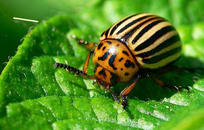
103 - 103
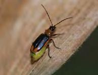
104

105 - - - Dankowska 2001) Ćwikliński and Ćwiklińska 2001; 105
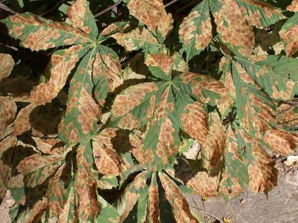

106 106


107 107

108
109 Bereś P.K., Sionek R Wybrane elementy biologii Diabrotica virgifera Le Conte na podstawie obserwacji w okolicy Rzeszowa ( ). Prog. Plant Prot./Post. Ochr. Roślin, 48(1): Ćwikliński L., Ćwiklińska J Nieco więcej o szrotówku kasztanowcowiaczku. Ochrona Roślin nr 5-6: Dankowska E Nowe zagrożenie dla kasztanowców. Szrotówek kasztanowcowiaczek (Cameraria ohridella). Aura Głowaciński, H. Okarma, J. Pawłowski, W. Solarz (red.) Gatunki obce w faunie Polski. Wyd. internetowe. [on-line]. Instytut Ochrony Przyrody PAN w Krakowie. [dostęp ]. 6. Lipa J.J Zachodnia kukurydziana stonka korzeniowa (Diabrotica virgifera virgifera) - nowy kwarantannowy szkodnik kukurydzy w Europie. Ochrona Roślin. 6: Pimentel D Biological invasions; economics and environmental costs of alien plant, animal, and microbe species. CRC Press, Boca Raton, London, New York, Washington DC:
110 110
111 Nieproszeni goście w naszych łóżkach Krzysztof Solarz Zakład Parazytologii, Śląski Uniwersytet Medyczny w Katowicach, ul. Jedności 8, Sosnowiec, solarzk@sum.edu.pl Unwelcome guests in our beds Abstract Some arthropods, mites or inects, may appear in our beds. The most frequent and most abundant guests in our beds are some species of pyroglyphids (Astigmatina, Pyroglyphidae) commonly known as the house dust mites. The three mite species, most often found in house dust throughout the world, are Dermatophagoides pteronyssinus, D. farinae and Euroglyphus maynei. These mites are the major sources of indoor inhalant allergens facilitating both the sensitization of atopic subjects and asthmatic attacks in patients. For the first time, in 1964, Voorhorst et al. reported that house dust contained mites of the genus Dermatophagoides and suggested that these were the source of the house dust allergen. Many-faceted studies of dust mites have been continued since 1964 in many countries of the world, including the surveys on their taxonomy and fauna, biology and ecology, epidemiology, allergenicity and control. The house dust mites have been reported from human dwellings and a wide variety of other habitats associated with man and his environment, both indoor and outdoor. Most often they are found in habitats intimately associated with man, such as beds, bed linen, couches, sofas, other upholstery furnitures, clothing, floors and carpets. These domestic environments are also very important locations for forensic investigations. This paper gives a review of literature on occurrence, medical importance and control of pyroglyphid dust mites, including own studies. An understanding of the biology and the life cycle of these mites, as well as environmental factors influencing mite populations, can be exploited in mite control. Typical species of the pyroglyphid dust mites Dermatophagoides pteronyssinus, D. farinae group and Euroglyphus maynei can develop in dwellings and they are not a contamination from outdoor sources. Wstęp Amerykańskie przysłowie mówi, że człowiek spędza swe życie w dwóch miejscach, w butach i w łóżku. Rzeczywiście od 1/4-1/3 swojego życia spędzamy w łóżku. Przy 8 godzinach snu daje to około 20 lat. W łóżkach mogą towarzyszyć nam jednak pewne stawonogi, w tym roztocze i owady, które są nieproszonymi gośćmi w miejscach do spania. Wśród owadów można wymienić pluskwy łóżkowe Cimex lectularius czy pchły ludzkie Pulex irritans (lub inne, np. psie lub kocie) krwiopijne, zewnętrzne, bezwzględne, czasowe, oligo- lub polikseniczne pasożyty ludzi i zwierząt. Do najważniejszych pod względem medycznym pasożytniczych roztoczy atakujących człowieka podczas odpoczynku nocnego należą obrzeżki gołębie Argas reflexus (Acari: Ixodida, Argasidae) i ptaszyńce Dermanyssus gallinae (Acari: Mesostigmata, 111
112 Dermanyssidae). Pasożyty te pojawiaja się w naszych łóżkach jednak incydentalnie, w zależności od różnych sprzyjających okoliczności. Typowymi mieszkańcami naszych łóżek i innych miejsc do spania są jednak roztocze kurzu domowego. Od połowy lat 60-tych ubiegłego stulecia coraz więcej uwagi poświęca się medycznemu znaczeniu tych roztoczy. Mianem roztocze kurzu domowego (roztocze kurzowe) określa się roztocze z rodziny Pyroglyphidae występujące w kurzu pochodzącym z łóżek, mebli tapicerskich oraz z podłóg w pomieszczeniach mieszkalnych. W środowisku tym stwierdza się, mniej lub bardziej powszechnie, także roztocze z innych grup systematycznych, w tym także niektóre gatunki związane z produktami spożywczymi, które łącznie z roztoczami kurzowymi określa się jako roztocze domowe. Jednak typowymi dla kurzu przedstawicielami są niektóre gatunki Pyroglyphidae, najszerzej rozprzestrzenione, najczęściej i najliczniej stwierdzane w mieszkaniach oraz najlepiej poznane pod względem szkodliwości medycznej, a mianowiecie Dermatophagoides pteronyssinus, Dermatophagoides farinae, Dermatophagoides microceras i Euroglyphus maynei. Ponadto udowodniono alergizujący wpływ na organizm człowieka kilku innych gatunków z tej rodziny, Dermatophagoides siboney, Malayoglyphus intermedius, Sturnophagoides spp. i Gymnoglyphus longior (Arlian 1991, 2001, 2002, Arlian i Platts-Mills 2001, Ferrandiz i wsp. 1997, Sewer i wsp. 2000). Roztocze te rozpowszechnione są na całym świecie; szczególnie licznie występują na bardziej wilgotnych obszarach umiarkowanej strefy klimatycznej (w Europie głównie - Holandia, Wielka Brytania, kraje skandynawskie, nie są rzadkie także w naszym kraju). W 1 gramie kurzu można znaleźć setki, tysiące, a nierzadko dziesiątki tysięcy okazów tych pajęczaków. Liczebność ich jest zwykle większa w próbach pochodzących ze Tabela 5. Alergeny roztoczy kurzowych i przechowalnianych. Grupa alergenowi Ciężar cząsteczkowy [kda] Grupa 1 Der p 1 Der f 1 Der m 1 Der s 1 Eur m 1 Blo t 1* Sui m 1* Grupa 2 Der p 2 Der f 2 Der m 2 Eur m 2 Lep d 2* Gly d 2* Sui m 2* Grupa 3 Der p 3 Der f 3 Blo t 3* Funkcja białka/ Homologia 25 proteinaza serynowa (np. katepsyna lub papaina) białko homologiczne proteinaza serynowa (trypsyna) 112
113 Sui m 3* Grupa 4 Der p 4 Eur m 4 Aca s 4* Blo t 4* Grupa 5 Der p 5 Lep d 5* Blo t 5* Sui m* Grupa 6 Der p 6 Der f 6 Blo t 6* Sui m 6* Grupa 7 Der p 7 Der f 7 Lep d 7* Amylaza 14 proteaza serynowa (chymotrypsyna) 25 proteaza serynowa (chymotrypsyna) nieznana Grupa 8 Der p S-transferaza glutationu Grupa 9 Der p 9 Blo t 9* Sui m 9* Grupa 10 Der p 10 Der f 10 Blo t 10* Grupa 11 Der p 11 Der f 11 Blo t 11* 23,8 proteaza serynowa (kolagenaza) 36 Tropomiozyna Paramiozyna Grupa 12 Blo t 12* 16 wiążące kwasy tłuszczowe Grupa 13 Aca s 13* Tyr p 13* Lep d 13* Blo t 13* 14 wiążące kwasy tłuszczowe Grupa 14 Der p 14 Grupa 15 Der p 15 Der f Apolipoforyna Der f Chitynaza Grupa 16 Der p wiążące aktynę cytoszkieletu komórek 113
114 Grupa 17 Der f 17? wiążące jony wapnia Grupa 18 Der f Chitynaza Grupa 19 Blo t 19* 9 ochrona przeciwbakteryjna roztoczy Grupa 20 Der p 20 Der f kinaza argininowa * alergeny dotyczące jedynie roztoczy przechowalnianych; Der p = Dermatophagoides pteronyssinus; Der f = D. farinae; Der m = D. microceras; Der s = D. siboney; Eur m = Euroglyphus maynei; Aca s = Acarus siro; Tyr p = Tyrophagus putrescentiae; Lep d = Lepidoglyphus destructor; Blo t = Blomia tropicalis; Sui m = Suidasia medanensis. starych zawilgoconych domów, ogrzewanych piecami. Występują często w domach jednorodzinnych, a także w blokach mieszkalnych. Często spotykamy nieprawidłowe określenia na te grupę stawonogów alergennych, szczególnie w literaturze medycznej i w mediach. Mianowicie używa się nazwy roztocza kurzu domowego (liczba mnoga) lub roztocze kurzu domowego (liczba pojedyncza). Terminy te sa prawidłowe, ale w odniesieniu do roślin cudzożywnych, saprofitów, nie zaś w odniesieniu do przedstawicieli podgromady pajęczaków Acari, które przecież noszą nazwy zoologiczne roztocze (liczba mnoga) oraz roztocz (liczba pojedyncza). Znaczenie medyczne Choroby alergiczne dotykają obecnie około 40% ludności świata (Arlian 2001, 2002). Około 25% wszystkich chorób alergicznych oraz 50% przypadków astmy powodowanych jest przez roztocze (Boczek i Błaszak 2005). Blisko 10% populacji ludnosci świata cierpi z powodu uczulenia na alergeny roztoczowe (Thomas 2011, Brewczyński i wsp. 2012). Alergogenne działanie kurzu domowego na organizm ludzki zostało udowodnione znacznie wcześniej, zanim powiązano je z obecnością w nim roztoczy. Polega ono na wywoływaniu u człowieka alergii atopowych - astmy oskrzelowej (asthma bronchiale), nieżytu nosa (rhinitis) oraz wyprysku atopowego (eczema atopicum). Spośród pospolitych wewnątrzmieszkalnych alergenów wziewnych, kurz domowy jest na ogół najczęstszą przyczyną atopowego nieżytu nosa i atopowej astmy oskrzelowej. Już w latach 2-tych XX wieku zasugerowano obecność specyficznego alergenu w kurzu domowym, jednak pochodzenie tego alergenu nie było wówczas znane. Dopiero w 1964 roku Voorhorst i wsp. stwierdzili poraz pierwszy w próbach kurzu z mieszkań alergików uczulonych na kurz domowy na terenie Lejdy w dużej liczebności roztocze Dermatophagoides pteronyssinus. Okazało się wkrótce, że pacjenci atopicy z alergią na kurz domowy w testach skórnych reagują nieomal identycznie na ekstrakty kurzu autologicznego (z ich mieszkań) oraz na ekstrakty z hodowli D. pteronyssinus. Następnie w 1969 Autorzy ci opublikowali monografię podsumowującą ich badania, w której starali się udowodnić, iż D. pteronyssinus jest źrodłem hipotetycznego alergenu kurzu domowego D. pteronyssinus, a silny alergen tego roztocza odpowiada w pełni wszystkim cechom charakteryzującym antygen kurzowy (Voorhorst i wsp. 1969). Według Arliana (2001) było to jedno z najważniejszych wydarzeń światowej alergologii. Spowodowało bowiem zainteresowanie roztoczami, a póżniej także owadami wystepującymi w pomieszczeniach mieszkalnych jako przyczyną schorzeń alergicznych 114
115 lokatorów. Wkrótce okazało się, że w kurzu z mieszkań występują także inne gatunki roztoczy z rodziny Pyroglyphidae głównie Dermatophagoides farinae i Euroglyphus maynei (Wharton 1976, van Bronswijk 1981, Fain i wsp. 1990, Colloff 1991). Obecnie wiadomo, iż kurz domowy zawiera różne źródła alergenów, oprócz roztoczy kurzowych z rodziny Pyroglyphidae sa to roztocze alergenne innych grup (np. rozkruszki, roztoczki), owady częste w mieszkaniach (np. rybiki cukrowe, niektóre chrząszcze magazynowe, mkliki, larwy ciem, karaczany), naskórek ludzki i zwierzęcy, sierść zwierząt, pióra ptaków, pyłki, zarodniki, mycelia grzybów pleśniowych i bakterie (van Bronswijk 1981, Fain i wsp. 1990, Pope i wsp. 1993, Arlian 2002, Piontek i wsp. 2011, Brewczyński i wsp. 2012). Mimo różnorodności antygenowej kurzu domowego trzeba stwierdzić, że roztocze z rodziny Pyroglyphidae (głównie D. pteronyssinus i D. farinae) są głównym źródłem alergenów oraz najczęstszą przyczyną uczulenia na kurz domowy. Dlatego też ostatnio uczulenia na kurz spowodowane przez roztocze Dermatophagoides, określa się mianem alergii na roztocze kurzu domowego, w celu odróżnienia ich od uczuleń na kurz spowodowanych przez inne alergeny w nim występujące (np. Arlian 1991, Platts-Mills i wsp. 1992, Pope i wsp. 1993, Solarz 1999). Alergia na roztocze kurzu domowego obejmuje może objawiać się w różny sposób, najczęściej jest to atopowa astma oskrzelowa (asthma bronchiale), alergiczne zapalenie spojówek (conjunctivitis), atopowy nieżyt nosa (rhinitis) oraz egzema (eczema). Źródłem alergenów roztoczowych są głównie drobiny kału roztoczy kurzu domowego, a także ich ciała, wylinki, wydzieliny, produkty rozrodcze oraz grudki pokarmu otoczone membraną (błoną) peritroficzną roztocza. Główny alergen kurzu domowego, określany jako antygen Der p 1, którego zawartość w kurzu waha się w szerokim zakresie od 100 do ng na 1 gram czystego kurzu, jest właśnie alergenem roztoczy D. pteronyssinus. Oprócz tego odkryto jeszcze co najmniej 19 innych rodzajów (grup) alergenów, których większość wykazuje aktywność proteolityczną, a jeden z możliwych mechanizmów ich działania uwzględnia osłabienie barier komórkowych nabłonka błony śluzowej dróg oddechowych (Aalberse 1998, Arlian 2001, 2002, Colloff 2009, Niesler i wsp. 2009, Asman i wsp. 2010, Thomas 2011). Niektóre z wymienionych alergenów roztoczy są enzymami proteolitycznymi (Robinson i wsp. 1997, Colloff 2009). Na nazwę alergenu zwierzęcego składają się trzy pierwsze litery nazwy rodzajowej zwierzęcia alergogennego, pierwsza litera nazwy gatunkowej oraz grupa antygenowa oznaczona cyfrą arabską (dawniej rzymską). Zgodnie z tym kodem najwczesniej opisany alergen Dermatophagoides pteronyssinus nosi nazwę Der p 1 (dawniej Der p I) (Arlian 2002). Najlepiej poznano alergeny grupy 1, 2 i 3 u roztoczy D. pteronyssinus, D. farinae, D. microceras i E. maynei (Tabela 1). Alergeny grupy 1 to glikoproteiny, o masie cząsteczkowej około 25 kda. W znacznym stopniu zbadano ich skład aminokwasowy. Alergeny te są syntetyzowane przez roztocze kurzu domowego, powstają w ich przewodzie pokarmowym, w tylnej części jelita środkowego, w miejscu gdzie odbywa się pozakomórkowe trawienie pokarmu. Białka te wykazują aktywność proteolityczną oraz duże podobieństwo sekwencji aminokwasowej do proteinazy cysteinowej (np. katepsyny albo papainy), oraz są enzymami odgrywającymi rolę w trawieniu u tych roztoczy. Największe stężenie alergenów grupy 1 obserwuje się w drobinach kału roztoczy oraz w ekstraktach z podłoża hodowlanego (culture medium extracts). Jak ustalono, pojedynczy okaz roztocza wydala w ciągu swego życia około 250 kulek kału, o średnicy od 10-40μm (w zależności od gatunku i stadium rozwojowego), w których zawarte jest nawet do 90% głównego alergenu kurzu domowego (Der p 1 lub Der f 1). Podobne proteolityczne własności mają alergeny grupy 3, białka o masie cząsteczkowej około kda, które są enzymami o aktywności proteinazy serynowej (np. trypsyna). Alergeny grupy 2 są z kolei łatwo rozpuszczalnymi 115
116 białkami, o masie cząsteczkowej około kda, znacznie mniej wrażliwymi na temperaturę. Funkcja alergenów grupy 2 nie została w pełni poznana. Wiadomo, że są rozmieszczone w różnych miejscach idiosomy roztocza, a największą ich koncentrację stwierdza się w wyciągach tzw. oczyszczonych ciał roztoczy (purified whole mite body extracts). Wykazano homologię alergenu Der f 2 z białkiem najądrzy naczelnych i białkiem linienia owadów. U Dermatophagoides farinae występuje on w wiekszej koncentracji w drogach wyprowadzających nasienie u samców i płynie wylinkowym (Arlian 1991, 2001, 2002, Hallas 1991, Korsgaard i Iversen 1991, Mosbech i wsp. 1991, Aalberse 1998, Colloff 1991, 2009, Tsai i wsp. 2000, Smith i wsp. 2001). Przypuszczać można, że pozostałe gatunki z rodziny Pyroglyphidae występujące w pomieszczeniach mieszkalnych także wytwarzają analogiczne alergeny. Ponieważ jednak są one rzadsze i mniej liczne, nie odgrywają poważniejszej roli w alergii na kurz domowy i roztocze kurzu domowego (Arlian i Platts-Mills 2001, Arlian i wsp. 2002). Alergia na roztocze kurzu domowego jest zatem obecnie diagnozowana i leczona przede wszystkim za pomocą wyciągów z oczyszczonych organizmów D. pteronyssinus, D. farinae i E. maynei. Reakcje na ekstrakty roztoczy kurzu domowego są najbardziej powszechne u dzieci, szczególnie u chłopców. Także podczas testowania alergologicznego dzieci, można łatwiej zademonstrować korelację między wyciągami z kurzu domowego oraz z roztoczy z rodzaju Dermatophagoides. Wydaje się, że narażenie na kontakt z silnymi alergenami w pierwszych 6 miesiącach życia dziecka ma związek z późniejszą nadwrażliwością w postaci astmy oskrzelowej albo nieżytu nosa i dotyczy to także uczulenia na roztocze kurzu domowego (Obuchowicz i wsp. 2011). Dlatego też trzeba chronić niemowlęta przed kontaktem z tymi roztoczami. Zdaniem badaczy amerykańskich pewne zespoły chorobowe u noworodków mogą być spowodowane szokiem anafilaktycznym na antygen roztoczy D. farinae. Podobnie w Japonii, zwraca się uwagę na możliwości udziału roztoczy Dermatophagoides w wywoływaniu tzw. choroby Kawasaki (od momentu jej opisania w roku 1967 do roku 1983 stwierdzono w tym kraju ponad zachorowań). Ponadto zasugerowano możliwość związku pomiędzy ekspozycją na alergeny roztoczy kurzu domowego (Dermatophagoides spp.) a wystąpieniem objawów wiosennego nieżytu spojówek połączonego z zapaleniem rogówki (keratoconiunctivitis vernalis). Coraz więcej danych świadczy także o indukującej roli uczulenia na roztocze D. pteronyssinus w patogenezie niektórych alergii pokarmowych (np. po spożyciu krewetek lub ślimaków winniczków). Na alergię na roztocze kurzu domowego przyjęło się w polskiej alergologii określenie roztoczyca. Określenie błędne z wielu powodów. Termin ten zaproponowano myśląc zapewne, iż roztocze (a najczęściej używa się terminu roztocza) to jedynie kilka gatunków występujących w kurzu domowym, odpowiedzialnych z awywoływanie chorób alergicznych, a głównie dwa gatunki D. pteronyssinus i D. farinae. Podczas gdy roztocze to podgromada pajęczaków obejmująca blisko gatunków różnych grup, w tym roztocze pasożytujące u ludzi i zwierząt, powodujące konkretne choroby takie jak świerzb czy nużyca, które to choroby również są roztoczycami. Ponadto znanych jest wiele grup roztoczy alergennych; oprócz roztoczy kurzu domowego można wymienić roztocze przechowalniane, przędziorki i inne pasożyty roślin, czy też alergizujące roztocze drapieżne i pasożytnicze. 116
117 Występowanie roztoczy Pyroglyphidae Rodzina Pyroglyphidae obejmuje 20 rodzajów i 48 gatunków roztoczy; podzielona jest na 5 wymienionych poniżej podrodzin (w nawiasach przedstawiam liczbę rodzajów/liczbe gatunków) (Fain i Bochkov 2003, Solarz 2012): 1. Pyroglyphinae (8/11) 2. Dermatophagoidinae (4/18) 3. Onychalginae (3/9) 4. Guatemalichinae (4/8) 5. Paralgopsinae (1/2) W kurzu domowym stwierdzono dotąd 15 gatunków roztoczy z tej rodziny (Tab. 2), 34 gatunki w gniazdach ptaków oraz 6 gatunków w środowisku gospodarstwa wiejskiego (lub farmy), tak więc niektóre gatunki występują w kurzu i gniazdach (D. pteronyssinus, Dermatophagoides evansi, Hirstia spp., Sturnophagides spp.), inne w kurzu i pomieszczeniach gospodarskich na wsi (G. longior, H. chelidonis, Dermatophagoides farinae). Roztocze G. longior i H. chelidonis spotykane były we wszystkich tych typach środowisk. Ogółem 6 gatunków, D. pteronyssinus, D. farinae, D. siboney, Hirstia domicola, Malayoglyphus intermedius i E. maynei, notowano w kurzu w sposób powtarzalny, bądź lokalnie bądź kosmopolitycznie (Tab. 2). Najpospolitsze gatunki roztoczy kurzu domowego z rodziny Pyroglyphidae, D. pteronyssinus, D. farinae i E. maynei, rozpowszechnione są na całym świecie. Szczególnie licznie występują na bardziej wilgotnych obszarach umiarkowanej strefy klimatycznej; w Europie głównie - Holandia, Wielka Brytania, kraje skandynawskie; są też liczne w naszym kraju (Turos 1979, Walshaw i Evans 1987, Fain i wsp. 1990, Arlian i Platts Mills 2001, Arlian i wsp. 2002, Racewicz 2001, Białkowska i Solarz 2008, Solarz 2010, Solarz i wsp. 2007, 2008, 2010a, b, Obuchowicz i wsp. 2011, Kosik-Bogacka i wsp. 2012, Ciechacka i wsp. 2013, Jakubas-Zawalska i wsp. 2013). Poza domami i mieszkaniami stwierdzano roztocze kurzu domowego w próbach kurzu z wielu innych miejsc. Były notowane z wielu innych miejsc związanych z obecnością lub działalnością człowieka, ze szpitali, bibliotek, szkół, przedszkoli, żłobków, hoteli, domów studenckich, domów opieki, sanatoriów, biur, różnego rodzaju miejsc pracy i odpoczynku, baraków wojskowych, magazynów i obiektów portowych, pomieszczeń na statkach dalekomorskich i okrętach wojennych, pasażerskich wagonów kolejowych, pomieszczeń gospodarczych w środowisku wiejskim, a nawet z bruków i chodników miejskich (Hewitt i wsp. 1973, Rao i wsp. 1975, Massey i wsp. 1988, Green i wsp. 1992, Babe i wsp. 1995, Janko i wsp. 1995, Zock i Brunekreef 1995, Racewicz 2001, Solarz 2001a, b, 2010, Skubała i wsp. 2005, Colloff 2009, Solarz i wsp. 2010a). Ponadto mogą występować w gniazdach ptaków, głównie Passeriformes (Chmielewski 1982, Fain i wsp. 1990, Colloff 1998, Solarz 2001a, Solarz i wsp. 2004b). O powszechności występowania roztoczy w biurach, świadczą wyniki badań przeprowadzonych w Portland (USA), gdzie połowa badanych pomieszczeń biurowych różnego rodzaju wykazywała obecność populacji roztoczy kurzu domowego, zaś miejscami szczególnie często zasiedlanymi były fotele tapicerowane (Janko i wsp. 1995). Także w Polsce w pomieszczeniach biurowych występują głównie na meblach tapicerskich (Solarz 2001a). W obrębie mieszkania roztocze występują w miejscach, które zapewniają im schronienie, a przede wszystkim pokarm i odpowiednią wilgotność, a więc w łóżkach i innych 117
118 miejscach do spania, maty tatami, dywany (długowłose), meble tapicerowane, zabawki pluszowe, zasłony i tekstylia obecne w domu. Są to miejsca, zatem, ściśle związane z działalnością człowieka, głównego źródła pożywienia, w postaci złuszczonego naskórka, łupieżu, grzybów pasożytujących na skórze. Człowiek może zapewnić także odpowiednią dla roztoczy wilgotność, poprzez swoją aktywność, działalność gotowanie, pranie, pocenie się (Walshaw i Evans 1987, Hart i Whitehead 1990, Harving i wsp. 1993, Hart 1998, Korsgaard 1988, 1998 a, b, Koorsgaard i Iversen 1991, Solarz 2001a, b, Solarz i wsp. 2010b). Ponadto włóknista i celularna struktura tych środowisk pozwala roztoczom gromadzić się i redukować utratę wody. Ich rozmieszczenie i liczebność są zmienne, w zależności od miejsca w obrębie mieszkania, pory roku, czy położenia geograficznego. Wiele czynników odgrywa tu rolę, przede wszystkim wilgotność powietrza i temperatura, dostępność pożywienia, ale także rodzaj, wiek i konstrukcja budynku, typ podłogi, typ mebli i miejsca do spania, piętro, częstość gotowania, prania, wietrzenia pomieszczeń, częstość i forma sprzątania, itp. Na ogół więcej roztoczy stwierdza się w sypialniach aniżeli w innych pomieszczeniach w obrębie mieszkania, najwięcej w łóżkach; więcej na podłogach pokrytych dywanami niż drewnianych lub wykładzinach typu linoleum, najwięcej na dywanach z długim włosem, które stanowią mikrośrodowisko sprzyjające gromadzeniu się naskórka oraz wilgoci, a także zapewniające pewną ochronę dla populacji roztoczy w trakcie odkurzania (Korsgaard 1988, Solarz 2001a, b). Występują także pewne odmienności w zależności od regionu, np. w Japonii roztoczy jest więcej w domach murowanych niż drewnianych, zaś w Rosji odwrotnie, najwięcej jest ich w starych domach z drewna (Uchiksohi i wsp. 1982, Yagofarov i Galikeev 1987). W Polsce występują na ogół liczniej w domkach jednorodzinnych niż w blokach mieszkalnych (Racewicz 2001, Solarz 2001a, b, 2010, Solarz i wsp. 2008, Jakubas-Zawalska i wsp. 2013, Ciechacka i wsp. 2013). Są one liczne w suterenach i mieszkaniach na parterze (Chmielewski 1995). Roztocze z rodziny Pyroglyphidae stanowią zwykle % roztoczy w kurzu domowym (Horak i wsp. 1995, Arlian 2001, 2002, Solarz 2010, Solarz i wsp. 2008, 2010a, b, Obuchowicz i wsp. 2011, Kosik-Bogacka i wsp. 2012). W Polsce do 85% roztoczy zebranych z mieszkania pochodzi z prób z miejsc do spania, podczas gdy tylko kilkanaście lub kilka procent z prób z podłóg i mebli tapicerowanych (Solarz 2001a, b, 2010, Obuchowicz i wsp. 2011, Kosik-Bogacka i wsp. 2012). W większości przypadków w miastach Górnego Śląska dominuje Dermatophagoides farinae, ale w innych regionach (Poznań, Warszawa, Łódź) może dominować D. pteronyssinus, zaś Euroglyphus maynei występuje sporadycznie, w małej liczebności (Chmielewski 1995, Samoliński i wsp. 1989, Horak i wsp. 1995, Solarz 2001b, 2010). W bibliotekach, szpitalach i biurach południowej części Polski, podobnie jak w mieszkaniach, dominował D. farinae. Obserwowano jednak znaczne zmiany w dominacji poszczególnych gatunków wraz ze zmianą warunków w mieszkaniach i szpitalach w Polsce, w latach (Ryc. 1-3; Solarz 2010). Poza mieszkaniami Pyroglyphidae występują w Polsce z różną dominacją i frekwencją w zależności od środowiska; są liczne i częste w gniazdach ptaków synantropijnych, gdzie zwykle dominują, ale ustępują innym roztoczom w pomieszczeniach na terenie gospodarstwa wiejskiego, w miejscach przebywania i odpoczynku górników w kopalniach węgla kamiennego, czy też w pomieszczeniach dla zwierząt w warunkach ogrodu zoologicznego, w których występują sporadycznie i są znacznie mniej liczne niż gatunki alergogenne z rodzin Acaridae i Glycyphagidae (Solarz 2001a, 2010, Solarz i wsp. 2004a, b, 2007, Solarz i Seńczuk 2003, Szilman i wsp. 2006, Meszyńska i wsp. 2008, Drelich i Solarz 2009, Pająk i Solarz 2013). 118


119 Dominacja (%) DF DP EM GL HC Ryc. 1. Zmiany dominacji poszczególnych gatunków kurzowych Pyroglyphidae w mieszkaniach z terenu Górnego Śląska i innych regionów południowej Polski w latach Dominacja (%) DF DP EM Ryc. 2. Zmiany dominacji poszczególnych gatunków kurzowych Pyroglyphidae w szpitalach na terenie Górnego Śląska w latach
120 Materace łóżek Tapczany/wersalki/sofy DF Ryc. 3. Średnia liczba roztoczy (w przeliczeniu na 1 gram kurzu) w miejscach do spania mieszkań Górnego Śląska w latach DP W 1g kurzu można znaleźć setki, tysiące, a nierzadko dziesiątki tysięcy okazów tych roztoczy (Tab. 3). W Europie łóżka i inne miejsca do spania są głównymi siedliskami populacji tych roztoczy, a kurz z materacy (i innych miejsc do spania) zawiera zwykle największe ich koncentracje (van Bronswijk 1981, Fain i wsp. 1990, Rao i wsp. 1975, Solarz 2001a, b, 2010). Średnia liczba roztoczy w próbach z miejsc do spania w mieszkaniach Górnego Śląska wynosiła około 73 okazów w przeliczeniu na gram czystego kurzu (Solarz 2001a) i wahała się na terenie Katowic i Sosnowca w zakresie 43,7-287,3. Ale w pewnych krajach naszej strefy, o bardziej wilgotnym klimacie (kraje skandynawskie, Wielka Brytania, Holandia), ta liczba może być krotnie wyższa (van Bronswijk 1981, Fain i wsp. 1990, Colloff 2009), podczas gdy, jak ustalono, ekspozycja na 100 okazów roztoczy w gramie kurzu jest wystarczająca do wywołania uczulenia (Platts-Mills i wsp. 1992, Pope i wsp. 1993). Liczba 100 okazów w gramie kurzu równoważna jest 2 g alergenu Der p 1/1gram kurzu; średnio ng tego alergenu zawiera pojedynczy okaz D. pteronyssinus, w zależności od stadium rozwojowego (Mosbech i wsp. 1991). Liczebność roztoczy, w przeliczeniu na gram kurzu, jest zwykle wyraźnie wyższa w próbach z mieszkań, w porównaniu ze szpitalami, sanatoriami i miejscami użyteczności publicznej (Rao i wsp. 1975, Massey i wsp. 1988, Fain i wsp. 1990, Colloff i wsp. 1991, Green i wsp. 1992, Babe i wsp. 1995, Janko i wsp. 1995, Zock i Brunekreef 1995, Racewicz 2001, Colloff 2009, Solarz 2010) (Tab. 3). Przypuszcza się, że częste sprzątanie, pastowanie podłóg, inne rutynowe zabiegi związane z utrzymaniem czystości, a także utrzymywanie niskiego poziomu wilgotności względnej powietrza, to zdaniem większości autorów, najważniejsze powody mniejszej liczebności roztoczy w szpitalach i miejscach publicznych (Colloff i wsp. 1991, Babe i wsp. 1995, Solarz 2001a, 2010). Występują jednak wyraźne różnice pomiędzy danymi literaturowymi. W Sztokholmie, na przykład, znajdowano pojedyncze roztocze tylko 120
121 w kurzu z podłóg, natomiast łóżka były zupełnie wolne od roztoczy (Turos 1979). Natomiast w naszym kraju, na terenie Górnego Śląska Pyroglyphidae ogółem występowały w zbliżonej liczebności na podłogach i w łóżkach pacjentów (Solarz 2001a). W Zachodniej Australii roztocze występowały w ponad dziesięciokrotnie mniejszej liczebności w sanatoriach niż w domach pacjentów (Colloff i wsp. 1991), podczas gdy w Czechach były one tylko dwukrotnie mniej liczne w szpitalach aniżeli w mieszkaniach dzieci z egzemą (Vobrázková i wsp. 1986). Ważny jest także typ umeblowania lub pokrycia podłóg. Kurz z podłóg klas szkolnych w Rotterdamie, pokrytych wykładzinami dywanowymi lub dywanami, zwierał więcej alergenu roztoczowego niż kurz z podłóg gładkich, ale znacznie mniej niż kurz z dywanów zebrany w pomieszczeniach mieszkalnych (Zock i Brunekreef 1995). Liczebność roztoczy Pyroglyphidae i innych roztoczy domowych (także w przeliczeniu na gram kurzu) jest zatem różna w różnych miejscach, zależy od miejsca poboru próby, w obrębie mieszkania, w różnych typach pomieszczeń, w różnych miejscach wewnątrz obiektów użyteczności publicznej, w różnych typach środowisk, gdzie są spotykane, czy to pomieszczenia gospodarstwa wiejskiego, czy też gniazda ptaków i ssaków (Solarz i wsp. 2004b). W badanych miastach południowej części Polski (Solarz 2001a, b, 2010, Obuchowicz i wsp. 2011) D. farinae występuje w większej koncentracji, w przeliczeniu na gram kurzu, we wszystkich badanych typach miejsc, ale w przypadku miejsc do spania, gatunek ten preferuje tapczany, sofy, wersalki, podczas gdy D. pteronyssinus materace łóżek. Około 66,5% badanych wówczas prób pozytywnych na roztocze zawierało jedynie roztocze z rodziny Pyroglyphidae, podczas gdy 27,8% zawierało obok Pyroglyphidae inne taksony roztoczy domowych. Jedynie 5,7% prób pozytywnych zawierało wyłącznie inne poza Pyroglyphidae taksony roztoczy. Te inne roztocze to przede wszystkim przedstawiciele rodzin Chortoglyphidae (Chortoglyphus arcuatus), Glycyphagidae (Glycyphagus domesticus, Gohieria fusca, Lepidoglyphus destructor), Acaridae (Tyrophagus putrescentiae i Acarus siro complex), Cheyletidae, Tetranychidae oraz Tarsonemida (Solarz 2001a, Piontek i wsp. 2011). Liczebność roztoczy (na 1 gram kurzu) wahała się od ,7 (Solarz 2001a, 2010). Łóżka i inne miejsca do spania zawierają największe koncentracje roztoczy. Ogólna liczba roztoczy w szpitalach jest niższa niż w mieszkaniach i bibliotekach. W bibliotekach na Górnym Śląsku najwyższą liczebność roztoczy na 1 gram kurzu stwierdzono na powierzchni regałów na książki oraz wykładzin dywanowych pokrywających podłogi pomieszczeń bibliotecznych i krzeseł tapicerowanych. Być może starsze woluminy (a przeto także półki i biurka, wykładziny) zawierają większą ilość naskórka lub łupieżu ludzkiego, głównego pożywienia tych roztoczy. Istotnym momentem może być zwyczaj czytania w łóżku, a także duża liczba osób odwiedzających pomieszczenia biblioteczne (Solarz 2001a, 2010). W biurach roztocze były liczne w próbach z mebli tapicerowanych i dywanów lub wykładzin dywanowych, zaś w pomieszczeniach komisariatu Policji w próbach z krzeseł tapicerskich (Solarz 2010). 121
122 Tabela 2. Stwierdzone dotąd w kurzu domowym gatunki roztoczy z rodziny Pyroglyphidae i ich występowanie w świecie. Gatunki roztoczy Dermatophagoides pteronyssinus Dermatophagoides farinae Dermatophagoides microceras Dermatophagoides evansi Dermatophagoides siboney Dermatophagoides neotropicalis Hirstia domicola Hirstia chelidonis Malayoglyphus intermedius Malayoglyphus carmelitus Sturnophagoides brasiliensis Euroglyphus maynei Gymnoglyphus longior Hughesiella africana Hughesiella valerioi Rozmieszczenie Gatunek kosmopolityczny Gatunek kosmopolityczny Belgia, Holandia, Anglia, Hiszpania, Włochy, Norwegia, Szwajcaria, USA, RPA Francja, Portugalia, Rosja, wschodnie regiony byłego ZSRR, Polska, USA (Kalifornia), Iran Kuba, Puerto Rico Indie, Brazylia, Surinam Francja, Hiszpania, Włochy, Węgry, Czechy, tereny byłego ZSRR, USA, Surinam, Kuba, Kolumbia, Indie, Tajlandia, Malezja, Singapur, Indonezja, Brunei, Chiny, Tajwan, Japonia, RPA Polska, Norwegia, obszar byłego ZSRR Tahiti, RPA, Nigeria, Indie, Tajlandia, Malezja, Singapur, Indonezja, Tajwan, Japonia, Kuba, Surinam, Kolumbia Hiszpania, Kolumbia, Zachodni Izrael, Indie, Włochy Francja, Brazylia, Malezja, Singapur, Indonezja Europa, wschodnie regiony byłego ZSRR, USA (w tym Hawaje), Brazylia, Kolumbia, Chile, Peru, Kostaryka, Portoryko, Izrael, Iran, Indie, Malezja, Chiny, Tajwan, Japonia, Papua-Nowa Gwinea, Australia, Algieria, RPA, Tristan da Cunha (Afryka) Włochy, Polska, Bułgaria, były ZSRR, Peru Brazylia, Kolumbia, Madagaskar Kostaryka Tabela 3. Porównanie liczebności roztoczy kurzu domowego (w przeliczeniu na gram kurzu), w mieszkaniach lub domach i miejscach użyteczności publicznej, w Polsce i na świecie, na podstawie danych literaturowych. Miejsce Liczba roztoczy/1 gram kurzu Mieszkanie/dom Miejsca użyteczności publicznej Australia Zachodnia (sanatorium; Colloff i wsp. 1991) Gdańsk i Gdynia 13,7 1,03 (szpitale; Racewicz 2001) Gdańsk i Gdynia 13,7 1,0 (hotele; Racewicz 2001) Cardiff 3, ,0 13,0-500,0 (szpitale; Rao i wsp. 122
123 1975) Sydney (różne miejsca 1 ; Green i wsp. 1992) Hawaje (domy studenta; Massey i wsp. 1988) Delaware (USA) Pojedyncze okazy, tylko latem (szpitale; Babe i wsp. 1995) Górny Śląsk 73,0 8,3 (szpitale; Solarz 2001a) Katowice i Sosnowiec 43,7-287,3 30,7 (biblioteki; Solarz 2001a) Katowice 287,3 23,5 (biurowiec; Solarz 2001a) 1 kina, szpitale, szkoły, kolejowe wagony pasażerskie, samoloty pasażerskie, poczekalnia lotniska, dom seniora, ogólne pomieszczenie uniwersyteckie, centra kształcenia dzieci i młodzieży. Najważniejszymi przenosicielami (wektorami) tych roztoczy są na pewno ludzie. Już od dawna stwierdzane były na skórze i we włosach, a nawet w moczu oraz w plwocinie pacjentów. Później wykazano także ich występowanie na odzieży personelu medycznego szpitala (Hewitt i wsp. 1973). Natomiast wewnątrz mieszkania rozprzestrzeniają się głównie z prądami powietrza, np. podczas manipulacji z pościelą. Okazy D. pteronyssinus były już izolowane z prób powietrza (Cunnington i Gregory 1968) z sypialni mieszkania w Wielkiej Brytanii. Wiedza na temat występowania, liczebności i biologii roztoczy kurzowych może być bardzo przydatna w różnego typu sprawach sądowych oraz dochodzeniach kryminalnych (tzw. akarologia sądowa) (Perotti i wsp. 2009, Solarz 2009). Biologia Cykl rozwojowy Pyroglyphidae obejmuje jajo, prelarwę, larwę, protonimfę, tritonimfę oraz formy dorosłe samice i samce (Tab. 4). Bezpośrednio po wylęgu samicy następuje kopulacja. Samice są płodne przez cały okres życia - dziennie składają od 1 do 6 jaj, średnio w ciągu życia jaj. Jaja składane są pojedynczo oraz z 1-2-dniowymi przerwami. Partenogeneza u tych roztoczy nie jest znana. Opisywane były natomiast przypadki żyworództwa u D. pteronyssinus i D. farinae, będące wynikiem rozwoju larw z jaj znajdujących się wewnątrz samic (zjawisko aparii) (Arlian 1989, Fain i wsp. 1990, Hallas 1991, Solarz i Brewczyński 1999, Colloff 2009). W optymalnych warunkach (odpowiedni pokarm, wilgotność względna powietrza 75-80% oraz temperatura C) pełny rozwój D. pteronyssinus zajmuje około 1 miesiąca. Postaci dorosłe żyją około 2-3 miesięcy. Czas rozwoju, długość życia postaci dorosłych zależą w dużym stopniu od temperatury powietrza (Tab. 4), i są na ogół najbardziej korzystne dla populacji w optymalnym zakresie temperatur dla danego gatunku. Optymalna temperatura dla rozwoju roztoczy kurzu domowego z rodziny Pyroglyphidae wynosi C. Rozwój ich zachodzi w zakresie od 17 do 32 C, przy czym gatunek E. maynei jest uważany za bardziej ciepłolubny od gatunków z rodzaju Dermatophagoides. Występują także pewne różnice w optimum temperatury pomiędzy D. 123
124 pteronyssinus a D. farinae. Ten drugi gatunek wymaga nieco wyższej temperatury od 25 do 30 C, podczas gdy pierwszy tylko około 25 C. Zakres optymalnej wilgotności wynosi dla większości gatunków od 75% do 85% wilgotności względnej, która odpowiada "aktywności pary wodnej" (a v ) od 0,75 do 0,85; dla D. farinae zakres ten wynosi 70-75% (a v = 0,70-0,75), dla D. pteronyssinus, D. evansi i E. maynei 75-80% (a v = 0,75-0,80) oraz dla H. chelidonis - 85% (a v = 0,85). Krytyczna wilgotność względna dla rozwoju D. pteronyssinus jest zatem wyższa niż w przypadku D. farinae. Aktywne stadia tych roztoczy, mogą jednak przeżywać przez pewien czas w warunkach wilgotności niższej od owego progu krytycznego, np. poniżej 60%, dzięki mechanizmowi adaptacyjnemu, pozwalającemu na ekstrahowanie przez roztocza wody bezpośrednio z nienasyconego powietrza, co umożliwia mu zrekompensowanie wszelkich strat wody. W środowisku kurzu domowego obserwowano już wzrost liczebności populacji D. farinae w okresie gdy średnia miesięczna wilgotność powietrza wynosiła od 47% do 50% wilg. wzgl., a więc była o wiele niższa od owej wartości krytycznej, a także od wyżej podanych wartości określających minimum wilgotnościowe na podstawie obserwacji hodowlanych (57-60%); dlatego też należy o tym pamiętać podejmując działania profilaktyczne, polegające na obniżeniu wilgotności wewnątrz pomieszczeń mieszkalnych. Wiadomo bowiem, że wilgotność w środowisku roztocza może być o wiele wyższa od wartości, którą można stwierdzić w wolnym powietrzu (Arlian i wsp. 1983). Na przykład 40% wilg. wzgl. powietrza może odpowiadać wilgotności rzędu od 80 do 100% na powierzchni wilgotnej podłogi, podczas gdy wilgotność kurzu na powierzchni podłogi suchej jest często równa wilgotności powietrza. Także w łóżkach zajętych przez człowieka wilgotność znacznie wzrasta osiągając poziom powyżej 60% wilg. wzgl. Można w ten sposób wyjaśnić liczne występowanie tych roztoczy w kurzu z łóżek i z innych miejsc do spania. Niektóre eksperymenty hodowlane wykazują, że optymalna wilgotność przy odpowiednim pokarmie może być niższa aniżeli wskazują wyżej cytowane prace. Na przykład Ree i wsp. (1998) uzyskali najszybszy wzrost populacji D. farinae w temperaturze 28 C i wilgotności względnej 64%, gdy jako podłoże stosowano mieszaninę pokarmu płatkowego dla ryb akwariowych oraz suszonych drożdży. Wszystkie aktywne stadia są keratynofagami (odżywiają się złuszczonym naskórkiem człowieka lub zwierząt) i żyją w jednym miejscu w kurzu domowym albo w gniazdach ptaków i ssaków. W ich odżywianiu dużą rolę, jak się wydaje, odgrywają grzyby z rodzaju Pityrosporum, które wstępnie nadtrawiają pokarm roztoczy. Uważa się, że tylko 150 mg ludzkiego naskórka wystarcza do zaspokojenia potrzeb pokarmowych populacji roztoczy obejmującej 3000 osobników w okresie 2-3 miesięcy. Okazało się, że w hodowli roztocze D. pteronyssinus ukrywają się pod substratem hodowlanym, natomiast okazy D. farinae są bardziej ruchliwe (Solarz i Brewczyński 1999). W warunkach laboratoryjnych roztocze D. pteronyssinus rozwijają się najlepiej, gdy hodowane są na suszonych rozwielitkach, płatkowym pokarmie dla tropikalnych ryb akwariowych lub na mieszance sproszkowanych drożdży piekarniczych i zarodków pszenicy. Pod tym względem (preferowanego pokarmu w warunkach laboratoryjnych) występują także różnice pomiędzy poszczególnymi gatunkami roztoczy kurzu domowego z rodziny Pyroglyphidae. Najkorzystniejszą dla rozwoju populacji D. pteronyssinus temperaturą jest 23 C (20-30 C), choć może on zachodzić w zakresie temperatur od 17 C do 30 C (Tab. 5). Głównym czynnikiem określającym rozwój i liczebność tych roztoczy jest wilgotność względna powietrza. Gatunek D. pteronyssinus wymaga 75-80% wilgotności względnej (optimum 80%); wilgotność względna 70% (w temp. 25 C) jest krytyczna dla równowagi wodnej tego roztocza. Jeśli wilgotność powietrza spada poniżej tej wartości, roztocze 124
125 oszczędnie gospodarują wodą lub nawet korzystają z wody metabolicznej, co nie wystarcza jednak do utrzymania właściwego bilansu wodnego na dłuższą metę, gdyż czerpią one wodę na drodze aktywnej sorpcji z otaczającej pary wodnej. Organizm ich jest jednak przystosowany do przeżywania przez pewien czas w niższych wilgotnościach, dzięki możliwości ograniczenia wyparowania wody z powierzchni ciała, a także do tolerowania wahań wilgotności powietrza w pewnym zakresie. Z drugiej strony, wilgotność powyżej 85% wilg. wzgl. jest już zbyt wysoka i niekorzystna dla omawianego gatunku. Sezonowym wahaniom wilgotności powietrza wewnątrz pomieszczeń mieszkalnych (i innych) towarzyszą zmiany liczebności roztoczy z rodzaju Dermatophagoides, głównie w charakterystycznych miejscach ich bytowania. W naszej strefie klimatycznej (o klimacie umiarkowanym), maksymalna liczba tych roztoczy przypada na lato oraz wczesną jesień (od czerwca do końca października), kiedy wewnątrz mieszkań panują dogodne dla ich rozwoju warunki wilgotności. Wtedy także wzrasta wyraźnie zapadalność na alergie atopowe spowodowane przez roztocze kurzu domowego (Colloff 2009, Solarz 2010). Natomiast w okresie zimowym, w strefie klimatu umiarkowanego, wilgotność powietrza wewnątrz pomieszczeń mieszkalnych jest niska (nawet w Holandii i Wielkiej Brytanii), utrzymując się poniżej owej krytycznej dla omawianych roztoczy wartości. Wtedy też liczba roztoczy w kurzu domowym znacznie się zmniejsza. Ich obecność świadczy jednak o tym, że mogą one funkcjonować w warunkach niskiej wilgotności względnej powietrza. Uważa się, że roztocze te przeżywają okres ogrzewania mieszkań głównie w postaci nieaktywnej protonimfy przetrwalnikowej (D. farinae) oraz, że wilgotność w mikrośrodowisku roztocza (np. w łóżku zajętym przez człowieka) musi być wyższa od wilgotności powietrza w pomieszczeniu (Wharton 1976, Arlian i wsp. 1983). Dlatego też, pomimo, że w warunkach laboratoryjnych roztocze D. pteronyssinus giną szybko w warunkach wilgotności względnej poniżej 65%, w mieszkaniach osób uczulonych konieczne jest obniżanie oraz utrzymywanie wilgotności na poziomie poniżej 45% (Arlian 1989, 1992, Arlian i wsp. 1998a, b, 1999 a, b, Solarz 2001a, b). Tabela 4. Czas trwania (w dniach) pełnego rozwoju postembrionalnego oraz poszczególnych stadiów młodocianych dla populacji hodowlanych Dermatophagoides pteronyssinus i D. farinae, w zależności od temperatury, w oparciu o zacytowane dane literaturowe. Gatunek Dermatophagoides farinae (według Furumizo 1975, Hallas 1991) Dermatophagoides pteronyssinus (według Arliana i wsp. 1990) C 15,6 21,1 26,6 32, Stadium Jajo 58,5 13,9 7,1 6,3 26,6 8,1 4,5 3,9 Larwa 28,3 6,8 3,5 3,0 37,4 10,4 6,4 4,2 125
126 aktywna Larwa 6,0 4,6 2,6 1,8 13,5 4,0 3,0 1,6 nieaktywna Protonimfa 24,5 15,7 2,5 2,7 28,8 6,9 4,4 3,4 aktywna Protonimfa 143,8 5,0 2,4 1,4 11,1 3,2 2,1 1,4 nieaktywna Tritonimfa 67,3 4,3 2,6 2,1 30,0 8,3 4,0 3,5 aktywna Tritonimfa 50,5 4,2 2,2 1,4 12,1 3,4 3,0 1,6 nieaktywna Pełny rozwój 388,8 54,5 23,2 18,8 122,8 34,0 19,3 15,0 Tabela 5. Wpływ diety i temperatury na długość czasu trwania poszczególnych stadiów młodocianych oraz pełnego cyklu życiowego, żywotność obu postaci dorosłych oraz płodność samic w rozwoju Dermatophagoides pteronyssinus (według Arliana i wsp. 1990, Saleha i wsp. 1991). Dieta Temperatura ( C) Wilgotność względna (%) Zarodki pszenicy Zarodki pszenicy i sproszkowane drożdze Mieszanina proteiny i białka zwierzęcego Mieszanina proteiny i białka zwierzęcego jajo 7,5 0,2 1,3 0,3 8,1 0,8 3,9 0,7 6,2 0,2 7,4 0,2 8,1 1,0 4,0 0,8 larwa 4,1 0,2 6,3 0,2 10,5 4,4 4,2 1,5 Średni czas trwania 3,5 0,6 3,7 0,2 10,4 2,2 4,2 1,0 126
127 proto- (w dniach) ( OS) 1 nimfa trito- nimfa pełny rozwój żywoność (imago) okres prereprodukcyj ny okres reprodukcyj ny okres postreprodukcyj ny Liczba składanych jaj / 1 3,5 0,2 2,3 0,1 9,6 0,4 6,3 0,2 24,6 0,3 18,4 0,3 32,9 0,3 22,8 0,4 3,1 0,1 16,2 0,2 13,8 0,2 7,4 0,3 Wylęgalność larw (%) 89,2 2,9 5,7 0,2 6,6 1,7 3,6 0,9 4,5 0,2 7,1 2,2 3,1 0,9 14,4 0,3 7,9 2,8 3,1 0,8 8,5 0,2 8,6 3,9 4,0 1,5 37,6 0,3 33,2 5,8 14,7 1,7 24,2 0,4 34,5 5,9 15,3 2,4 47,1 0,4 31,2 11,1 15,5 9,6 34,3 0,3 brak danych 4,3 0,1 brak danych brak danych brak danych 22,3 0,2 26,2 10,7 11,6 6,4 20,4 0,2 brak danych brak danych 9,4 0,3 68,4 30,4 48,0 29,6 92,5 2,3 brak danych brak danych Objaśnienia: 1 Średnia arytmetyczna odchylenie standardowe; = w rozwoju samicy; = w rozwoju samca Na liczebność roztoczy, poza wilgotnością i temperaturą, mogą mieć wpływ także inne czynniki, jak na przykład: częstość przewietrzania mieszkań, ich czystość, wiek i stan techniczny budynku lub poziom socjalno-ekonomiczny zamieszkującej rodziny (Hart 1998). Zwykle częściej i w większej liczbie stwierdzano te roztocze w kurzu z rzadko sprzątanych, nie wietrzonych mieszkań - w starych zawilgoconych domach, ogrzewanych piecami (Wharton 127
128 1976, van Bronswijk 1981, Fain i wsp. 1990, Korsgaard 1988, Fain i wsp. 1990, Korsgaard i Iversen 1991, Colloff 2009). Ich obecności sprzyja wystrój mieszkania, pluszowe meble tapicerowane, długowłose dywany, zasłony, kilimy, story, kapy na łóżkach, inne przedmioty gromadzące kurz, obecność zwierząt, których naskórek stanowi pokarm dla roztoczy, głównie psów i kotów, a ponadto wszelka aktywność i nawyki mieszkańców powodujące wzrost wilgotności w pomieszczeniach mieszkalnych (Walshaw i Evans 1987, Hart 1998, Korsgaard 1988, Korsgaard i Iversen 1991, Solarz 2001a, b, 2003, 2010). Czy populacje roztoczy kurzowych mogą rozwijać się w mieszkaniach? Hallas (2010) przedstawił dyskusyjną, w świetle tego co napisano powyżej, hipotezę, iż populacje roztoczy kurzu domowego z rodziny Pyroglyphidae nie rozwijaja się w mieszkaniach, nie rozmnażają się tam, są jedynie kontaminacją ze środowiska zewnątrzmieszalnego. Aby sprawdzić czy koncepcja ta jest słuszna dokonano analizy wyników przeprowadzonych wcześniej badań, wymienionych poniżej: 1. Analiza serii muzealnych gatunków z rozdziny Pyroglyphidae głównie Bruksela (Królewski Belgijski Instytut Nauk Przyrodniczych, 381 preparatów), Beltsville, USA (kolekcja akarologiczna Smithsonian Institutions w Waszyngtonie, 80 preparatów), Columbus, Ohio, USA (kolekcja akarologiczna Stanowego Uniwersytetu Ohio, 79 preparatów), Londyn (Zakład Entomologii, Muzeum Historii Naturalnej), 135 preparatów). 2. Badania akarologiczne prób kurzu domowego na terenie południowej części Polski (analiza 600 prób kurzu z mieszkań i domów). 3. Analiza wyników badań materiału akarologicznego uzyskanego z 69 gniazd ptaków synantropijnych na ternie Polski. 4. Badania 150 prób z gospodarstw wiejskich w Polsce na obecność roztoczy alergogennych. 5. Analiza występowania roztoczy w synantropijnych środowiskach zewnątrzmieszkalnych w miastach Górnego Śląska (ściółka zanieczyszczona odpadami komunalnymi, 160 prób). 6. Analiza występowania roztoczy (szczególnie Pyroglyphidae) w synantropijnych środowiskach zewnątrzmieszkalnych na terenach podmiejskich i wiejskich województw małopolskiego, śląskiego i świętokrzyskiego (ściółka, 150 prób). Z powyższych obserwacji wyciagnięto następujące konkluzje: 1. Część gatunków Pyroglyphidae to roztocze występujące stale w kurzu domowym, gdzie rozmnażają się głównie w miejscach do spania, wykorzystując przede wszystkim naskórek ludzki jako źródło pożywienia. Obserwowano roztocze w trakcie linienia, aparię, larwy i nimfy faratowe co świadczy o tym, iż populacje typowych kurzowych roztoczy z rodziny Pyroglyphidae rozwijają się w mieszkaniach. Dotyczy to głównie gatunków D. pteronyssinus, D. farinae, w mniejszym stopniu E. maynei. 2. W środowisku zewnętrznym, naturalnym wystepują inne gatunki roztoczy z rodziny Pyroglyphidae w gospodarstwach wiejskich H. passericola i G. longior, a w gniazdach ptaków oprócz tych dwu gatunków często dodatkowo jeszcze Dermatophagoides evansi. Roztocze te w środowisku farmy wystepują w niewielkiej liczebności, natomiast masowe populacje tworza w gniazdach ptaków synantropijnych. 128
129 3. Nie stwierdza się obecności kurzowych roztoczy z rodziny Pyroglyphidae w ściółce w środowiskach zewnątrzmieszkalnych na terenie miasta (ściółka zanieczyszczona odpadami komunalnymi), jak też na terenach podmiejskich i wiejskich. 4. Kolekcje muzealne pokazują iż roztocze D. pteronysinus, D. farinae, a także E. maynei i H. domicola na ogół opisywano z mieszkań, natomiast inne gatunki, np. H. chelidonis (=H. passericola, Dermatophagoides passericola), G. longior, G. osu i D. evansi - z gniazd ptaków lub środowiska farmy. Literatura Aalberse R.C Allergens from mites: implications of cross-reactivity between antigens. Allergy 53 (Suppl. 48): Arlian L.G Biology and Ecology of House Dust Mites, Dermatophagoides spp. and Euroglyphus spp. (Airborne allergens). Immunol. Allergy Clin. North Am. 9: Arlian L.G House-dust-mite allergens: a review. Exp.Appl. Acarol. 10: Arlian L.G Water balance and humidity requirements of house dust mites. Exp. Appl. Acarol. 16: Arlian L.G Dust mites: update on their allergens and control. Curr. Allergy Asthma Rep. 1: Arlian L.G Arthropod allergens and human health. Annu. Rev. Entomol. 47: Arlian L.G., Platts-Mills T.A.E The biology of dust mites and the remediation of mite allergens in allergic disease. J. Allergy Clin. Immunol. 107: Arlian L.G., Morgan M.S., Neal J.S Dust mite allergens: ecology and distribution. Curr. Allergy Asthma Rep. 2002, 2: Arlian L.G., Rapp C.M., Ahmed S.G Development of Dermatophagoides pteronyssinus (Acari: Pyroglyphidae). J. Med. Entomol. 27: Arlian L.G., Neal J.S., Vyszenski-Moher D.A.L. 1999a. Fluctuating hydrating and dehydrating relative humidities effects on the life cycle of Dermatophagoides farinae (Acari: Pyroglyphidae). J. Med. Entomol. 36: Arlian L.G., Neal J.S., Vyszenski-Moher D.A.L. 1999b. Reducing relative humidity to control the house dust mite Dermatophagoides farinae. J. Allergy Clin. Immunol. 104: Arlian L.G., Confer P.D., Rapp C.M., Vyszenski-Moher D.L., Chang J.S.C. 1998a. Population dynamics of the house dust mites Dermatophagoides farinae, D. pteronyssinus, and Euroglyphus maynei (Acari: Pyroglyphidae) at specific relative humidities. J. Med. Entomol. 35: Arlian L.G., Neal J.S., Bacon S.W. 1998b. Survival, fecundity, and development of Dermatophagoides farinae (Acari: Pyroglyphidae) at fluctuating relative humidity. J. Med. Entomol. 35: Arlian L.G., Woodford P.J., Bernstein I.L., Gallagher J. S Seasonal population structure of house dust mites, Dermatophagoides spp. (Acari: Pyroglyphidae). J. Med. Entomol. 20: Asman M., Solarz K., Szilman E. Szilman P Analysis of expression and amino acid sequence of the allergen Mag 3 in two species of house-dust mites Dermatophagoides farinae and D. pteronyssinus (Acari: Astigmata: Pyroglyphidae). Ann. Agric. Environ. Med. 17: Babe K.S., Arlian L.G., Confer P.D., Kim R House dust mite (Dermatophagoides farinae and Dermatophagoides pteronyssinus) prevalence in the rooms and hallways of a tertiary care hospital. Clinical aspects of allergic disease. J. Allergy Clin. Immunol. 95:
130 Białkowska M., Solarz K Ochrona przed alergenami roztoczy kurzu domowego [Pro tection against the house-dust-mite allergens]. Farm. Przegl. Nauk. 5 (7-8): Brewczyński P., Asman M., Cieplik K., Szilman P., Bronder A., Góra M., Solarz K., Szilman E., Dudek-Bogacka B., Hom A Astma oskrzelowa i alergiczny nieżyt błony ślu zowej nosa pochodzenia zawodowego wywołane antygenami rozwielitki [Occupational asthma and allergic rhinitis caused by Daphnia pulex]. Alergia 2 (52): Boczek J., Błaszak C Roztocze (Acari) Znaczenie w życiu i gospodarce człowieka. SGGW, Warszawa Bronswijk J. E. M. H. Van House dust biology (for allergists, acarologists and mycolo gists). NIB Publishers, Zoelmond. Ciechacka M., Pająk C., Solarz K., Szilman P., Szilman E., Włodarz-Ulman I Abun dance allergenic mites in bed dust samples from one-family houses in the territory of Żywiec district. W: Buczek A., Błaszak Cz. (red.), Stawonogi: Aspekty medyczne i weterynaryjne. Koliber, Lublin: Chmielewski W Roztocze (Acarina) zamieszkujące gniazda wróbla domowego (Passer domesticus L.). Wiad. Parazytol. 28: Chmielewski W Mites (Acarina) in house dust of a basemat flat. Proceedings of the Symposium on "Advances of Acarology in Poland", Siedlce, TN, September 26-27, Plant Protection Committee and Polish Academy of Science Press. Drukarnia ISK, Siedlce: Colloff M.J A review of the biology and allergenicity of Euroglyphus maynei (Cooreman, 1950) (Acari: Pyroglyphidae). Exp. Appl. Acarol. 11 (2-3): Colloff M.J Taxonomy and identification of dust mites. Allergy 53 (Suppl. 48): Colloff M.J., Stewart G.A., Thompson P.J House dust acarofauna and Der p I equivalent in Australia: the relative importance of Dermatophagoides pteronyssinus and Euroglyphus maynei. Clin. Exp. Allergy 21: Cunnington A.M., Gregory P.H Mites in bedroom air. Nature 217: Drelich A., Solarz K Środowisko naturalne jako potencjalne źródło występowania Roztoczy alergennych w mieszkaniach i pomieszczeniach gospodarstw wiejskich. Farm. Przegl. Nauk. 6 (12): Fain A., Guerin B., Hart B. J Mites and allergic disease. Allerbio, Varennes en Argonne. Ferrandiz R., Casas R., Dreborg S Purification and IgE binding capacity of Der s 3, a major allergen from Dermatophagoides siboney. Clin. Exp. Allergy 27: Green W.F., Marks G.B., Tovey E.R., Toelle B.G., Woolcock A.J House dust mites and mite allergens in public places. J. Allergy, Clin. Immunol. 89: Hallas T.E The biology of mites. Allergy 46 (Suppl. 11): 6-9. Hallas T.E House-dust mites in our homes are a contamination from outdoor sources. Medical Hypotheses 74: Hart B.J Life cycle and reproduction of house-dust mites: environmental factors influencing mite populations. Allergy 53 (Suppl. 48): Hart B. J., Whitehead L Ecology of house dust mites in Oxfordshire. Clin. Exp. Aller gy, 20: Harving H., Korsgaard J., Dahl R House-dust mites and associated environmental con ditions in Danish homes. Allergy 48: Hewitt M., Barrow G.I., Miller D.C., Turk F., Turk S Mites in the personal environment and their role in skin disorders. Br. J. Dermatol. 89: Horak B., Dutkiewicz J., Solarz K Microflora and acarofauna of house dust from 130
131 homes in Upper Silesia, Poland. Ann. Allergy Asthma Immunol. 76: Jakubas-Zawalska J., Siuda K., Solarz K Studium porównawcze akarofauny kurzu domowego mieszkań i domów jednorodzinnych z terenów zurbanizowanych i wiejskich południowej Polski. W: Buczek A., Błaszak Cz. (red.), Stawonogi: Aspekty medyczne i weterynaryjne. Koliber, Lublin: Janko M., Gould D.C., Vance L., Stengel C.C., Flack J Dust mite allergens in the office environment. Am. Ind. Hyg. Assoc. J. 56: Korsgaard J Mite asthma and building construction in Denmark. Allergologie 11: Korsgaard J. 1998a. Epidemiology of house-dust mites. Allergy 53 (Suppl. 48): Korsgaard J. 998b. House-dust mites and asthma. A review on house-dust mites as a domestic risk factor for mite asthma. Allergy 53 (Suppl. 48): Korsgaard J., Iversen M Epidemiology of house dust mite allergy. Allergy 46 (Suppl. 11): Kosik-Bogacka D. I., Kalisińska E., Henszel Ł., Kuźna-Grygiel W Seasonal dynamics of house dust mites in dust samples collected from sleeping places in North-Western Po land. Zoon. Pub. Health 59: Massey D.G., Furumizo R.T., Fournier-Massey G. Kwock D., Harris S.T House dust mites in university dormitories. Ann. Allergy 61: Meszyńska E., Kwapuliński J., Solarz K., Szilman P., Wiechuła D Pracownicy zoo ocena stopnia narażenia na alergeny roztoczowe [Zoo workers - evaluating the degree of exposure to mite allergens]. Post. Dermatol. Alergol. 25: Mosbech H., Gravesen S., Heinig J.H., Korsgaard J., Schou C Diagnostic procedures exposure and environment. Allergy 46 (Suppl. 11): Niesler A., Górny R.L., Solarz K., Złotkowska R., Basznia-Kocot R., Łudzień-Izbińska R., Ścigała G., Sąkol G Der p 1 allergen concentrations In Upper Silesian day care centers, kindergartens and dwellings preliminary results. In: Buczek A., Błaszak Cz. [eds.]. Arthropods. Invasions and their control. Akapit, Lublin: Obuchowicz A., Solarz K., Nowak W., Szilman E., Pietrzak J., Marek M., Cuber P Bronchial asthma in children and teenagers and their exposition to house dust mites in urban area in the Upper Silesia. A preliminary study. Ann. Acad. Med. Siles. 65: Pająk C., Solarz K Allergenic mites from selected farming environments in the south Poland. W: Buczek A., Błaszak Cz. (red.), Stawonogi: Aspekty medyczne i weterynaryj ne. Koliber, Lublin: Perotti M.A., Goff M.L., Baker A.S., Turner B.D., Braig H. R Forensic acarology: an introduction. Exp. Appl. Acarol. 49: Piontek M., Łuszczyńska K., Solarz K The occurence of mites (Acari) on walls infested with moulds in dwelling houses. Civil Environ. Eng. Rep. 66: Platts-Mills T.A.E., Thomas W.R., Aalberse R.C., Vervloet D., Chapman M.D Dust mite allergens and asthma: Report of a Second International Workshop. J. Allergy Clin. Immunol. 1992, 89: Pope A.M., Patterson R., Burge H Indoor allergens. Assessing and controlling adverse health effects. National Academy Press, Washington. Racewicz M House dust mites (Acari: Pyroglyphidae) in the cities of Gdańsk and Gdynia (Northern Poland). Ann. Agric. Environ. Med. 8: Rao V.R.M., Dean B.V., Seaton A., Williams D.A A comparison of mite populations in 131
132 mattress dust from hospital and from private houses in Cardiff, Wales. Clin. Allergy 5: Ree H.I., Lee I.Y., Kim T.E., Jeon S.H., Hong C.S Mass culture of house dust mites, Dermatophagoides farinae and D. pteronyssinus (Acari: Pyroglyphidae). Med. Entomol. Zool. 48: Robinson C., Kalshrker N.A., Srinivasan N., King C.M., Garrod D.R., Thompson P.J., Stewart G.A On the potential significance of the enzymatic activity of mite allergens to immunogenicity. Clues to structure and function revealed by molecular characterization. Review. Clin. Exp. Allergy 27: Saleh S.M., Abdel-Hamid M.M., Rezk H.A Biology of the European House Dust Mite, Dermatophagoides pteronyssinus (Trouessart). Acarologia 32: Samoliński B., Zawisza E., Wasylik A Akarofauna domowa na terenie Warszawy w mieszkaniach chorych z alergią wziewną. Mat. VI Symp. Akaroentomol. Med. Wet., Gdańsk, września 1989: 37. Sewer M., Uyema K., Labrada M., Gonzales M., Coca M Monoclonal antibodies against Der s 1, a major allergen of Dermatophagoides siboney. Int. Arch. Allergy Immunol. 123: Skubała P., Madej G., Solarz K., Kłys G Old mine underground galleries as the habitat for mites (Acari). [In:] Tajovsky K., Balik V., V. Pizl V. (eds), Studies on Soil Fauna in Central Europe. Institute of Soil Biology, Academy of Sciences of the Czech Republic, Česke Budejovice: Smith A.M., Benjamin D.C., Hozic N., Derewenda U. Smith W.A., Thomas W.R., Gafvelin G., van Hage-Hamsten M., Chapman M.D The molecular basis of antigenic crossreactivity between the group 2 mite allergens. J. Allergy Clin. Immunol. 107: Solarz K Roztocze kurzu domowego (Acari: Pyroglyphidae) jako częsta przyczyna alergii u ludzi [House dust mites (Acari: Pyroglyphidae) as the frequent cause of allergies in humans]. Wiad. Parazytol. 45: Solarz K. 2001a. Pyroglyphidae (Acari: Acaridida) in Poland: Distribution, biology, population ecology and epidemiology. Acta Zool. Cracov. 44: Solarz K. 2001b. Risk of exposure to house dust pyroglyphid mites in Poland. Ann. Agric. Environ. Med. 8: Solarz K Pyroglyphidae (Acari: Astigmata) of Poland: Distribution, biology, population ecology and epidemiology. Risk of exposure to house dust pyroglyphid mites in Poland. Ann. Acad. Med. Siles. Suppl. 52: Solarz K Indoor mites and forensic acarology. Exp. Appl. Acarol. 49: Solarz K Temporal changes in the composition of house-dust-mite fauna in Poland. Acta Zool. Cracov. 53B: Solarz K House Dust Mites and storage Mites (Acari: Oribatida: Astigmatina). Identification Keys. Institute of Systematics and Evolution of Animals, Polish Academy of Sciences, Cracow, Poland: Solarz K., Brewczyński P Biologia roztoczy kurzu domowego (Acari: Astigmata: Pyroglyphidae) [Biology of house dust mites (Acari: Astigmata: Pyroglyphidae)]. Wiad. Parazytol., 45, Solarz K., Seńczuk L Allergenic acarofauna of synanthropic outdoor environments in a densely populated urban area in Katowice, Upper Silesia, Poland. Internat. J. Acarol. 29:
133 Solarz K., Szilman P., Szilman E. 2004a. Occupational exposure to Allergenic Mites in a Polish ZOO. Ann. Agric. Environ. Med. 11: 1 7. Solarz K., Krajewska-Siuda I., Krajewski-Siuda K., Asman M Allergenic mites in house dust from one-family houses in one subagricultural settlement of southern Poland. In: Buczek A., Błaszak Cz. [eds.]. Arthropods. Influence on host. Akapit, Lublin: Solarz K., Pompa O., Asman M., Szilman E. 2010a. Ryzyko ekspozycji na roztocze kurzu domowego i inne stawonogi w trzech górnośląskich szpitalach. Alergoprofil 6: Solarz K., Asman M., Kucharzewski M., Szilman E., Sadowski T. 2010b. A survey of house dust mites In flats In Upper Silesia (Poland) during a winter heating season. A preliminary study. In: Buczek A., Błaszak Cz. [red.]. Arthropods. Ecological and pathological aspects of parasite-host relationships. Akapit, Lublin: Solarz K., Seńczuk L., Maniurka H., Cichecka E., Peszke M Comparisons of the allergenic mite prevalence in dwellings and certain outdoor environments of the Upper Silesia (Southwest Poland). Int. J. Hyg. Environ. Health 210: Solarz K., Szilman P., Szilman E., Krzak M., Jagła A. 2004b. Some allergenic species of astigmatid mites (Acari, Acaridida) from different synanthropic environments in southern Poland. Acta Zool. Cracov. 47: Szilman P., Szilman E., Szilman M., Meszyńska E., Maniurka H., Solarz K., Sieroń A.L Occupational exposure to allergenic mites among workers of the Silesian Zoo. Biological Lett. 43: Thomas W.R The advent of recombinant allergens and allergen cloning. J. Allergy Clin. Immunol. 127: Tsai L.C., Chao P.L., Hung M.W., Sun Y.C., Kuo I.C., Chua K.Y., Liaw S H., Chua K.Y., Kuo I.C Protein sequence analysis and mapping of IgE and IgG epitopes of an allergenic 98- kda Dermatophagoides farinae paramyosin, Der f 11. Allergy 55: Turos M Mites in house dust in the Stockholm area. Allergy 34: Uchiksohi S., Kimura H., Nomura K. Chien C., Iida M., Miyake H A study of the ecology of the house dust mite in dwelling houses. Tok. J. Exp. Clin. Med. 7: Vobrázková E., Kasiakova A., Samšiňák K Analysis of dust samples from the clinical environmet of children with eczemas. Angew Parasitol. 27: Voorhorst R., Spieksma-Boezeman M.I.A., Spieksma F.T.M Is a mite (Dermatophagoides sp.) the producer of the house-dust allergen? Allerg. Asthma 10: Voorhorst R., Spieksma F.T.M., Varekamp H House-dust atopy and the house-dust mite Dermatophagoides pteronyssinus (Trouessart, 1897). Stafleu's Scientific Publ.Co., Leiden. Walshaw M.J., Evans C.C The effect of seasonal and domestic factors on the distribution of Euroglyphus maynei in the homes of Dermatophagoides pteronyssinus allergic patients. Clin. Allergy 17: Wharton G.W House dust mites: review article. J. Med. Entomol. 12: Yagofarov F.F., Galikeev Kh.L Some data on the ecology of house dust mites in the conditions of the town of Semipalatinsk. Parazitologiya 21: Zock J.P., Brunekreef B House dust mite allergen levels in dust from schools with smooth and carpeted classroom floors. Clin. Exp. Allergy 25:
134 134
135 Problem obecności Arthropoda w przestrzeni placówki opieki społecznej Dorota Wodzisławska-Czapla 1,2, Grzegorz Hudzik 1, Krzysztof Solarz 2 1 Wojewódzka Stacja Sanitarno-Epidemiologiczna w Katowicach, ul. Raciborska 39, Katowice ; z-capwis@wsse.katowice.pl 2 Zakład Parazytologii, Wydział Farmaceutyczny z Oddziałem Medycyny Laboratoryjnej w Sosnowcu, Śląski Uniwersytet Medyczny w Katowicach, ul. Jedności 8, Sosnowiec The problem of the presence of Arthropoda in the space of social care services Abstract Polish legal system treats social care separately from the health care. Social care centres lack legal regulations of hygienic requirements, or infection monitoring. The research intended to define potential impact of Arthropoda living in social care centres on infection transmissions. It was undertaken in the randomly chosen Community Self-Help Centre for mentally disabled people. Centres were established to develop their wards self-reliance and social skills by coaching them the basic household activities, such as cooking and cleaning. The glue traps were placed in the building (marked as Z) to detect the presence of Arthropoda. Caught animals were identified and checked for the microorganisms on their shells. Significant number of Arthropoda found in the building was vector for Ectobius, previously undetected in public buildings. The level of microbiological cleanliness of the insects indicates potential phoretic transmission via caught Arthropoda, regardless their species. Wstęp Pod względem wymagań sanitarnych placówki opieki społecznej są traktowane inaczej niż placówki lecznicze. Nie posiadają prawnych rygorów higienicznych, ani prawnego nakazu monitorowania zakażeń zakładowych. Teoretycznie przebywa w nich zdrowy pensjonariusz. Należy zauważyć, iż wpływ na odporność na zakażenia ma znaczna ilość czynników (sposób odżywiania, choroby współistniejące, przyjmowane terapie, stres), na które narażeni są również podopieczni placówek opieki społecznej. Aby doszło do zakażenie musi nastąpić w przestrzeni i czasie współistnienie mikroorganizmu drogi transmisji wrażliwego na zakażenie człowieka. Jakie znaczenie w tym układzie mają stawonogi tak bytujące w przestrzeni placówki opieki społecznej, jak i znajdujące się tam przypadkowo. Szkodliwość Arthropoda w przestrzeni placówek leczniczych (Adamek 2007, Czajka 2003, Dembińska-Krzemińska 2001, Salehzadeh 2007, Sawicka 1993, Sawicka 2005, Stypułkowska-Misiurewicz 2006, Tatfeng 2005) jest obiektem zainteresowania chociażby 135


136 części Komitetów i Zespołów ds. Zwalczania Zakażeń Szpitalnych. W domach opieki społecznej nie funkcjonują analogiczne struktury. Czy nie jest to błędem? Czy na pewno osoby korzystające z takich form pomocy nie stanowią populacji w sposób szczególny narażonej na zakażenia? Odpowiedzi na tak postawione pytanie zaczęto szukać w losowo wybranym Środowiskowym Domu Samopomocy, z którego korzystają osoby z upośledzeniem umysłowym, które same sprzątają, same przygotowują sobie posiłki w ramach rozwijania samodzielności i aktywności społecznej. Do zadań Zespołu Terapeutycznego zatrudnionego w Środowiskowym Domu Samopomocy, należy ponadto kształtowanie umiejętności współżycia i współdziałania w zespole osób korzystających z pomocy, prowadzenie wszechstronnej aktywizacji wszystkich uczestników, a także podejmowanie szeroko rozumianej współpracy z innymi podmiotami, instytucjami o charakterze zdrowotnym, oświatowym, zawodowym i społecznym w celach służącym osobom korzystającym z Środowiskowego Domu Samopomocy. Podjęto zatem współpracę w celu oszacowania zagrożenia transmisji zakażeń przez Arthropoda. Materiał i metody Obiekty pomocy społecznej nie mają wymogu rejestracji zakażeń zakładowych i, co z tym związane, monitorowania obecności Arthropoda. W obiekcie funkcjonującym w jednym z miast na terenie województwa śląskiego, oznaczonym literą alfabetu Z, zaproponowano wyłożenie pułapek lepowych detektorów obecności stawonogów. Pułapki na Prusaki i karaczany detektor Panko, powszechnie wykorzystywane w placówkach leczniczych, wykładano w pomieszczeniach kuchni, warsztacie terapii, sanitariatach, szatniach, czyli tam gdzie przebywały osoby obejmowane opieką i terapią. Cztery razy wykładano pułapki i po stosownym czasie sprawdzano ich zawartość (Z1: ; Z2: ; Z3: ; Z4: ). Podczas wizytacji placówki pozyskiwano pułapki lepowe zawierające odłowione osobniki Arthropoda. Określano taksony Arthropoda występujące w placówce Z. Zinewntaryzowanie stawonogów w poszczególnych pułapkach utrudniał stan złapanych osobników i samych pułapek lepowych. Oznaczanie gatunków, a nawet rodzajów najczęściej było niemożliwe zwłaszcza biorąc pod uwagę, iż miały służyć dalszym badaniom mikrobiologicznym. Sporządzono dokumentację fotograficzną. Osobniki Arthropoda z pozyskiwanych pułapek lepowych, po oznaczeniu stanu ilościowego i taksonomicznego, w warunkach laboratoryjnych WSSE w Katowicach, z zachowaniem warunków czystości mikrobiologicznej, umieszczano w probówkach z płynną pożywką TSB, wytrząsano i inkubowano w temperaturze 37 o C przez 24 godziny. Po stwierdzeniu wzrostu bakterii na podłożu TSB wdrażano postępowanie zgodne z Procedurami Badawczymi i Instrukcjami Ryc. 1. Pułapka Panko. Ryc. 2. Zawartość pułapki Panko wyłożonej w obiekcie Z. 136
137 Roboczymi oraz Procedurami Systemu Zarządzania obowiązującymi w Dziale Laboratoryjnym Wojewódzkiej Stacji Sanitarno-Epidemiologicznej w Katowicach. Dla pałeczek z rodziny Enterobacteriaceae wykonuje się preparaty barwione metodą Grama. Wykonanie preparatu przedstawia Instrukcja Robocza Nr CHZZ/IR-08 Wykonanie i ocena preparatów mikroskopowych [Procedury badawcze i Instrukcje Robocze obowiązujące w WSSE w Katowicach]. Wysuszone preparaty mikrobiologiczne analizowano przy użyciu mikroskopu optycznego Olympus BX60 wyposażonego w aparat cyfrowy XC50 i standardowym oprogramowaniem Olympus Cell Sens. Wyniki Lustracja placówki i przegląd pułapek lepowych na owady pozwoliły stwierdzić obecność znacznego odsetka detektorów wskazujących obecność stawonogów w obiekcie Z. Działania naprawcze i edukacyjne przyczyniły się do spadku liczby pułapek z osobnikami Arthropoda. Liczby te można porównać do stanu intestacji w placówkach leczniczych nie prowadzących monitoringu, jednak daleko odbiegają do placówek leczniczych prowadzących ocenę obecności stawonogów (Tabela 1.). W pułapkach lepowych detektorach obecności owadów odławiano w placówce Z znaczne liczby osobników Arthropoda (797 osobników w 41 pułapkach; średnio 19,44 osobnika na 1 pułapkę) przez cały czas trwania eksperymentu (Tabela 2.). Zauważyć należy znaczne liczby Arthropoda odławiane w kuchniach obiektu. Średnio 47 osobników w pułapce jest ilością alarmującą w sytuacji samodzielnego przygotowywania posiłków przez podopiecznych obarczonych niepełnosprawnością umysłową czy psychiczną. Tabela 1. Wyniki monitoringu obecności stawonogów w placówce opieki społecznej w porównaniu do średnich wyników placówek leczniczych prowadzących monitoring i bez monitoringu oznaczane podczas pierwszej (1) i kolejnej (2) lustracji r. % pułapek z Arthropoda wg dokumentacji placówki % pułapek z Arthropoda wg lustracji własnej % brak pułapek, brak lokalizacji % pułapek z niepr. dot. lepu % pułapek z niepr. dot. tabletki Śr. placówki prowadzące monitoring 1 / 2 Śr. placówki bez monitoringu 1 / 2 Obiekt Z 1 / 2 0% / 21,7% Nie dotyczy Nie dotyczy 24,3% / 23,5% 96,7% / 72,8% 94,1% / 86,7% 19,2% / 7,4% 1,7% / 4,3% 0% / 0% 13,2% / 5,2% 9,3% / 2,7% 0% / 6,7% 13,2% / 5,2% 1,7% / 2,7% 0% / 6,7% Tabela 2. Liczba odłowionych Arthropoda w pułapkach lepowych (N liczba pułapek) w zależności od lokalizacji w placówce opieki społecznej Z podczas kolejnych lustracji. 137

138 2014 r. warsztat sanitariaty kuchnie pralnia inne Z1 N=1; 7 N=8; 70 N=3; 114 N=1; 11 N=4; 8 N=17; 210 Z2 N=1; 7 N=7; 86 N=3; 185 N=1; 8 N=2; 19 N=14; 305 Z3 N=0 N=2; 78 N=2; 47 N=0 N=1; 10 N=5; 135 Z4 N=0 N=2; 19 N=2; 124 N=0 N=1; 4 N=5; 147 N=2; 14 N=19; 253 N=10; 470 N=2; 19 N=8; 41 N=41; 797 Wśród taksonów Arthropoda odławianych w placówce opieki społecznej Z podczas czterech kolejnych lustracji w 2014 roku w sposób istotny dominowały muchówki, w tym przedstawiciele rodziny ćmiankowatych. W sumie odłowiono 490 osobników Diptera (średnio 12/pułapkę) oraz 208 osobników Formicidae (średnio 5/pułapkę). W dalszej kolejności należy wymienić mrówki, pajęczaki i inne Arthropoda - chrząszcze i motyle (Ryc. 3.). Należy zauważyć obecność zadomki Ectobius z rodziny Ectobiidae w obrębie rzędu (Błaszak 2012), do tej pory nie notowanej w siedlisku obiektu użyteczności publicznej. Z1 Z2 Z3 Z4 100% 80% 60% 40% 20% 0% Ryc. 3. Taksony Arthropoda odławiane w placówce Z w 2014 roku podczas 4 kolejnych lustracji w 2014 r. 138
139 250% Z1 Z2 Z3 Z4 200% 150% 100% 50% 0% warsztat terapii sanitariaty kuchenki pralnia inne Ryc. 4. Stan zainwestowania poszczególnych pomieszczeń w placówce Z stwierdzane podczas 4 kolejnych lustracji w 2014 r. Najbardziej zainwestowanymi pomieszczeniami okazały się kuchenki i sanitariaty. Ryc. 4. W jednym przypadku (Z3) znaczna liczba Diptera spowodowana była obecnością rozkładającego się, padłego w przewodach wentylacji grawitacyjnej szczura. Usunięcie przyczyny poprawiło sytuację podczas kolejnego przeglądu pułapek (Z4). W badaniu czystości mikrobiologicznej pozyskanych do badania osobników Arthropoda z placówek leczniczych i placówki opieki społecznej w 2014 roku stwierdzono, że dominują Enterobacteriaceae (34,7%) oraz laseczki saprofityczne Gram-dodatnie (34,1%). Zwraca uwagę ilość gronkowców i innych ziarniaków Gram-dodatnich (16,2%). Pozostałe stwierdzane mikroorganizmy to pałeczki Gram-ujemne i niefermentujące bliżej nie określone (BNO) (6,0%), pleśnie BNO (1,2%). Posiewy jałowe stanowiły jedynie 4,8% z wszystkich 167 prób Arthropoda pobranych do tej pory z placówek biorących udział w badaniu. Proporcje występujących grup mikroorganizmów są porównywalne w obu typach obiektów bakterie saprofityczne bakterie chorobotwórcze pleśnie BNO posiewy jałowe pajęczaki rybiki karaczanychrząszczebłonkówki motyle muchówki inne Ryc. 5. Struktura mikroorganizmów hodowanych na taksonach Arthropoda pozyskanych z pułapek lepowych rozmieszczonych w badanych placówkach województwa śląskiego w 2014 r. 139

 - 5 Klebsiella oxytoca - 2 Enterobacter cloacae 4")
140 Stan czystości mikrobiologicznej pozyskanych Arthropoda przedstawiają Rycina 3 i 4. Po odrębnym zbadaniu muchówek ćmiankowatych, komarów i pozostałych Diptera należy zauważyć potencjalne możliwości transmisji foretycznej zakażeń przez wszystkie odławiane Arthropoda niezależnie od przynależności taksonomicznej i cech morfologicznych. Tabela 3. Mikroorganizmy hodowane z pozyskanych Arthropoda z placówki Z podczas czterech kolejnych lustracji Z1 Z2 Z3 Z4 Laseczki saprofityczne Gram (+) - 5 Klebsiella oxytoca - 2 Enterobacter cloacae 4 Pleśnie BNO Laseczki saprofityczne Gram (+) - 4 E. coli Staphylococcus warneri Staphylococcus epidermidis Staphylococcus agglomerans Enterobacter cloacae Hafnia alvei Pałeczki niefermentujące BNO Pleśnie BNO Laseczki saprofityczne Gram (+) - 5 Staphylococcus equorum 2 Enterobacter agglomerans 2 Staphylococcus warneri Paciorkowce zieleniejące E. coli Klebsiella pneumoniae Leclercia adecarboxylata Ryc. 6. Mikroorganizmy hodowane z pozyskanych z pułapek lepowych taksonów Arthropoda z placówki Z podczas czterech kolejnych lustracji w 2014 r. 140

.")
141 Ryc. 7. Mikroorganizmy hodowane z pozyskanych z pułapek lepowych taksonów Diptera z placówki Z podczas czterech kolejnych lustracji w 2014 r. Dyskusja W wielu publikowanych doniesieniach wskazujących obecność Arthropoda w przestrzeni zarówno placówek leczniczych jak i innych obiektów użyteczności publicznej czy w domach mieszkalnych nie znaleziono wzmianki o bytowaniu zadomki Ectobius (Gliniewicz 2002, 2003, 2004, 2006, Wodzisławska-Czapla 2005, 2005, 2006). Jedynie ten rodzaj karaczanów ma zdolność lotu (a nie jedynie lotu szybowcowego z wyżej położonych powierzchni). Ułatwia mu to zajmowanie nowych siedlisk, w tym obiektów użyteczności publicznej czy placówek leczniczych, gdzie również był znajdowany (badania własne niepublikowane). Stąd wydaje się zasadne zabezpieczanie otworów okiennych siatkami o odpowiedniej wielkości oczek. Siatki w oknach, jak i zabezpieczenie siatkami otworów wentylacji grawitacyjnej ograniczyłoby również liczebność Diptera w placówce opieki społecznej Z. Specyfika obiektu jakim jest dom opieki społecznej oraz specyfika pensjonariusza przebywającego w przestrzeni tego domu, jego obniżona odporność na zakażenia, ale i zachowania predysponujące do transmisji zakażeń powinny wymuszać na zarządzających placówkami opieki społecznej stworzenie bezpiecznego pod względem mikrobiologicznym środowiska dla zminimalizowania liczby potencjalnych zakażeń. Elementem wspomagającym uzyskanie pożądanego efektu mogłoby być środowisko z wyeliminowaną, co jest niemożliwe, a zatem jedynie ograniczoną populacją Arthropoda. Brak podstaw prawnych nie sprzyja osiągnięciu celu. Może jednak chociaż wykorzystanie istniejących zasad chociażby higienicznego przygotowywania posiłków byłoby krokiem w pożądanym kierunku. Nie można także zapominać, że placówka opieki społecznej jest 141
142 niewątpliwie również zakładem zbiorowego żywienia i obowiązują ją również przepisy prawa żywieniowego. Stosowanie jedynie chemicznych środków biobójczych w momencie krytycznym nie jest korzystnym rozwiązaniem. Nadmierna, a zbędna czasami chemizacja środowiska ma negatywny wpływ nie tylko na ludzi, ale również generuje powstawanie oporności na środki biobójcze wśród Arthropoda (Gliniewicz 1996, 2000, Kleinrok 2001, Krzemińska 1994). Nie można zapominać o wpływie obecności stawonogów na występowanie reakcji alergicznych u pensjonariuszy, zazwyczaj obarczonych chorobami współistniejącymi (Rudzki 2004, Stelmach 2003). Propagowanie, tak jak w placówkach leczniczych, zintegrowanej metody monitorowania obecności stawonogów polegającej na diagnozie problemu, udokumentowanym monitorowaniu, a tylko w razie obecności Arthropoda celowej dezynsekcji, wydaje się jedynym racjonalnym rozwiązaniem. Pamiętać należy, że pułapki lepowe, składające się z tekturowego pudełeczka z powierzchnią klejącą i substancji wabiącej stawonogi, pełnią funkcję nie tylko detekcyjną obecności stawonogów, ale są również mechanicznym sposobem dezynsekcji, co w sytuacji niewielkiej intestacji obiektu może całkowicie wyeliminować szkodliwe osobniki. Wnioski Wstępne wnioski prezentują potrzebę zracjonalizowania eliminowania Arthropoda jako wektorów transmisji patogenów. 1. Bezspornie stwierdzono, że wszystkie Arthropoda mogą być szkodnikami sanitarnymi. 2. Rola Arthropoda w transmisji zakażeń zależy nie od pozycji taksonomicznej, ale od czystości mikrobiologicznej pomieszczeń, w których bytują. 3. Wydaje się konieczne rozszerzenie wymagań prawnych odnośnie eliminacji Arthropoda o inne rodzaje obiektów poza placówkami leczniczymi, w tym opieki społecznej. Podziękowania Serdecznie dziękujemy Kadrze Zarządzającej Środowiskowego Domu Samopomocy, która z pełnym zrozumieniem istoty problemu podjęła trud współpracy przy realizacji badania. Spis literatury 1. Adamek B., Wiczkowski A., Spausta G., Szczerba Sachs A., Wodzisławska-Czapla D., Zalewska-Ziob M Cockroaches bacterial flora In hospital environment the phenomenon or the risk factor? In: Buczek A., Błaszak Cz. (eds.), Stawonogi. Środowisko, patogeny i żywiciele. Koliber, Lublin: Czajka E., Pancer., Kochman M., Gliniewicz A., Sawicka B., Rabczanko D., Stypułkowska- Misiurewicz A Charakterystyka bakterii wyizolowanych z powierzchni karaczanów prusaków występujących w środowisku szpitalnym. Przegl. Epidemiol. 57 (4): Dembińska-Krzemińska A., Gliniewicz A., Sawicka B Dezynsekcja i deratyzacja jako niezbędne elementy higieny szpitalnej. In: Kanclerski K., Kuszewski K., Tadeusiak B. (eds.), Wybrane zasady ochrony pracowników medycznych przed zawodowymi zagrożeniami biologicznymi. Wydawnictwo Lekarskie PZWL, Warszawa: Gliniewicz A Pełzajaca epidemia. Menadżer Zdrowia 1: Gliniewicz A., Krzemińska A., Sawicka B Oporność prusaków Blattella germanica L. odłowionych w szpitalach na wybrane insektycydy pyretroidowe i karbaminianowe. Rocz. PZH 47 (3):
143 6. Gliniewicz A., Mikulik E., Sawicka B Karaczany w środowisku życia człowieka. Możliwe zagrożenia dla zdrowia i znaczenie ekonomiczne. In: Buczek A., Błaszak Cz. (eds.), Stawonogi. Interakcje pasożyt-żywiciel. Liber, Lublin: Gliniewicz A., Sawicka B Wrażliwość prusaków Blattella germanica L. pochodzących ze szczepów terenowych na chlorpiryfos. In: Buczek A., Błaszak Cz. (eds.), Stawonogi pasożytnicze i alergogenne. KGM, Lublin: Gliniewicz A., Sawicka B., Czajka E Owady szkodniki sanitarne w środowisku miejskim. Zagrożenia i możliwości zwalczania. In: Buczek A., Błaszak Cz. (eds.), Stawonogi w medycynie. Liber, Lublin: Gliniewicz A., Sawicka B., Czajka E Ocena zagrożenia i możliwości zwalczania owadów szkodników sanitarnych w szpitalach w Polsce. Przegl. Epidemiol. 57: Instrukcja Robocza Nr ChZZ/IR-08 Wykonanie i ocena preparatów mikroskopowych. 11. Instrukcja Robocza Nr ChZZ/IR-09 Oznaczanie wrażliwości bakterii na antybiotyki metodą krążkową. 12. Instrukcja Robocza Nr ChZZ/IR-12 Dokumentacja przebiegu badań diagnostyki ogólnej 13. Instrukcja Robocza Nr PPP/IR- 20 Przygotowanie pożywek i odczynników do badań mikrobiologicznych. 14. Kleinrok Z., Kozicka M., Nazimek T., Turski W Toksyczne działanie insektycydów u ludzi i zwierząt. In: Buczek A., Błaszak Cz. (eds.), Stawonogi. Pasożyty i nosiciele. KGM, Lublin: Krzemińska A., Gliniewicz A., Styczyńska B Badania oporności na permetrynę prusaków (Blattella germanica L.) odłowionych z terenu Polski. Rocz. PZH 45 (1/2): Procedura Badawcza Nr ChZZ/PB 01 Badanie pałeczek Salmonella i Shigella. 17. Procedura badawcza Nr ChZZ/PB-04 Wykrywanie i identyfikacja ziarniaków z rodzaju Staphylococcus. 18. Procedura badawcza Nr ChZZ/PB-11 Diagnostyka pałeczek z rodziny Enterobacteriaceae. 19. Procedura Systemu Zarządzania Nr PSZ -06 Działania korygujące. 20. Procedura Systemu Zarządzania Nr PSZ-14/2 Zapewnienie jakości wyników badania. 21. Rudzki E Alergeny. Odcinek 98: Karaczany (karaluchy). Med. Prakt. 11: Salehzadeh A., Tavacol P., Mahjub H Bacterial, fungal and parasitic contamination of cockroaches in public hospitals of Hamadan, Iran. J Vect Borne Dis 44: Sawicka B Owady szkodniki sanitarne jako przenosiciele patogennych mikroorganizmów w obiektach służby zdrowia. Przegl. Epidemiol. 4: Sawicka B., Gliniewicz A., Mikulak E Występowanie i zwalczanie szkodników sanitarnych w szpitalach w Polsce. In: Buczek A., Błaszak Cz. (eds.), Stawonogi. Różnorodność form i oddziaływań. Koliber, Lublin: Stelmach I Alergia na karaluchy nowy alergen, stare problemy. Terapia 11: Stypułkowska-Misiurewicz H., Pancer K.W., Gliniewicz A., Mikulik E., Laudy A., Podsiadło B., Robczenko D Synantropijne karaczany (Blattella germanica L.) w środowisku szpitalnym narażenie mikrobiologiczne pacjentów i oszacowanie ryzyka zakażeń szpitalnych.. Przegl. Epidemiol. 60 (3): Tatfeng Y. M., Usuanlele M. U., Orukpe A., Digban A. K., Okodua M., Oviasogie F., Taray A. A Mechanical transmission of pathogenic organisms: the role of cockroaches. J Vect Borne Dis 42:
144 28. Wodzisławska-Czapla D Karaluchy do poduchy szpitalnej, czyli ocena zakaraluszenia stacjonarnych zakładów opieki zdrowotnej województwa śląskiego w latach Biuletyn PSP DDD 2/2005: Wodzisławska-Czapla D Ocena zakaraluszenia stacjonarnych zakładów opieki zdrowotnej województwa śląskiego w latach Pielęgniarka Epidemiologiczna 2 (25): Wodzisławska-Czapla D., Adamek B., Szczerba-Sachs A., Wiczkowski A., Krzanowska E Owady szkodniki sanitarne w szpitalach województwa śląskiego w latach In: Buczek A., Błaszak Cz. (eds.), Stawonogi. Różnorodność form i oddziaływań. Koliber, Lublin: Zoologia. Stawonogi. Tchawkodyszne. Redakcja naukowa Czesław Błaszak. PWN Warszawa 2012: 6-7;
145 POWIĄZANIA ROZTOCZY Z OWADAMI (ACARI INSECTA) UTRWALONE W BURSZTYNIE PRZED MILIONAMI LAT Wit Chmielewski Zakład Pszczelnictwa, Instytut Ogrodnictwa, ul. Kazimierska 2, Puławy, Polska; wit.chmielewski@man.pulawy.pl Associations of mites with insects (Acari Insecta) fixed as amber inclusions some millions years ago Abstract Results of the study based on analyses of amber samples from own author s collection and some museum deposits show that some specimens of fossilized mites discovered in amber have the certain features such as massive chelicerae and pedipalps very near to these mouthparts of nowadays predatory mites (Anystidae, Bdellidae, Cheyletidae, Smariidae, Teneriffiidae), which determine their feeding behavior. Gnathosoma of many fossil specimens (e.g. larvae of Erythraeidae, Trombidiidae, Trombiculidae) is typical for presentday mites parasitic and/or phoretic on insects, various invertebrates and other animals. Especially interesting are the combined or mixed acaro-entomological inclusions (syninclusions), which contain fossils of mites and insects together. Exhibits of fossilized mites attached to the body surface of insects are rare. This kind of fossils gives outlook on prehistory of the mode of life of these two groups of arthropods and are evidence of close associations of mites with their hosts in the past such as commensalism, parasitism, phoresy and predatism. These types of relations are often observed between mites and other groups of animals presently living in nature. Keywords: amber, associations, fossils, insects, mites, parasitism, phoresy, predatism, syninclusions. Wstęp Skamieliny stawonogów spotykane w skałach z dawnych okresów geologicznych są na ogół trudnymi obiektami badawczymi. Problem ten dotyczy głównie roztoczy, drobnych owadów i innych przedstawicieli mikro- i mezo-fauny ze względu na małe wymiary i delikatne struktury ciała, zwykle zmienione lub całkiem zniszczone w procesie fosylizacji, co utrudnia lub nawet uniemożliwia dokładną ich identyfikację. Wyjątek stanowią jednakże inkluzje spotykane w skamieniałych żywicach drzew (Agathis, Pinus i in.), w których fosylia stawonogów należą do najczęściej i najliczniej spotykanych skamielin zwierzęcych. Różne rodzaje bursztynu (birmański, dominikański, libański), pochodzące z różnych okresów geologicznych - Eocen, Oligocen (sprzed 33,9-23,3 mln lat), a nawet znacznie starsze (Kreda mln lat temu), okazały się doskonałym medium konserwującym mikro- 145
 datowanym na 53-28 mln lat.")
146 skamieniałości, które zachowały się do czasów dzisiejszych w stosunkowo dobrym stanie. Skamieliny roztoczy jak i owadów należą do dosyć często i licznie spotykanych stawonogów zwłaszcza w bursztynie bałtyckim (sukcynit) datowanym na mln lat. Natomiast najstarszymi fosyliami stawonogów odkrytymi dotychczas w bursztynie są inkluzje dwóch gatunków roztoczy z grupy szpecieli (Eriophyoidea), a mianowicie Triasacarus fedelei i Ampezzoa triassica (Schmidt et al.), które opisane zostały przez Schmidta i współautorów jako nowe dla nauki, a z owadów okaz muchówki (Diptera) z okresu Triasu (wiek ok. 230 mln lat) (Schmidt i in. 2006, 2012) (Ryc. 1). Te odkryte niedawno skamieliny szpecieli w żywicach kopalnych drzew iglastych sugerują, że roztocze te były prawdopodobnie z nimi związane jako z roślinami żywicielskimi. Potomkowie tych kopalnych roztoczy, żyjące obecnie gatunki Eriophyoidea, są podobnymi i bardzo zbliżonymi do nich morfologicznie, powszechnie spotykanymi szkodnikami wielu roślin uprawnych i dziko rosnących w przyrodzie. Skamieliny muchówek należą z kolei do najczęściej i najliczniej znajdywanych inkluzji entomologicznych w bursztynie, a obecnie żyjące gatunki grupy Diptera, podobnie jak kiedyś ich przodkowie, należą do najpospolitszych przedstawicieli świata owadów. Ryc. 1 - Najstarsze stawonogi, przedstawiciele owadów i roztoczy znalezione w bursztynie z okresu triasowego (południowo-wschodnie Włochy): (fot. od lewej do prawej) muchówka długo-czułkowa (Insecta, Diptera, Nematocera) i dwa gatunki roztoczy z grupy szpecieli (Acari, Eriophyoidea): Triasacarus fedelei (fot. w środku), oraz (fot. na prawo) Ampezzoa triassica (Schmidt et al.) (Museum of Geology and Paleontology / University of Padova). Materiał i metody Celem obecnych studiów jest prezentacja mikro-skamieniałości roztoczy pod kątem ich powiązań z owadami utrwalonych w bursztynie. Kolekcje inkluzji tych stawonogów w próbkach bursztynu zebranego w rejonie polskiego wybrzeża Bałtyku stanowiły materiał do analiz mikroskopowych i badań laboratoryjnych. W badaniach porównawczych korzystano też częściowo z okazów i depozytów muzealnych, a także z opisów i ilustracji w piśmiennictwie z tego zakresu. Szczegóły metodyki podano we wcześniejszych publikacjach o podobnej tematyce (Baker i in. 2002, 2003, Chmielewski 2011). Wyniki i dyskusja Wyniki studiów wykazały, że narządy gębowe (szczękoczułki, nogogłaszczki) niektórych okazów roztoczy znalezionych w bursztynie mają cechy zbliżone do odpowiednich cech współczesnych gatunków roztoczy drapieżnych (Anystidae, Bdellidae, Cheyletidae, 146
 innych skamieniałych okazów (Erythraeidae, Trombiculidae, Trombidiidae) jest typowy dla współczesnych pasożytów owadów, innych zwierząt i ludzi (Ryc.")
. Fosylia te wskazują na drapieżny tryb życia i inne, mniej lub bardziej ścisłe powiązania roztoczy z ich gospodarzami, np.")
 Teneriffidae i Trombidiidae (fot. na prawo) wyposażone w aparaty gębowe (gnatosoma) charakterystyczne dla współczesnych, drapieżnych przedstawicieli tych grup pajęczaków. Ryc.")
147 Smariidae, Teneriffiidae). Z kolei kłująco-ssący aparat gębowy (gnatosoma) innych skamieniałych okazów (Erythraeidae, Trombiculidae, Trombidiidae) jest typowy dla współczesnych pasożytów owadów, innych zwierząt i ludzi (Ryc. 2-5). Szczególnie interesujące okazały się mieszane inkluzje wspólne (syninkluzje), zawierające skamieniałe owady z przyczepionymi do powierzchni ich ciał roztoczami, zwłaszcza w stadium larwy, w miejscach i w sposób charakterystyczny dla współczesnych pasożytów (Ryc. 6-9). Fosylia te wskazują na drapieżny tryb życia i inne, mniej lub bardziej ścisłe powiązania roztoczy z ich gospodarzami, np. typu komensalizmu, forezy czy też pasożytnictwa, które to relacje obserwuje się często u przedstawicieli współczesnych grup tych zwierząt (Ryc. 10).. Ryc. 2 Inkluzje roztoczy z rodzin (fot. na lewo) Teneriffidae i Trombidiidae (fot. na prawo) wyposażone w aparaty gębowe (gnatosoma) charakterystyczne dla współczesnych, drapieżnych przedstawicieli tych grup pajęczaków. Ryc. 3 Inkluzje drapieżnych i pasożytniczych roztoczy w bursztynie: (u góry na lewo) imago (Fot. W. Krzemiński) i (na prawo) larwa Trombidiidae (Krzemińska i in. 1993); (u dołu) formy larwalne zbliżone morfologicznie do niektórych współczesnych przedstawicieli Parasitengonae, pasożytów owadów i innych stawonogów (na lewo Fot. D. Teper; na prawo Fot. P. J. Evennett). 147

 Weitschat, Wichard 1998;")


; (fot.")
; współczesne gatunki z")
148 Ryc. 4 Inkluzje roztoczy z rodziny Bdellidae w bursztynie: (fot. na lewo) Baker i in. 2003; (fot. na prawo) Weitschat, Wichard 1998; roztocze te są drapieżcami innych stawonogów. Ryc. 5 Inkluzje drapieżnych roztoczy w bursztynie: (fot. na lewo) przedstawiciel rodziny Anystidae (Fot. E. Sontag); (fot. na prawo) Smariidae (Fot. J. Kupryjanowicz); współczesne gatunki z tych rodzin są wrogami naturalnymi owadów i innych drobnych bezkręgowców. 148
 na powierzchni ciała owada (Insecta, Chironomidae); na prawo ten sam okaz w")
 z przyczepionymi larwami roztoczy w")

149 Ryc. 6 Inkluzje wspólne (syninkluzje) w bursztynie: fot. na lewo - roztocz (Acari) na powierzchni ciała owada (Insecta, Chironomidae); na prawo ten sam okaz w powiększeniu (Fot. J. Evennett). Ryc. 7 Syninkluzje w bursztynie zawierające skamieniałości muchówek (Diptera) z przyczepionymi larwami roztoczy w różnych okolicach ich ciał (odwłok, tułów); umiejscowienie i sposób ich przyczepienia na żywicielach są typowe dla pasożytniczych gatunków reprezentujących rodziny Erythraeidae i Trombidiidae. Są to przykłady powiązań typu forezy i pasożytnictwa utrwalone przed milionami lat, które obserwuje się często u współczesnych przedstawicieli tych grup stawonogów. 149
 w bursztynie: (fot.")

 roztocz przyczepiony do powierzchni ciała muchówki z grupy")
150 Ryc. 8 Syninkluzje roztoczy (Acari) i owadów z grupy muchówek (Insecta, Diptera) w bursztynie: (fot. u góry ) larwy pasożytniczych roztoczy przypominających współczesne ektopasożyty z rodzin Erythraeidae i Trombidiidae, umiejscowione w różnych okolicach ciała żywicieli; (fot. u dołu) roztocz przyczepiony do powierzchni ciała muchówki z grupy długoczułkowych (Diptera, Nematocera) (Fot. D. Azar). 150
 larwa roztocza z rodzaju Leptus (Erythraeidae) na muchówce krótko-czułkowej z rodziny Dolichopodidae (Diptera, Brachycera); (fot. u góry, na prawo) Leptus sp.")
 foretyczne nimfy roztoczy z rodziny Uropodidae (Mesostigmata, Uropodina) na chrząszczu z rodziny przekraskowatych (Coleoptera, Cleridae) (Weitschat, Wichard 1998).")


151 Ryc. 9 Powiązania roztoczy z owadami (Acari Insecta) typu pasożytnictwa i forezy utrwalone w bursztynie: (fot. u góry, na lewo) larwa roztocza z rodzaju Leptus (Erythraeidae) na muchówce krótko-czułkowej z rodziny Dolichopodidae (Diptera, Brachycera); (fot. u góry, na prawo) Leptus sp. na muchówce długo-czułkowej z rodziny Limoniidae (Diptera, Nematocera); (fot. u dołu, na lewo) Leptus sp. na muchówce z rodziny Mycetophilidae (Diptera, Nematocera); (fot. na prawo) foretyczne nimfy roztoczy z rodziny Uropodidae (Mesostigmata, Uropodina) na chrząszczu z rodziny przekraskowatych (Coleoptera, Cleridae) (Weitschat, Wichard 1998). W niektórych publikacjach autorów krajowych i zagranicznych, z różnego okresu (Wheeler 1915, Ewing 1937, Kethley 1982, Kosmowska-Ceranowicz, Konart 1989, Koteja 1989, Kuśka 1992, Magowski 1995, Ross 1998, Kosmowska-Ceranowicz 2001, Baker i in. 2003, Dunlop i in. 2012, Wappler i in. 2012) na temat inkluzji stawonogów w żywicach kopalnych, spotyka się informacje o współwystępowaniu roztoczy i owadów w tych samych bryłkach bursztynu; m.in. stwierdzono skamieliny owadów (głównie muchówek i chrząszczy) z przyczepionymi do ich ciała pasożytniczymi i foretycznymi formami roztoczy (głównie larwy Trombidiidae, nimfy Uropodidae). Wyniki obecnie prezentowanych studiów i obserwacji są zbliżone do danych w piśmiennictwie na temat relacji roztoczy z owadami utrwalonymi w bursztynie i skłaniają do formułowania podobnych wniosków. Mianowicie potwierdzają one tezę o bogatej prehistorii i różnorodności powiązań tych grup stawonogów (drapieżnictwo, foreza, komensalizm, pasożytnictwo) w minionych okresach geologicznych. Tego typu powiązania można często 151

 samice brzuchacza, Pyemotes ventricosus (Newp.")

 foretyczne nimfy szypułkowe przyczepione w różnych okolicach ciała")
 foretyczne formy (hypopusy) rozkruszków Acaridae przyczepione na odwłoku chrząszcza")
152 obserwować w świecie współcześnie żyjących gatunków, potomków roztoczy i owadów spotykanych w formie inkluzji w skamieniałych żywicach kopalnych. Ryc Przykłady relacji współcześnie żyjących gatunków roztoczy z owadami: (fot. u góry, od lewej do prawej) samice brzuchacza, Pyemotes ventricosus (Newp.) pasożytujące na larwie trojszyka ulca, Tribolium confusum (Du Val); samice roztocza, Varroa destructor Anderson et Truemann na poczwarce pszczoły miodnej, Apis mellifera L.; drapieżny sierposz rozkruszkowiec, Cheyletus eruditus (Schr.) atakujący swoją ofiarę; (fot. u dołu, na lewo i w środku) foretyczne nimfy szypułkowe przyczepione w różnych okolicach ciała koprofagicznego chrząszcza (Coleoptera, Histeridae); (fot. na prawo) foretyczne formy (hypopusy) rozkruszków Acaridae przyczepione na odwłoku chrząszcza (Coleoptera, Dermestidae) i roztocze z grupy Histiostomatidae u nasady skrzydła muchy domowej, Musca domestica L. (Fot. Autor). 152
153 Podziękowanie Autorom publikacji i zdjęć cytowanych w prezentowanym opracowaniu, którzy byli uprzejmi przesłać i udostępnić ich odbitki, składam serdeczne podziękowanie. Literatura 1. Baker R. A., Chmielewski W., Evennett P. J Photographs of acaralogical inclusions in amber. (in:) Postępy polskiej akarologii (ed:) Ignatowicz S., Wydawnictwo SGGW, Warszawa: Baker R. A., Chmielewski W., Evennett P. J Amber inclusions of arthropods (particularly insects and mites) in European museums - documentation and photography. Acta zoologica cracoviensia, 46 (suppl. - Fossil Insects): Chmielewski W Skamieniałości stawonogów (Arthropoda) w bursztynie bałtyckim. 4. (w:) Zwierzęta w życiu człowieka publikacje pozjazdowe: Ogólnopolska Konferencja oraz XX Jubileuszowy Zjazd P. T. Zool., Szczecin: Dunlop. J. A., Wirth S., Penny D., McNeil A., Bradley R. S., Withers P. J., Preciosi R. F A minute fossil phoretic mite recovered by phase-contrast X-ray computed tomography. Biology Letters 8(3): Ewing H. E Arachnida: order Acarina. In: Insects and arachnids from Canadian amber. Univ. Toronto Stud., Geo. Ser., 40: Kethley J. B Acariformes: Prostigmata. Synopsis and Classification of Living Organisms, vol. 2 (ed. by S. Parker), pp McGraw-Hill, New York, 8. Kosmowska-Ceranowicz B. (red.) Bursztynowy skarbiec. Cz. I. 9. (pr. zbiorowa) Muzeum Ziemi, Warszawa, 97ss. 10. Kosmowska-Ceranowicz B., Konart T Tajemnice bursztynu. Wyd. Sport i Turystyka, Warszawa, 231ss. 11. Koteja J Inka minuta gen. et sp. n. (Homoptera, Coccinea) from Upper Cretaceous Taymyrian amber. Annales Zoologici 43: Krzemińska E., Krzemiński W., Haenni J.-P., Dufour C W bursztynowej pułapce. Muzeum Przyrodnicze ISiEZ PAN, Kraków, 141pp 13. Kuśka A Three new species of Beetles (Coleoptera: Cantharidae, Anobiidae, Curculionidae) from the Baltic amber. Annals of the Upper Silesian Museum in Bytom, Entomology 3: Magowski W. Ł Fossil Heterostigmatid mites in amber 85 million year-old a Arthropod mite relationships. (in.:) The Acari, physiological and ecological aspects of Acari-host relationships, (eds.:) D. Kropczyńska, J. Boczek, A. Tomczyk, DABOR, Warszawa 1995: Ross A Amber the natural time capsule. The Natural History Museum, London, 73pp. 16. Schmidt A. R., Ragazzi E., Coppellotti O., Roghi G A microworld in Triassic amber. Nature 444: Schmidt A. R., Jancke S., Lindquist E. E., Ragazzi E., Roghi G., Nascimbene P. C., Schmidt K., Wappler T., Grimaldi D. A Arthropods in amber from the Thriassic Period. Proc. Nat. Acad. Sci. USA, 109: Wheeler W. M [1914]. The ants of the Baltic Amber. Schr. Phys.- Ökon. Ges. Königsb. 55:
154 154
155 Mono-infestacja jeża wschodniego (Erinaceus roumanicus) pchłami (Archaeopsylla erinacei) na terenach rekreacyjnych Poznania* Sylwia Bialik, Jerzy Michalik Zakład Morfologii Zwierząt, Wydział Biologii, Uniwersytet im. Adama Mickiewicza, ul. Umultowska 89, Poznań, * Praca finansowana z grantu MNiSW N N Mono-infestation of the northern white-breasted hedgehog (Erinaceus roumanicus) with fleas (Archaeopsylla erinacei) on recreational areas of the city of Poznań Abstract In Poland, the Northern white-breasted hedgehogs Erinaceus roumanicus, like the western hedgehogs (E. europaeus) are being progressively of synanthropisation. They are infested by hematophagous arthropods, especially by the ticks Ixodes ricinus and I. hexagonus and fleas. Hedgehogs were examined on recreational areas of the city of Poznan in years 2012 and 2014, during the period of their activity (May - September). In total, 86 animals were trapped, 64 (74.4%) of which hosted 1272 fleas, with a mean intensity of 19.9 individuals per infested host. Only Archaeopsylla erinacei fleas were identified. In the case of infested males (29/35) and females (35/51), the intensity of infestation was 15,7 and 23,3 respectively. The highest infestation indexes were recorded in September: the average intensity was 26,2 specimens per infested host. The role of Archaeopsylla erinacei in the transmission of zoonotic agents remains poorly known. European field studies show that this flea may be involved in the circulation of Bartonella spp. and Rickettsia spp. Therefore data on the occurrence of this species fleas in populations of wild mammals and domestic animals are necessary to evaluate the significance of this species in ecology of vector-borne pathogens. Wstęp Zachodnia Polska leży w zasięgu sympatrycznego występowania dwóch gatunków jeży Erinaceus europaeus (jeż zachodni) i E. roumanicus (jeż wschodni) (Dziemian i wsp. 2014). Pierwszy z nich rozprzestrzenia się na wschód od Odry. Swoim zasięgiem obejmuje: Sudety Zachodnie i Wschodnie, Dolny Śląsk, Wzgórza Trzebnickie, Pojezierze Pomorskie, Pobrzeże Bałtyku i zachodnie części Niziny Wielkopolosko-Kujawskiej. Ten ostatni w Polsce 155
156 zamieszkuje obszary całego kraju, aż do granic zachodnich wzdłuż rzeki Odry, między Nysą Łużycką, Odrą i Wartą, a także na wyspie Uznam (Pucek 1984). U obu gatunków jeży widać wzmożoną migracje z ekosystemów naturalnych do antropogenicznych, których udział wzrasta m.in. w rezultacie urbanizacji, intensywnego wylesiania i fragmentacji siedlisk. W Europie zmiany te obserwuje się w ciągu ostatnich 50 lat. Zwierzęta te wykazują synantropijny trend zamieszkując ekosystemy podmiejskie i miejskie (Egli 2004, Riber 2006). W szczególności bytują na brzegach lasów liściastych i mieszanych, a także w parkach i ogródkach przydomowych (Pucek 1984). Jeże w trakcie aktywnego poszukiwania pokarmu (bezkręgowców i małych kręgowców) w warstwie ściółki są w naturalny sposób narażone na częsty kontakt z hematofagicznymi ektopasożytami: kleszczami z rodzaju Ixodes oraz pchłami. Te dzikie ssaki są powszechnie infestowane przez wszystkie stadia rozwojowe (larwy, nimfy, samice) dwóch różnych ekologicznie gatunków kleszczy: Ixodes ricinus, który jest typowym przedstawicielem kleszczy poza-gniazdowych oraz Ixodes hexagonus przedstawiciel grupy gniazdowo-norowych i zasiedlający gniazda i nory (Smith 1968, Siuda 1993). Jeże są także silnie infestowane przez pchły reprezentowane wyłącznie przez Archaeopsylla erinacei (pchła jeżowa) należące do rodziny Pulicidae (Dziemian i wsp. 2015). Pchły (Siphonaptera) są hematofagicznymi ektopasożytami lądowych kręgowców (ssaków i ptaków) o ważnym znaczeniu w medycynie i weterynarii. Tylko osobniki dorosłe prowadzą tryb pasożytniczy; larwy są wolno żyjące i żywią się szczątkami organicznymi. Podczas pobierania krwi mogą wektorować wiele patogenów m.in. bakterie i wirusy (Beugnet i Mariѐ 2009). Istotną rolę w utrzymywaniu krążenia czynników chorobotwórczych przypisuje się europejskim gatunkom jeży, które mogą decydować o pojawianiu się nowych ognisk zoonoz (Hornok i wsp. 2014). U pchły jeżowej stwierdzono występowanie bakterii Rickettsia felis, Bartonella clarridgeiae i Bartonella elizabethae, które wykryto u okazów zebranych z jeża algierskiego (Atelerix algirus), zachodniego (E. europaeus) i wschodniego (E. roumanicus) (Bitam i wsp. 2006, Gilles i wsp. 2009, Bitam i wsp. 2010, Hornok i wsp. 2014). Dane literaturowe dotyczące infestacji jeża wschodniego przez A. erinacei są nadal nieliczne. Ograniczają się one w większości do dwóch gatunków jeży: A. algirus i E. europaeus. Mimo szerokiego występowania E. roumanicus rola tego gatunku, jako gospodarza dla pcheł była badana tylko na Węgrzech i w Polsce (Hornok i wsp. 2014, Dziemian i wsp. 2015). W niniejszej pracy przedstawiamy wstępne wyniki dotyczące występowania pcheł A. erinacei na jeżach E. roumanicus zasiedlających zalesione tereny rekreacyjne miasta Poznania. Materiał i metody Teren badań. Badania zostały wykonane na zalesionych terenach rekreacyjnych miasta Poznania (52 "I7'34" N, 16o44'08''E) m.in. nad Jeziorem Rusałka, w Ogrodzie Botanicznym, na Cytadeli i Łęgach Dębińskich. Są to tereny zielone wykorzystywane w celach rekreacyjnych przez mieszkańców miasta Poznania. Jeże były odławiane w okresie ich aktywności (maj-wrzesień) w latach 2012 i Odłowy prowadzono wiosną i jesienią między 21:00-24:00 oraz w okresie letnim od 22:00 do 01:00. Każdy obszar był przeszukiwany raz w tygodniu. Jeże odławiano ręcznie i przewożono do laboratorium. Każdy okaz umieszczany był w osobnym plastikowym pojemniku i przebywał zwykle 24 godziny do momentu wypuszczenia na wolność. Zwierzętom dostarczano wodę oraz karmę dla kotów. 156
157 Następnego dnia jeże były ważone, oznaczane do gatunku i płci oraz przeglądane pod względem ektopasożytów występujących na nich. Zbiór materiału. Pchły zbierane były za pomocą pęsety bezpośrednio z ciała, a także z białej flaneli, którą jeż został okryty (z ominięciem pyska i nozdrzy) na około 3-5 minut po uprzednim spryskaniu jego brzucha i grzbietu płynem Frontline zalecanym przez weterynarzy do wypłaszania pasożytów. Zebrane ektopasożyty zostały utrwalone w 75% alkoholu etylowym i przechowywane w probówkach do celów identyfikacyjnych. Wszystkie pchły oznaczano, liczono i segregowano (w zależności od płci) pod mikroskopem za pomocą klucza Skuratowicz (1964). Odłowione jeże były znakowane indywidualnym numerem znajdującym się na dwóch plastikowych rurkach przyklejanych na kolce tylnej części grzbietu z prawej i z lewej strony. Każde badanie trwało od 10 do 60 minut w zależności od intensywności infestacji. Po wszystkich czynnościach wykonanych w laboratorium jeże były odwożone i wypuszczane w miejscu odłowu. Procedury dotyczące odłowów uzyskały stosowne zgody Generalnego Dyrektora Ochrony Środowiska (DOPozgiz-4200/IV-36/2359/10/km) oraz Lokalnej Komisji Etycznej (Uchwała nr 17/2010). Parametry infestacji. Do oceny infestacji żywicieli przez pchły zastosowano poniższe parametry: ekstensywność (prewalencję) infestacji (%) procent żywicieli infestowanych przez pchły (minimum 1 okaz) abundancja (wskaźnik infestacji) średnia liczba pcheł na żywicielu intensywność infestacji (egz.) średnia liczba pcheł na infestowanym okazie żywiciela (Bush i wsp. 1997). Wyniki Ogółem odłowiono 86 okazów jeży E. roumanicus. Wśród odłowionych zwierząt dominowały samice (n=51; 59,3%). Obecność pcheł stwierdzono na 64 (74.4%) zwierzętach: w tym na 35 (40,7%) samicach i 29 (33,7%) samcach. Łącznie zebrano 1272 okazów pcheł, reprezentowanych wyłącznie przez Archaeopsylla erinacei (Ryc. 1). Średnia intensywność infestacji wyniosła 19,9 (zakres 1-105) pcheł, a abundancja 14,8 (±25,1) okazów (tab. 1). Na siedmiu z 64 (10,9%) infestowanych jeży odnotowano występowanie ponad 50 okazów A. erinacei. Wśród zebranych pcheł odnotowano preferencję w wyborze płci żywiciela. W przypadku infestowanych samic (35/51) i samców (29/35) intensywność infestacji wyniosła odpowiednio 23,3 i 15,7. Średnia liczba występujących pcheł na samicach wynosiła 9,5 (±29,9), natomiast na samcach 5,3 (±19,1). 157
 n (%) infest.")
 1 ekstensywność (prewalencja), 2 abundancja średnia liczba pcheł na żywicielu, 2 abundancja (wskaźnik infestacji) średnia liczba pcheł na żywicielu 3 intensywność infestacji średnia")
158 A B \ Ryc. 1. Archaeopsylla erinacei samica (A) i samiec (B) (fot. S. Bialik) Tabela 1. Występowanie Archaeopsylla erinacei na jeżu wschodnim E. roumanicus z uwzględnieniem płci żywicieli Płeć jeża n pcheł (%) n (%) infest. żywicieli 1 Abundancja 2 ± SD Intensywność infestacji 3 (zakres) Samice 817 (64,2) 35 (40,7) 9,5 ± 29,9 23,3 (1-105) Samce 455 (35,8) 29 (33,7) 5,3 ± 19,1 15,7 (1-74) ŁĄCZNIE (74,4) 14,8 ± 25,1 19,9 (1-105) 1 ekstensywność (prewalencja), 2 abundancja średnia liczba pcheł na żywicielu, 2 abundancja (wskaźnik infestacji) średnia liczba pcheł na żywicielu 3 intensywność infestacji średnia liczba pcheł na infestowanym okazie żywiciela Najwyższe wartości abundancji i intensywności odnotowano w sierpniu i we wrześniu (ryc. 1). Na wszystkich odłowionych w tym okresie zwierzętach (n=33) występowały pchły. Najsilniej infestowane były okazy badane we wrześniu, z których zebrano łącznie 455 pcheł. Na samicy odłowionej w tym miesiącu wystąpiła rekordowa liczba 105 okazów A. erinacei. Najniższą wartość abundancji i intensywności infestacji odnotowano w czerwcu (7,4 pchły na infestowanym jeżu). 158
159 średnia liczba pcheł na żywicielu ,7 Abundancja Intensywność Prewalencja 13,3 9,3 0,6 0,7 7,4 22,1 18,5 4,1 4,2 18,6 22,7 19,8 5,2 V VI VII VIII IX 26, % osobników z pchłami (n = 6) (n = 13) (n = 27) (n = 21) (n = 19) Ryc. 1. Sezonowe zmiany infestacji A. erinacei na jeżu wschodnim. W nawiasach podano liczbę zbadanych okazów. Dyskusja Niniejsza praca przedstawia sezonową dynamikę występowania pcheł A. erinacei na jeżach wschodnich, które były badane przyżyciowo na terenach rekreacyjnych miasta Poznania. Taki wybór stanowisk pozwolił na analizę parametrów infestacji jeży żyjących w bezpośrednim kontakcie z człowiekiem. Przedstawione wyniki dokumentują ważną rolę tego gatunku ssaka, jako żywiciela dla pchły jeżowej. Aktywnie pasożytujące osobniki dorosłe tych stawonogów (1272 okazów) infestowały aż 74,4% z 86 odłowionych osobników E. roumanicus. Najwyższe wartości abundancji i intensywności infestacji odnotowano w sierpniu i we wrześniu. W tym okresie na wszystkich zwierzętach żerowały pchły. Najsilniej infestowane były okazy badane we wrześniu. Wyraźny spadek infestacji był zauważalny w czerwcu. Dziemian i wsp. (2015) badając dziko żyjące populacje jeża wschodniego, na terenach trzech dużych osiedli znajdujących się w Poznaniu, również odnotowali najwyższe wartości parametrów infestacji A. erinacei we wrześniu i skrajnie niskie w miesiącach wiosennych. Badania dotyczące układu pasożyt-żywiciel w odniesieniu do pchły jeżowej i jeża wschodniego były przeprowadzane po raz pierwszy w Polsce w roku 1953 przez Janusza Złotorzyckiego, który prowadził swoje obserwacje od czerwca do września we Wrocławiu i jego okolicach. W ciągu miesiąca zbierał 10 jeży. Znalezionego osobnika umieszczał w 8 litrowym słoju, do którego wrzucał kawałek waty nasączonej eterem dwuetylowym i zamykał słój pokrywą. Z uśpionego jeża wybierał pchły (również uśpione). Po przebudzeniu jeża, wypuszczał go na wolność. Unikając ponownego odłowu tego samego osobnika, uwalniał go na teren, z którego nie zbierał jeży. Rejony badań (Poznań i Wrocław) to tereny 159
160 zurbanizowane. Metody, które stosował Złotorzycki diametralnie różnią się od metod, na podstawie których powstała niniejsza praca. Samych badań nie sposób porównać, natomiast wnioski, które autor wyciągnął ze swoich obserwacji warto skonfrontować. Pierwszym istotnym spostrzeżeniem jest fakt, iż dominującą pchłą jest A. erinacei, która jest opisywana jako charakterystyczna pchła dla jeży. W artykule Złotorzyckiego (1953) stwierdzone zostało, iż samice jeża są silniej atakowane przez pchły niż samce. Te dane potwierdzają wyniki uzyskane w tej pracy. Ponadto badana sezonowość zarażenia pasożytami również wskazuje, iż we wrześniu przed okresem snu zimowego jeży liczba pcheł rośnie, a wartości skrajnie niskie są zauważalne w czerwcu. Przedstawione wyniki badań wskazują na mono-infestację A. erinacei, która wykazuje silną specyficzność hostalną wobec jeży. Transmisja patogenów odzwierzęcych przenoszonych przy udziale pcheł może powodować pojawianie się nowych ognisk zoonoz, które mogą prowadzić do poważnych konsekwencji epidemiologicznych. Stąd ważne są dalsze badania dotyczące występowania tego gatunku pchły w populacjach dzikich ssaków oraz wśród zwierząt domowych. Podziękowania Serdeczne podziękowania składamy wolontariuszom pomagającym w odławianiu jeży. Za nieustającą wiarę we mnie i wsparcie dziękuję moim wspaniałym Rodzicom i Przyjaciołom. Literatura 1. Beugnet F., Mariѐ J.L Emerging arthropod-borne diseases of companion animals in Europe. Vet. Parasitol. 163: Bitam I., Parola P., Dittmar de la Cruz K., Matsumoto K., Baziz B., Rolain J.M., Belkaid M., Raoult D First molecular detection of Rickettsia felis in fleas from Algeria. Am. J. Trop. Med. Hyg. 74: Bitam I., Dittmar K., Parola P., Whiting M.F., Raoult D Fleas and flea-borne diseases. Int. J. Infect. Dis. 14: e667 e Bush A.O., Lafferty K.D., Lotz J.M., Shostak A.W Parasitology meets ecology on its own terms: Margolis et al. revisited. J. Parasitol. 83: Dziemian S., Michalik J., Piłacińska B., Bialik S., Sikora B., Zwolak R Infestation of urban populations of the Northern white breasted hedgehog, Erinaceus roumanicus, by Ixodes spp. ticks in Poland. Med. Vet. Entomol. 28: Dzieman S., Sikora B., Piłacińska B., Michalik J., Zwolak R Ectoparasite loads in sympatric urban populations of the northern white-breasted and the European hedgehog. Parasitol. Res. 436 (in press) 7. Egli, E Comparison of physical condition and parasite burdens in rural, suburban and urban hedgehogs Erinaceus europaeus: implications for conservation. MSc Thesis. Bern University. 8. Gilles J., Silaghi C., Just F.T., Pradel I., Pfister K Polymerase chain reaction detection of Rickettsia felis-like organism in Archaeopsylla erinacei (Siphonaptera: Pulicidae) from Bavaria, Germany. J. Med. Entomol. 46:
161 9. Hornok S., Földvári G., Rigó K., Meli M.L., Tóth M., Molnár V., Gönczi E., Farkas R., Hofmann-Lehmann R Vector-borne agents detected in fleas of the northern white-breasted hedgehog. Vector-Borne Zoonotic Dis. 14: Pucek Z Klucz do oznaczania ssaków Polski. PWN. Warszawa Riber A.B Habitat use and behaviour of European hedgehog Erinaceus europaeus in a Danish rural area. Acta Theriol. 51: Siuda K Kleszcze Polski (Acari: Ixodida). II. Systematyka i rozmieszczenie. Monografie Parazytologiczne 12. PTW. Warszawa. 13. Skuratowicz W. (1967) Pchły Siphonaptera (Aphaniptera): Klucze do oznaczania owadów Polski 29. Państwowe Wydawnictwo Naukowe, Warszawa 14. Smith J.M.B Diseases of hedgehogs. Vet. Bull. 38: Złotorzycki J O pchłach jeża (Erinaceus). Acta Parasitol. Polon. 3:
162 162
163 II Stawonogi i patogeny - wpływ na inne organizmy 163
164 164
165 Candidatus Neoehrlichia mikurensis: nowe szerzące się infekcje i ich eko-epidemiologia Edward Siński Zakład Parazytologii, Wydział Biologii, Uniwersytet Warszawski, ul. Miecznikowa 1, Warszawa; esinski@biol.uw.edu.pl Candidatus Neoehrlichia mikurensis: a new emerging infections and their ecoepidemiology Abstract Candidatus Neoehrlichia mikurensis is a newly discovered coccoid Gram-negative bacterium belonging to the family Anaplasmataceae. Ca. Neoehrlichia mikurensis as obligate intracellular pathogen infects both: vectors - Ixodidea ticks and reservoir hosts - humans, rodents, dogs. Ixodes ricinus is most likely the vector in Europe, and this pathogen could be transmitted transovarially from one generation of ticks to another. Till now, the range of reservoir hosts is not fully known, but some studies suggested wild rodents (Apodemus, Myodes) as natural reservoir. It was proved in a xsenodiagnostic tests. Also, a recent study has documented in rodents the transplacental transmission of infection to fetuses and newborn pups. In Europe, the human infections with manifestation of a severe form of neoehrlichiosis were mostly in immunocompromised patients due to splenectomy, hematologic and autoimmune diseases or immunosuppressive therapy. However, Ca. Neoehrlichia mikurensis infections was also reported in immunocompetent persons. Since Ca. Neoehrlichia mikurensis as intracellular bacterium has not been possible to cultivate in vitro neoehrlichiosis is still underdiagnosed. Also, the eco-epidemiology of this new zoonotic tick-borne disease is quite complex. It depends on the several biotic and abiotic factors, which are vary in space and time. Especially, environmental factors (changing of microclimate, agricultural and wildlife management, urbanization, forest fragmentation) may modulate epidemiology of neoehrlichiosis. In Poland, in endemic regions for I. ricinus, neoehrlichiosis can create an increasing concern for public health. 1. Biologia Candidatus Neoehrlichia mikurensis Candidatus Neoehrlichia mikurensis jest nowoodkrytym gatunkiem α-proteobakterii należących do rodziny Anaplasmataceae, rząd Rickettsiales. Obecnie do rodziny Anaplasmataceae, poza rodzajem Neoehrlichia, zaliczane są bakterie z rodzajów: Ehrlichia, Anaplasma, Neorickettsia, Aegyptianella i Wolbachia. Przed opublikowaniem w 2004 roku wyników badań przeprowadzonych w Japonii przez Kawahara i wsp. (2004), bakterie Ca. 165
166 Neoehrlichia mikurensis były opisywane pod innymi nazwami, takimi jak: Ehrlichia-like, Ehrlichia-like sp. Schott (Schouls i wsp. 1999, Alekseev i wsp. 2001). Analiza filogenetyczna wykazała, że Ca. Neoehrlichia mikurensis jest gatunkiem blisko spokrewnionym z Candidatus Neoehrlichia lotoris, który był stwierdzany u szopa pracza (Procyon lotor) oraz z Ehrlichia ruminantium, Ehrlichia chaffeensis i Anaplasma phagocytophilum (Kawahara i wsp. 2004, Rar i wsp. 2011). Candidatus Neoehrlichia mikurensis jest obligatoryjnie wewnątrzkomórkowym patogenem u ssaków (ludzie, gryzonie, psy). Podobnie jak inne Anaplasmataceae, Ca. Neoehrlichia mikurensis jest Gram-ujemną bakterią, która infekuje komórki śródbłonka naczyń krwionośnych (endothelium), granulocyty i inne komórki układu immunologicznego (Fehr i wsp. 2010). Jednak brak jest wyczerpujących wiadomości dotyczących morfologii, tropizmu do komórek żywiciela oraz cyklu rozwojowego tego gatunku. Wyniki badań w mikroskopie elektronowym śledziony zakażonego szczura wskazują na to, że są to zaokrąglone, pleomorficzne struktury o średnicy od 0,5 do 1,2 µm, które zawierają DNA i rybosomy (Kawahara i wsp. 2004). Daje się jednak zauważyć duże podobieństwo tego gatunku do innych bakterii z rodziny Anaplasmataceae. Wykorzystując transmisyjny mikroskop elektronowy wykazano kokoidalne struktury tych bakterii w granulocytach u zakażonego człowieka, co może wskazywać na możliwość fagocytowania tych bakterii przez neutrofile (Pekova i wsp. 2011). Nie udało się jednak do tej pory wykazać w zakażonych leukocytach człowieka typowej struktury moruli, która jest charakterystyczna dla A. phagocytophilum i E. chaffeensis. Podobnie, w dwóch przypadkach ludzi zakażonych z fibrozą nie stwierdzono tych bakterii wewnątrz ściany naczyń krwionośnych (von Loewenich i wsp. 2010). 2. Epidemiologia i koinfekcje u kleszczy, gryzoni i ludzi Bakterie Ca. Neoehrlichia mikurensis są przenoszone przez kleszcze z rodzajów Ixodes, Haemaphysalis, Rhipicephalus i Dermacentor. Są szeroko rozprzestrzenione w Europie i Azji, a ostatnio były stwierdzone również w Afryce. W Europie występują w 17 krajach (Austria, Belgia, Czeska Republika, Dania, Francja, Hiszpania, Holandia, Mołdawia, Niemcy, Norwegia, Rosja, Rumunia, Polska, Słowacja, Szwajcaria, Szwecja, Włochy). W Azji wykryto je w Chinach, Japonii i Mongolii. Natomiast w Afryce opisano w 2013 roku po raz pierwszy Ca. Neoehrlichia mikurensis u kleszczy (Rhipicephalus sanguineus i Heamaphysalis leachi) oraz u psów (Kamani i wsp. 2013). Jednakże, bakterii tych nie stwierdzono dotychczas w Wielkiej Brytanii, mimo podjętych pod tym kątem badań (Tijsse-Klasen i wsp. 2013, Hansford i wsp. 2014). Brak jest również doniesień o ich występowaniu w USA i Australii. Ca. Neoehrlichia mikurensis jest trzecim, po Borrelia spp. i Rickettsia spp., najczęściej występującym patogenem w Europie, i również często występuje w koinfekcji z Borrelia, Babesia, Anaplasma. Stosunkowo duży współczynnik koinfekcji stwierdzany u kleszczy, szczególnie z Borrelia sp., może wynikać ze współżerowania różnych stadiów rozwojowych kleszczy na dziko żyjących gryzoniach (Richter i Matuschka 2012, Andersson i wsp. 2013, Coipan i wsp. 2013). Prewalencja Ca. Neoehrlichia mikurensisu u kleszczy ciągle wzrasta, w odróżnieniu od dość stabilnej prewalencji takich patogenów jak: R. helvetica, Borrelia spp., Babesia spp., i A. phagocytophilum. Od 2010 roku na świecie opisano łącznie 26 przypadków zakażenia ludzi przez Ca. Neoehrlichia mikurensis, w tym 19 przypadków w Europie (Szwecja, Niemcy, Szwajcaria, Czeska Republika, Polska) oraz 7 przypadków w Chinach. Większość z nich (16 z 26) stwierdzano u ludzi immunokompetentnych (Li i wsp. 2012, Falęciak i wsp. 2014), pozostałe 166
167 10 zakażeń dotyczyło ludzi z niedoborami immunologicznymi, w tym 8 asplenicznych osób leczonych immunosupresyjnie, 6 z chorobami autoimmunologicznymi oraz 6 leczonych rituximabem (Grankvist i wsp. 2014). 3. Źródła infekcji i drogi transmisji W Europie głównym wektorem Ca. Neoehrlichia mikurensis o największej prewalencji zakażenia są kleszcze Ixodes ricinus, natomiast naturalnym rezerwuarem zwierzęcym są dziko żyjące gryzonie (Derdáková i wsp. 2014, Welc-Falęciak i wsp. 2014). Inne gatunki kleszczy, które mogą przenosić te bakterie to: I. persulcatus (Rar i wsp. 2011), I. ovatus (Kawahara i wsp. 2004), I. frontalis (Movila i wsp. 2013), I. hexagonus (Krucken i wsp. 2013), Dermacentor reticulatus (Silaghi i wsp. 2012), Haemaphysalis concinna (Li i wsp. 2012), H. leachi i Rhipicephalus sanguineus (Kamani i wsp. 2013). Na podstawie dotychczasowych badań nie można jednoznacznie wskazać, które ze stadiów rozwojowych kleszcza odgrywa istotną rolę w transmisji tych bakterii. Średnia prewalencja Ca. Neoehrlichia mikurensis u kleszczy z rodziny Ixodidae w Europie wynosi ok. 3%. Bakterie Ca. Neoehrlichia mikurensis stwierdzano również u kleszczy zebranych z ptaków, głównie z kosa (Turdus merula) i zięby (Fringilla coelebs) oraz ssaków, w tym z ludzi, psów, gryzoni, dzików, saren, jeleni, muflonów, owiec i bydła. Jednakże, aby udowodnić, że zwierzęta te są naturalnym źródłem Ca. Neoehrlichia mikurensis konieczne są dalsze badania. Główną drogą zakażenia ludzi przez Ca. Neoehrlichia mikurensis jest transfer bakterii w czasie żerowania kleszcza na żywicielu. Dowodem na to jest fakt, że u zakażonych ludzi stwierdzano ten sam genotyp bakterii, który występował w środowisku, z którego pochodziły żerujące na ludziach kleszcze (von Loewenich i wsp. 2010, Li i wsp. 2012, Maurer i wsp. 2013). Ponadto, w badaniach przeprowadzonych w Holandii (Tijsse-Klasen i wsp. 2011) i w Niemczech (Richter i Matuschka 2012) wykazano jednoznacznie, że zebrany z człowieka co 8 lub 12 kleszcz I. ricinus był zakażony przez te bakterie. Stanowi to dość duże ryzyko zakażenia dla ludzi. Występowanie tych bakterii stwierdzano również u kleszczy I. ricinus i wolno żyjących gryzoni w parkach miejskich i biotopach leśnych w sąsiedztwie kilku dużych miast w Europie, takich jak Berlin, Budapeszt, Lipsk, Warszawa, Zurych (Silaghi i wsp. 2012, Krucken i wsp. 2013, Maurer i wsp. 2013, Foldvar i wsp. 2014, Kowalec i wsp. 2014). Świadczy to o prawdopodobnym podwyższonym ryzyku zakażenia ludzi nie tylko w naturalnych, endemicznych rejonach występowania kleszczy I. ricinus, ale również na terenach zalesionych w dużych aglomeracjach miejskich. Bakterie Candidatus Neoehrlichia mikurensis mogą być przekazywane z jednej generacji kleszczy na drugą w drodze trasowarialnej transmisji. Wskazuje na to stosunkowo duży odsetek zakażonych larw I. ricinus. Dla przykładu, w Austrii (Kundl) bakterie te były wykrywane u co 4 larwy na 10 badanych (Derdáková i wsp. 2014). Zostało to również potwierdzone doświadczalnie z wykorzystaniem ksenotestu (Burri i wsp. 2014). Ponadto, autorzy tych badań wykazali podwójną transmisję patogenów. Stwierdzono bowiem, że zakażony żywiciel może równocześnie przekazać w ksenoteście krętki Borrelia afzelii i bakterie Ca. Neoehrlichia mikurensis. Większość gatunków wolno żyjących gryzoni, w tym myszy (Apodemus), nornice (Myodes), wiewiórki (Neotamios) i szczury (Rarttus) stanowią naturalny, zoonotyczny rezerwuar dla tych bakterii. W Europie prewalencja zakażenia gryzoni jest ściśle skorelowana z sezonem; nie stwierdza się zakażenia gryzoni w miesiącach wczesno wiosennych, od marca do maja, natomiast najwyższą prewalencję odnotowano późnym latem i jesienią, od września do listopada (Silaghi i wsp. 2012, Coipan i wsp. 2013, Andersson i wsp. 2014). 167
168 Potencjalnym rezerwuarem tych bakterii mogą być również jeże (Foldvari i wsp. 2014). W Niemczech nie stwierdzano zakażenia tymi bakteriami u ryjówek (Soricidae) (Silaghi i wsp. 2012). Nie wiadomo czy inne gatunki ssaków (pies, dzik, sarna, jeleń, muflon, owca, bydło) pełnią rolę właściwego rezerwuaru tych bakterii, chociaż zakażenie tymi bakteriami stwierdzono u kleszczy zebranych z tych zwierząt. Natomiast udowodniono doświadczalnie, że wolno żyjące gryzonie są oporne na zakażenie blisko spokrewnionym gatunkiem bakterii Ca. Neoehrlichia lotoris (Yabsley i wsp. 2008). Doświadczalnie infekowane szczury (Kawahara i wsp. 2004) oraz naturalnie zakażane gryzonie nie wykazują żadnych objawów chorobowych, w odróżnieniu od zakażonych psów (Diniz i wsp. 2011). Wykazano możliwość zakażenia gryzoń gryzoń przez dootrzewnowe podanie szczurom wolnym od tych bakterii homogenatu śledzony naturalnie zakażonych szczurów. W tym doświadczeniu bakterie były wykrywane metodą nested PCR w śledzionie szczurów w trzecim tygodniu po iniekcji (Kawahara i wsp. 2004). U naturalnie zakażonych gryzoni bakterie Ca. Neoehrlichia mikurensis lokalizują się głównie w śledzionie, wątrobie i nerkach, ale DNA tych bakterii był stwierdzany również w mózgu i skórze (Szekeres i wsp. 2014, Jahfari i wsp. 2012, Silaghi i wsp. 2012). Co ważne, DNA tych bakterii wykrywano w płodach i u noworodków wolno żyjących gryzoni. Jest to pierwszy, mocny dowód na to, że u gryzoni transmisja bakterii Ca. Neoehrlichia mikurensis może się odbywać poprzez łożysko (Obiegala i wsp. 2014). Należy jednak bardzo wyraźnie podkreślić, że sugerowane przez badaczy dwie drogi transmisji: transowarialna u kleszczy i transplacentalna u gryzoni wymagają potwierdzenia w dalszych badanych doświadczalnych i środowiskowych. Obie te drogi, warunkując wertykalną transmisję, mogą w znacznym stopniu przyczyniać się do powszechnego występowania tych bakterii w środowisku naturalnym. 4. Wykrywanie DNA bakterii Ca. Neoehrlichia mikurensis i ich genotypy Bakterie Ca. Neoehrlichia mikurensis były 15 lat temu wykryte u kleszczy I. ricinus w Holandii, jednakże do 2010 roku nie opisano w Europie żadnego przypadku zakażenia tymi bakteriami u ludzi. Obecnie diagnostyka zakażonych kleszczy, gryzoni, psów i ludzi oparta jest na badaniach molekularnych, głównie PCR i sekwencjonowaniu produktów tej reakcji. W przypadku zakażenia u ludzi przydatne są trzy odmiany PCR: tzw. pan-bakteryjny PCR wykrywający gen 16S rrna (rrs) (von Loewenich i wsp. 2010, Welinder-Olsson i wsp. 2010, Fehr i wsp. 2010); nested PCR specyficzny dla 16S rrna Anaplasmataceae (Li i wsp. 2012) oraz Neoehrlichia-specyficzny RT-PCR dla dwóch genów: 16S rrna (rrs) i groel (Maurer i wsp. 2013, Andréasson i wsp. 2014, Grankvist i wsp. 2014). U ludzi najczęściej PCR wykonuje się w próbach DNA izolowanego z krwi (pełna krew, surowica, plazma) lub ze szpiku kostnego. W przypadkach podejrzanych o neuroinfekcje nie udało się wykryć tych bakterii w płynie mózgowo-rdzeniowym (Grankvist i wsp. 2014). Pomimo licznych prób, do tej pory bakterie Ca. Neoehrlichia mikurensis nie są hodowane in vitro, ich genom nie jest zsekwencjonowany, nieznane są również antygeny powierzchniowe, stąd brak jest testów serologicznych w diagnostyce noeoehrlichiozy u ludzi (Raoult 2014). Są to poważne ograniczenia w ostatecznym poznaniu epidemiologii tych bakterii, co wpływa niewątpliwie na niedoszacowanie prewalencji tych infekcji u ludzi. Podobnie jak u innych gatunków bakterii z rodziny Anaplasmataceae, również u Ca. Neoehrlichia mikurensis występuje duża zmienność w sekwencjach najczęściej badanych genów 16S rrna (rrs) i groel. Wykazano, że trzy różne gatunki kleszczy (I. ricinus, I. persulcatus, I. ovatus) są zakażone różnymi genotypami tych bakterii. Również istotną zmienność genetyczną tych bakterii wykazano w różnych odległych regionach u kilku badanych gatunków dziko żyjących gryzoni. Sugeruje to, 168
169 że niektóre gatunki żywicieli mogą utrzymywać enzootyczne genotypy Ca. Neoehrlichia mikurensis i przyczyniać się do ich transmisji w środowisku (Li i wsp. 2013). Natomiast u zakażonych ludzi są stwierdzane trzy zoonotyczne genotypy tych bakterii (Li i wsp. 2012, von Loewenich i wsp. 2010). Eko-epidemiologa tej nowej choroby odkleszczowej w dużej mierze skorelowana jest z szeregiem czynników biotycznych i abiotycznych manifestowanych w różnym czasie i środowisku. Do czynników tych należy zaliczyć zmiany w mikroklimacie, gospodarce i zarządzaniu środowiskiem naturalnym, urbanizacji, fragmentacji kompleksów leśnych. Wstępne badania prowadzone w południowych regionach Szwecji, gdzie stwierdzono większość opisanych przypadków tej choroby u ludzi (Grankvist 2014) oraz opisane pierwsze przypadki zakażenia Ca. Neoehrlichia mikurensis w północno-wschodnich regionach Polski (Welc-Falęciak i wsp. 2014) wskazują na to, że neoerlichioza u ludzi występuje w zwartych, mało pofragmentowanych biotopach lasów liściastych i mieszanych (Siński i wsp. 2014). Biorąc pod uwagę dotychczasową wiedzę dotyczącą biologii i epidemiologii bakterii Ca. Neoehrlichia mikurensis, wydaje się koniecznym diagnozowanie w Polsce zakażeń tymi bakteriami u ludzi w przypadku stwierdzenia ogólnych symptomów charakterystycznych dla chorób odkleszczowych. Podziękowania Artykuł ten, z okazji 20-lecia pracy nad kleszczami i patogenami odkleszczowymi, dedykuję wszystkim moim współpracownikom, z którymi miałem przyjemność pracować w tym czasie. Literatura 1. Alekseev A.N., Dubinina H.V., Van De Pol I., et al Identification of Ehrlichia spp. and Borrelia burgdorferi in Ixodes ticks in the Baltic regions of Russia. J. Clin. Microbiol. 39: Andersson M., Bartkova S., Lindestad O., et al Co-infection with 'Candidatus Neoehrlichia mikurensis' and Borrelia afzelii in Ixodes ricinus ticks in southern Sweden. Vector Borne Zoonotic Dis. 13: Andersson M., Scherman K., Raberg L Infection dynamics of the tick-borne pathogen"candidatus Neoehrlichia mikurensis" and coinfections with Borrelia afzelii in bank voles in Southern Sweden. Appl. Environ. Microbiol. 80: Andréasson K., Jönsson G., Lindell P., et al Recurrent fever caused by Candidatus Neoehrlichia mikurensis in a rheumatoid arthritis patient treated with rituximab. Rheumatology Epub ahead of print Nov Burri C., Schumann O., Schumann C., et al Are Apodemus spp. mice and Myodes glareolus reservoirs for Borrelia miyamotoi, Candidatus Neoehrlichia mikurensis, Rickettsia helvetica, R. monacensis and Anaplasma phagocytophilum? Ticks Tick Borne Dis.5: Coipan E., Jahfari S., Fonville M., et al Spatiotemporal dynamics of emerging pathogens in questing Ixodes ricinus. Front. Cell Infect. Microbiol. 3: Derdáková M., Vaclav R., Pangracova-Blanarova L., et al Candidatus Neoehrlichia mikurensis and its co-circulation with Anaplasma phagocytophilum in Ixodes ricinus ticks across ecologically different habitats of Central Europe. Parasit. Vectors. 7:
170 8. Diniz P.P., Schulz B.S., Hartmann K., et al "Candidatus Neoehrlichia mikurensis" infection in a dog from Germany. J. Clin. Microbiol. 49: Fehr J.S., Bloemberg G.V., Ritter C., et al Septicemia caused by tick-borne bacterial pathogen Candidatus Neoehrlichia mikurensis. Emerg. Infect. Dis. 16: Foldvari G., Jahfari S., Rigo K., et al Candidatus Neoehrlichia mikurensis and Anaplasma phagocytophilum in urban hedgehogs. Emerg. Infect. Dis. 20: Grankvist A., Andersson P.O., Mattsson M., et al Infections with the tick-borne bacterium "Candidatus Neoehrlichia mikurensis" mimic noninfectious conditions in patients with B cell malignancies or autoimmune diseases. Clin. Infect. Dis. 58: Hansford K.M, Fonville M., Jahfari S., et al Borrelia miyamotoi in host-seeking Ixodes ricinus ticks in England. Epidemiol. Infect. 143: Jahfari S., Fonville M., Hengeveld P., et al Prevalence of Neoehrlichia mikurensis in ticks and rodents from North-west Europe. Parasit. Vectors 5: Kamani J, Baneth G, Mumcuoglu K.Y., et al Molecular detection and characterization of tick-borne pathogens in dogs and ticks from Nigeria. PLoS Negl. Trop. Dis. 7: e Kawahara M., Rikihisa Y., Isogai E., et al Ultrastructure and phylogenetic analysis of 'Candidatus Neoehrlichia mikurensis' in the family Anaplasmataceae, isolated from wild rats and found in Ixodes ovatus ticks. Int. J. Syst. Evol. Microbiol. 54: Kowalec M., Welc-Falęciak R., Szewczyk T., et al NIP jako wskaźnik ryzyka TBD: infekcje Rickettsiales u kleszczy Ixodes ricinus z siedlisk na terenach miejskich i niezurbanizowanych Polski. XVI Międzynarodowe Sympozjum Stawonogi Pasożytnicze, Alergogenne i Jadowite Znaczenie Medyczne i Sanitarne, Kazimierz Dolny, 2-4 Czerwca, 2014, abstrakt, str Krucken J., Schreiber C., Maaz D., et al A novel high-resolution melt PCR assay discriminates Anaplasma phagocytophilum and "Candidatus Neoehrlichia mikurensis". J. Clin. Microbiol. 51: Li H., Jiang J.F., Liu W., et al Human infection with Candidatus Neoehrlichia mikurensis, China. Emerg. Infect. Dis. 18: Li H., Jiang J., Tang F., et al Wide distribution and genetic diversity of "Candidatus Neoehrlichia mikurensis" in rodents from China. Appl. Environ. Microbiol. 79: von Loewenich F.D., Geissdorfer W., Disque C., et al Detection of "Candidatus Neoehrlichia mikurensis" in two patients with severe febrile illnesses: evidence for a European sequence variant. J. Clin. Microbiol. 48: Maurer F.P., Keller P.M., Beuret C., et al Close geographic association of human neoehrlichiosis and tick populations carrying "Candidatus Neoehrlichia mikurensis" in eastern Switzerland. J. Clin. Microbiol. 51: Movila A., Toderas I., Uspenskaia I., et al Molecular detection of tick-borne pathogens in Ixodes ricinus from Moldova collected in Ticks Tick Borne Dis. 4: Obiegala A., Pfeffer M., Pfister K., et al Candidatus Neoehrlichia mikurensis and Anaplasma phagocytophilum : prevalences and investigations on a new transmission path in small mammals and ixodid ticks. Parasit. Vectors. 7:
171 24. Pekova S., Vydra J., Kabickova H., et al Candidatus Neoehrlichia mikurensis infection identified in 2 hematooncologic patients: benefit of molecular techniques for rare pathogen detection. Diagn. Microbiol. Infect. Dis. 69: Rar V.A., Epikhina T.I., Livanova N,N., et al The study of heterogeneity of 16S rrna gene and groesl operone in DNA samples of Anaplasma phagocytophilum, Ehrlichia muris, and "Candidatus Neoehrlichia mikurensis" determined in Ixodes persulcatus ticks on the territory of Ural, Siberia and Far East of Russia. Mol. Gen. Mikrobiol. Virusol. 2: Richter D., Matuschka F.R "Candidatus Neoehrlichia mikurensis," Anaplasma phagocytophilum, and lyme disease spirochetes in questing european vector ticks and in feeding ticks removed from people. J. Clin. Microbiol. 50: Raoult D Uncultured Candidatus Neoehrlichia mikurensis. Clin. Infect. Dis. 59: Schouls L.M., Van De Pol I., Rijpkema S.G., et al Detection and identification of Ehrlichia, Borrelia burgdorferi sensu lato, and Bartonella species in Dutch Ixodes ricinus ticks. J. Clin. Microbiol. 37: Silaghi C., Woll D., Mahling M., et al Candidatus Neoehrlichia mikurensis in rodents in an area with sympatric existence of the hard ticks Ixodes ricinus and Dermacentor reticulatus, Germany. Parasit Vectors. 5: Siński E., Welc-Falęciak R., Łącki M., et al Borelioza z Lyme (BL): lokalne ryzyko choroby związane z ekosystemami leśnymi północno-wschodniej Polski. W: Stawonogi. Zagrożenie zdrowia człowieka i zwierząt. Koliber, Lublin 2014, Szekeres S., Coipan C.E., Rigo K., et al Candidatus Neoehrlichia mikurensis and Anaplasma phagocytophilum in natural rodent and tick communities in Southern Hungary. Ticks Tick Borne Dis. 6: Tijsse-Klasen E., Jacobs J.J., Swart A., et al Small risk of developing symptomatic tick-borne diseases following a tick bite in The Netherlands. Parasit. Vectors. 4: Tijsse-Klasen E., Hansford K., Jahfari S., et al Spotted fever group rickettsiae in Dermacentor reticulatus and Haemaphysalis punctata ticks in the UK. Parasit. Vectors. 6: Welc-Faleciak R., Kowalec M,, Karbowiak G., et al Rickettsiaceae and Anaplasmataceae infections in Ixodes ricinus ticks from urban and natural forested areas of Poland. Parasit. Vectors. 7: Welc-Faleciak R., Sinski E., Kowalec M., et al Asymptomatic "Candidatus Neoehrlichia mikurensis" infections in immunocompetent humans. J. Clin. Microbiol. 52: Welinder-Olsson C., Kjellin E., Vaht K., et al First case of human Candidatus Neoehrlichia mikurensis infection in a febrile patient with chronic lymphocytic leukemia. J. Clin. Microbiol. 48: Yabsley M.J., Murphy S.M., Luttrell M.P., et al Raccoons (Procyon lotor), but not rodents, are natural and experimental hosts for an ehrlichial organism related to "Candidatus Neoehrlichia mikurensis". Vet. Microbiol. 131:
172 172
173 Bartonella spp. infections in bats and their hematophagous ectoparasitic Acari* Jerzy Michalik 1, Agnieszka Szubert-Kruszyńska 2, Justyna Liberska 3, Mirosława Dabert 3, Joanna Stańczak 4 1 Department of Animal Morphology, Faculty of Biology, Adam Mickiewicz University, Umultowska 89, Poznan, Poland, michalik@amu.edu.pl 2 Evolutionary Biology Group, Faculty of Biology, Adam Mickiewicz University, Umultowska 89, Poznan, Poland 3 Molecular Biology Techniques Laboratory, Faculty of Biology, Adam Mickiewicz University, Umultowska 89, Poznan, Poland 4 Departtment of Tropical Parasitology, Institute of Maritime and Tropical Medicine, Medical University of Gdansk, Powstania Styczniowego 9B, Gdynia, Poland *This study was supported by the Ministry of Science and Higher Education (grant no. N N ) Abstract Bartonella spp. are intracellular parasites associated with erythrocytes and endothelial cells of mammals. These bacteria are considered to be emerging pathogens and are highly adapted to a wide variety of reservoir mammals e.g. rodents, ungulates, carnivores, and also bats. The latter are capable of an active flight and often harbor high loads of haematophagous ectoparasites, i.e. ticks and spinturnicid mites. Literature and our preliminary data on the involvement of these mammals and their blood-feeding ectoparasitic Acari were presented and discussed. Introduction Bacteria of the genus Bartonella are hemotropic intracellular agents that infect erythrocytes and endothelial cells of a wide range of mammalian hosts including humans, and domestic animals (dogs, cats, horses, and cows). These agents are considered to be highly adapted to a large number of reservoir mammals in which induce long-lasting or persistent intraerythrocytic infections (Chomel et al. 2009). Bartonella infections have been identified in members of seven different mammalian orders including carnivores, primates, ungulates, rodents, lagomorphs, insectivores, and bats (Britles 2005). The colonization of erythrocytes as well as prolonged bacteremia clearly facilitate acquisition of these vector-borne blood bacteria by various groups of hematophagous arthropods, and subsequent transmission to other hosts through their bites or feces after superficial scratching of the host skin. Mostly fleas, lice, sandflies, ked flies, bat flies, mites, and presumably ticks play a key role in the 173
174 natural cycle of these agents by acting as vectors. However, the range of vectors involved in the transmission of Bartonella species has not been finally identified (Billeter et al. 2008). In naturally infected reservoir hosts of Bartonella spp. these agents persist without pathophysiological manifestations, though in accidental hosts may induce illness (bartonellosis) with a wide range of acute clinical symptoms, including fever, myocarditis, endocarditis, lymphadenitis (Kaiser et al. 2011). Since the discovery of B. bacilliformis in 1905 by Alberto Barton Thompson (a Peruvian microbiologist), over 30 Bartonella species or subspecies were described. At least half of them are known as emerging zoonotic pathogens causing potentially life-threatening infections in humans or domestic animals (Breitschwerdt et al. 2010, Kosoy et al. 2012). Ecological studies have demonstrated that these bacteria exhibit preference to limited host species groups (host adaptation) and additionally to specific blood-feeding arthropods serving as vectors and possibly as reservoirs (Chomel et al. 2009, Vayssier-Taussat et al. 2009). Bartonella spp. exhibit extremely high genetic diversity in certain mammalian orders, especially among rodent and bat species (Jardine et al. 2005, Bai et al. 2011). Such vector preference appears to be responsible for more frequent association of particular Bartonella species with a specific host. For instance humans are believed to be competent reservoir hosts for only two species which are classified as specialists : B. bacilliformis and B. quintana, the causative agents of Oroya fever and Trench fever, respectively. The former induces hemolytic anemia and is transmitted exclusively by the sandfly, Lutzomyia verrucarum among the inhabitants of the Andes region of South America (Ihler, 1996). The latter is responsible for cyclical fever, headaches, endocarditis, and leg pain and is vectored by the human body louse, Pediculus humanus corporis. Trench fever was first described in soldiers during World War I. However, recently, this pathogen has re-emerged among homeless people which are naturally exposed to frequent lice infestation (Foucault et al. 2002). Some Bartonella spp. are associated with wild and pet carnivores. Domestic cats are main reservoirs for B. henselae and B. clarridgeiae which are the etiological agents of cat scratch disease (CSD) associated with a regional lymphadenopathy and ocular infections. Ctenocephalides felis fleas are necessary for the maintenance of the infection among cat populations. Currently, CSD has become the most common Bartonella zoonotic infection worldwide. The bacterium is present in the flea feces that are inoculated by cat claws. However, specificity of B. henselae appears to be not limited to the cat flea, and has also been identified in Ixodes ricinus ticks sampled in several European countries including Poland (e.g. Sanogo et al. 2003, Sytykiewicz et al. 2012). Dogs and coyotes have been implicated as specific hosts for Bartonella vinsonii subsp. berkhoffii, though domestic dogs similarly as humans can develop pathophysiological consequences (endocarditis) (Chomel et al. 2006, Chomel and Kasten, 2010). However, a potential vector (s) of the pathogen is still unknown. Some Bartonella spp. that have been considered as human pathogens are linked to rodent species. These species include B. elizabethae, B. grahamii, B. washoensis, and B. vinsonii subsp. arupensis (Kosoy et al. 2012). Moreover, B. schoenbuchensis and B. capreoli, have been detected in roe deer (Dehio et al. 2001, Welc-Falęciak et al. 2013), but so far little is known about the risk of human infection with cervid associated Bartonella species. Bats (Chiroptera) are the second most diverse order of mammals. They constitute onefourth of the world s species of mammals, with over 1,100 species and high degree of ecological diversification (Simmons 2005). They are known reservoirs of a large number of zoonotic viruses including rabies, lyssaviruses, severe acute respiratory syndrome, Nipah virus, coronaviruses, but their involvement in the maintenance of human pathogenic 174
175 bacteria has not been studied in detail. Whether these mammals may carry medically significant bacterial agents is unknown (Calisher et al. 2006, Wong et al. 2007). Bats are widely geographically dispersed and are the only mammals capable of acrobatic flight that makes them very mobile animals. They use echolocation which is an unique biological sonar system that make them able to navigate and hunt during flight in total darkness. As nocturnal animals they usually spend the daylight hours roosting in caves, rock crevices, trees (e.g. woodpecker cavities), and various anthropogenic locations including attics of houses, churches or stables, cellars and cavity walls. Most of bat species are found in colonies that may include over a million individuals. It should be noted, that these mammals can live for >20 years, whereas small rodents usually live no longer than 1 2 years (Calisher et al. 2006). Moreover, bats are frequently infested by various specialized groups of blood-feeding ectoparasitic arthropods including bat flies (Nycteribiidae, Streblidae), fleas (Ischnopsyllidae), bugs (Cimicidae), and some Acari. Ticks (Argasidae and Ixodidae) and mites, mostly Spinturnicidae, Macronyssidae and Trombiculidae are the most frequent acarine ectoparasites associated with these hosts. Given the behavior of bats such as communal roosting (close physical contact during breeding seasons), longevity, mobility, and heavy loads with blood-feeding Acari, these mammals as well as their ectoparasites seem to be naturally exposed to various blood-borne pathogens, especially those associated with ticks. Bartonella spp infection in bats. First data on molecular detection of Bartonella spp. DNA in bats were reported by Concannon et al. (2005) from England. The authors found the infection in the blood of 5 (8%) of 60 tested insectivorous bats from the family Vespertilionidae. The Bartonella-infected bats belonged to four different host species (Table 1). Alignment of partial citrate synthase gene sequences (327 bp glta) from the bat-associated bartonellae revealed 5 distinct genotypes. They were most similar to that of B. vinsonii, sharing 94.1% similarity. More recently field studies conducted in eastern Africa (Kenya) revealed high diversity of Bartonella strains in communities of frugivorous bat species (Kosoy et al. 2010). The positive isolates were derived from the blood of 30.2% (106/331) of the bat tested. Bartonella isolates derived from six bat species (Table 1). Sequence analyses of DNA from the 94 isolates revealed 58 glta genotypes that represented 11 novel genogroups. This survey demonstrated a clear host-specificity for Bartonella strains circulating in African bat species. The isolates obtained from Rousettus aegyptiacus, Coleura afra, and Triaenops persicus bats belonged to the specific Bartonella spp. group found exclusively in the particular bat species. By contrast, Eidolon helvum and Miniopterus spp. yielded strains that clustered in several groups. In a study by Bai et al. (2011) conducted in Guatemala, Bartonella spp. DNA was identified in blood of 39 (33.1%) of the 118 bats. The bacterium was cultured in blood isolates obtained from 8 of 15 bat species tested (Table 1). Sequencing analyses revealed 21 genetic variants that clustered into 13 genogroups. Some genogroups were detected in isolates derived from different bat species suggesting that bats of different species may share the same Bartonella strain. The authors suggest that depending on the bat species, Bartonella spp. may display high, low, or no infectivity, which may explain the variation in Bartonella spp. prevalence. Unlike the study conducted in Kenya (Kosoy et al. 2010), host specificity of Bartonella spp. was not confirmed in bats in Guatemala. The lack of this specificity might be partially attributed to a large spectrum of various blood-feeding vectors infesting bats. 175
176 Lin et al. (2012) cultured Bartonella spp. isolates from the blood of 42.9% (6/14) of the Miniopterus schreibersii in Taiwan. Sequence analysis of these isolates revealed an unique clade that could be considered as a new Bartonella species. A novel Bartonella species has been recently identified in blood isolates of Myotis daubentonii, and Eptesicus nilssonii sampled in Finland (Veikkolainen et al. 2014). Two of these isolates (from M. daubentonii and E. nilssonii) belong to the Candidatus status species B. mayotimonensis, a recently identified etiologic agent of endocarditis in humans (Lin et al. 2010), and one isolate derived from M. daubentonii represents a new Bartonella species (B. naantaliensis sp. nov.) on the basis of the glta sequences. Moreover detection of B. mayotimonensis in bat flies and fleas collected from these bat species indicate that they may serve as reservoir hosts of human pathogenic B. mayotimonensis. In Poland, a fragment of Bartonella glta gene was amplified in 15 of 91 blood samples obtained from Myotis myotis, and in 2 of 17 obtained from Nyctalus noctula bats (Szubert- Kruszyńska et al and Table 1). Alignment of some sequences amplified from M. myotis showed 99.8% identity with sequences of unknown pathogenicity recently isolated from forest workers in Poland (Podsiadły et al. 2010). In Europe there are currently 51 bat species (Dietz et al. 2006), whereas the bat fauna in Poland comprises 25 species belonging to two families: Rhinolophidae and Vespertilionidae (Sachanowicz et al. 2006, Niermann et al. 2007). Vespertilionid bats are most prevalent and include 23 species which occur in almost all regions of Poland. Detection of bat-associated Bartonella glta sequences nearly identical (99.8%) to those isolated from Polish forest workers may imply that M. myotis bats could act as hosts for Bartonella genetic variants potentially infectious to humans. Our findings support data from Finland and demonstrate that European bats of the family Vespertilionidae may maintain circulation of Bartonella strains as reservoir hosts. Bartonella spp infection in acarine ectoparasites associated with bats Ticks (Argasidae and Ixodidae) and mites mostly from the families: Spinturnicidae, Macronyssidae and Trombiculidae are frequent acarine ectoparasites infesting bats. The haematophagous mode of feeding of non-tick acarines and relatively frequent co-infestation of bats with ticks implies that bat-associated mites potentially have opportunities to encounter and acquire tick-borne pathogens during a bacteremic period of the host. Bartonella spp infection in bat-associated macronyssid mites. Revees et al. (2006) first demonstrated the presence of undescribed Bartonella spp. DNA in a pool of Steatonyssus spp. macronyssid mites (Mesostigmata: Macronyssidae). These mites were collected from an Egyptian tomb bat (Taphozous perforates) sampled in Tanzania. The sequence of the 16S rrna gene from this Bartonella was 96% similar to B. grahamii associated with rodents. So far, this is the only report on the presence of this bacterium in mites from the family Macronyssidae parasitizing bats. Bartonella spp infection in bat-associated spinturnicid mites. Spinturnicid mites are the most common and important hematophagous acarines group associated with bats. They belong to the group of obligatory, permanent, host-specific ectoparasites restricted only to bats. They live on bare elements of wing and tail membranes of the host and all developmental stages (protonymphs, deutonymphs and adult mites) feed on hosts blood (Rudnick 1960). Their life cycle is completely synchronized with the reproductive cycle of their hosts. Therefore, most European bat species carry the highest loads of spinturnicid mites during the bat s summer breeding season (Estrada-Pena 1991, Zahn and Rupp 2004) when female bats gather in breeding colonies where efficient vertical (among individuals) 176
177 and horizontal (from mother to offspring) transfer of parasites is facilitated (Dusbábek 1972). Spinturnicid mites are the most common and important hematophagous acarines group associated with bats. They are spread world-wide matching the distribution of their hosts. To date, in Europe Spinturnicidae comprises 15 species (Deunff et al. 1997, 2004). Among them 11 have been reported from Poland (Ferenc and Skoracki 2000, Ferenc and Mysłajek 2003, Hatlinger and Piksa 2012, Kristofik et al. 2012) (Table 2). The bacterium was detected for the first time in spinturnicid mites by our research team (Szubert-Kruszyńska et al. 2009, 2011). To date four mite species (Spinturnix myoti, S. andegavinus, S. mystacinus and S. kolenati) were tested by PCR assay targeting a fragment of Bartonella glta gene. So far, the bacterium was amplified in 56 (24.3%) of 230 mite pools (both nymphs and adults). All positive amplicons derived from S. myoti mites were collected from Myotis myotis bats. Sequencing of these products showed 96.35%-99.77% identity with Bartonella spp. sequences isolated from forest workers in Poland (Podsiadły et al. 2010). The presence of the same sequences in PCR-positive bats (M. myotis) as well as in spinturnicid mites collected from these hosts suggests that they could function as a vector or at least as a specific biological marker of current or past Bartonella infections in bat hosts. Bartonella spp infection in bat-associated ticks. Bats are among the wide range of wildlife hosting ticks mostly from the family Argasidae. However, data on the prevalence of Bartonella spp. infections in ticks feeding on bats are very scarce. Loftis et al. (2005) found Bartonella DNA in one of the 31 live-collected Carios kelleyi ticks from the family Argasidae. The Bartonella ITS amplicon proved to be 100% homologous to a published sequence for B. henselae. This argasid tick species is found on bats and in bat-infested buildings. Moreover, in a recent French study, none of the 5 Argas vespertilions ticks tested, yielded the bacterium (Socolovschi et al. 2012). In Europe and in the majority of the Old World two ixodid hard ticks were known to be highly specialized to bats:. the long-legged bat tick (Ixodes vespertilionis) and the bat tick (Ixodes simplex). More recently, a new tick species, Ixodes ariadnae sp. nov. has been described and reported from Hungary (Hornok et al. 2014). Analysis of ninety I. vespertilionis collected in Pilis Mountains, Hungary from cave walls showed the presence of Bartonella DNA in a single female tick, implying an association of bartonellae with Rhinolophus spp. and/or Myotis myotis inhabiting these caves (Hornok et al. 2012). This is the first detection of bartonellae in I. vespertilionis, and in any members of the Ixodes genus except for the Ixodes ricinus complex. This tick is a typical cave-dwelling species and occurs also in similar environments such as mines and cellars but humans can also act as accidental hosts (Piksa et al. 2013). Our preliminary screening of 32 engorged I. vespertilionis ticks collected from M. myotis, showed the presence of Bartonella DNA in a single female tick. The glta gene sequence of this PCR-positive female was 99.3% homologous to the sequences detected in M. daubentonii from the UK and Finland. Conclusion Recent phylogenetic analyses of Bartonella strains derived from bats identified several distinct genogroups indicating the presence of a variety of novel Bartonella species associated with these mammals which presumably might be transmitted by bat-associated ticks and spinturnicid mites. Further ecological studies are necessary to understand the complexity of the ecological diversity of members of the Bartonella genus associated with 177
178 bats. Moreover, an understanding of the modes of transmission and suspected hematophagous vectors involved in these infections is extremely important. Table 1. Prevalence of Bartonella spp. in bats. In bold species infected with the bacterium. Hosts species No. tested/ Source Country References positive Myotis daubentonii 1/1 heart Cornwall, UK Concannon et al Myotis 2/1 heart Cornwall, UK mystacinus Nyctalus noctula 1/1 heart Cornwall, UK Pipistrellus spp. 36/1 heart Cornwall, UK Myotis nattereri 1/0 heart Cornwall, UK Barbastella 1/0 heart Cornwall, UK barbastellus Plecotus auritus 15/0 heart Cornwall, UK Myotis nattereri 1/0 heart Cornwall, UK Rhinolophus hipposideros 3/0 heart Cornwall, UK Miniopterus spp. 87/49 blood Kenya Kosoy et al Eidolon helvum 88/23 blood Kenya Rousettus 105/22 blood Kenya aegyptiacus Coleura afra 9/4 blood Kenya Triaenops persicus 8/7 blood Kenya Hipposideros commersoni 4/1 blood Kenya Desmodus 31/15 blood Guatemala Bai et al rotundus Carollia 14/4 blood Guatemala perspicillata Glossophaga 52/2 blood Guatemala soricina Micronycteris 3/1 blood Guatemala microtis Phyllostomus 9/8 blood Guatemala discolor Pteronotus davyi 10/7 blood Guatemala Sturnira lilium 12/1 blood Guatemala Artibeus toltecus 1/1 blood Guatemala Miniopterus schreibersii Pipistrellus abramus 14/6 blood Taiwan Lin et al /0 blood Taiwan 178
179 Hipposideros terasensis Rhinolophus monoceros 1/0 blood Taiwan 4/0 blood Taiwan Myotis daubentoni 10/5 blood Finland Veikkolainen et al Eptesicus nilssonii 5/1 blood Finland Myotis mystacinus 3/0 blood Finland Myotis brandtii 2/0 blood Finland Myotis myotis 91/15 blood Poland Our data including Nyctalus noctula 17/2 blood Poland Szubert-Kruszyńska et al Table 2. Species of Spinturnicid mites noted in Poland and their specific host bat species. Spinturnicidae Genus Species Specific host species Eyndhovenia euryalis oudemani Rhinolophus sp. Spinturnix acuminatus andegavinus bechsteini emarginata helvetiae kolenati myoti mystacinus brandti mystacinus mystacinus plecotinus punctata Nyctalus noctula Myotis daubentonii Myotis bechsteinii Myotis emarginatus Nyctalus leisleri Eptesicus serotinus Myotis myotis Myotis brandtii Myotis mystacinus Plecotus auritus Barbastella barbastellus References 1. Bai Y, Kosoy M, Recuenco S, Alvarez D, Moran D, Turmelle A, Ellison J, Garcia DL, Estevez A, Lindblade K, Rupprecht C Bartonella spp. in Bats, Guatemala. Emerg Infect Dis. 17: Billeter S.A., Levy M.G., Chomel B.B., Breitschwerdt E.B Vector transmission of Bartonella species with emphasis on the potential for tick transmission. Medical and Veterinary Entomology 22:
180 3. Birtles, R.J Bartonellae as elegant hemotropic parasites. Ann. NY Acad. Sci. 1063: Calisher CH, Childs JE, Field HE, Holmes KV, Schountz T Bats: important reservoir hosts of emerging viruses. Clin Microbiol Rev. 19: Chomel B.B., Boulouis H.J., Maruyama S., Breitschwerdt E.B Bartonella spp. in pets and effect on human health. Emerg Infect Dis. 12: Chomel, B.B., Boulouis, H.J., Breitschwerdt, E.B., Kasten, R.W., Vayssier-Taussat, M., Birtles, R.J., Koehler, J.E., Dehio, C Ecological fitness and strategies of adaptation of Bartonella species to their hosts and vectors. Veterinary Research 40: Chomel, B.B., Kasten, R.W Bartonellosis, an increasingly recognized zoonosis. Journal of Applied Microbiology. 109: Dehio C, Lanz C, Pohl R, Behrens P, et al Bartonella schoenbuchii sp. nov., isolated from the blood of wild roe deer. Int J Syst Evol Microbiol. 51: Deunff J., Keller A., Aellen V Redescription of Spinturnix punctata (Sundevall, 1833) (Acari, Mesostigmata, Spinturnicidae), a specific parasite of Barbastella barbastellus (Chiroptera, Vespertilionidae). Revue suisse de Zoologie, 104: Deunff J., Walter G., Bellido A., Volleth M Description of a cryptic species, Spinturnix bechsteini n. Sp. (Acari, Mesostigmata, Spinturnicidaae), parasite of Myotis bechsteinii (Kuhl, 1817) (Chiroptera, Vespertilionidae) by using ecoethology of host bats and statistical methods. Journal of Medicall Entomology 41: Dusbábek F The zone of bat acarinia in Central Europe. Folia Parasitologica, 19: Estrada-Peña A., Peribáñez M.A., Serra-Cobo J The life cycle of Spinturnix psi (Mesostigmata: Spinturnicidae) on Miniopterus schreibersi (Mammalia: Chiroptera). Modern Acarology. 2: Ferenc H., Mysłajek R Spinturnix helvetiae Deunff, Keller, Aellen, 1986 (Acari: Mesostigmata: Spinturnicidae) - a new species in the Polish fauna. Acta Zoologica Cracoviensia, 46: Foucault C, Barrau K, Brouqui P, Raoult D Bartonella quintana bacteremia among homeless people. Clin Infect Dis. 35: Haitlinger R., Piksa K First record of Spinturnix bechsteini (Acari: Mesostigmata: Spinturnicidae) from Poland with remarks on the diagnostic value of some characters. Annals of Parasitology 58: Hornok S, Kovács R, Meli ML, Gönczi E, Hofmann-Lehmann R, Kontschán J, Gyuranecz M, Dán A, Molnár V First detection of bartonellae in a broad range of bat ectoparasites. Veterinary Microbiology. 159: Hornok S, Kontschán J, Kováts D, Kovács R, Angyal D, Görföl T, et al Bat ticks revisited: Ixodes ariadnae sp. nov. and allopatric genotypes of I. vespertilionis in caves of Hungary. Parasit Vectors. 7: Ihler GM. Bartonella bacilliformis: dangerous pathogen slowly emerging from deep background FEMS Microbiol Lett. 144: Jardine C., Appleyard G., Kosoy M., McColl D., Chirino-Trejo M., Wobeser G., Leighton D Rodent-associated Bartonella in Saskatchewan, Canada. Vector-Borne Zoonotic Dis. 5:
181 20. Kaiser P.O., Riess T., O Rourke F., Linke D., Kempf V.A Bartonella spp.: throwing light on uncommon human infections. Int. J. Med. Microbiol. 301: Kosoy M., Bai Y., Lynch T., Kuzmin I., Niezgoda M., Franka R., Agwanda B., Breiman R., Rupprecht R New Bartonella species in bats from Kenya. Emerg. Inf. Dis. 16: Kosoy M., Hayman D.T.S.,e, Kung-Sik Chan K-S Bartonella bacteria in nature: Where does population variability end and a species start? Infection, Genetics and Evolution 12: Kristofik J., Piksa K., Sachanowicz K Two spinturnicid mites new to the fauna of Poland (Acari: Spinturnicidae). Polish Journal of Entomology 81: Lin EY, Tsigrelis C, Baddour LM, Lepidi H, Rolain JM, Patel R, et al Candidatus Bartonella mayotimonensis and endocarditis. Emerg Infect Dis. 16: Lin J.-W., Y.-M. Hs, B. B. Chomel, L.-K. Lin, J.-C. Pei, S.-H. Wu, C.-C. Chang Identification of novel Bartonella spp. in bats and evidence of Asian gray shrew as a new potential reservoir of Bartonella. Vet. Microbiol. 156: Loftis AD, Gill JS, Schriefer ME, Levin ML, Eremeeva ME, Gilchrist MJ, et al Detection of Rickettsia, Borrelia, and Bartonella in Carios kelleyi (Acari: Argasidae). J Med Entomol. 42: Niermann I., Biedermann M., Bogdanowicz W., Brinkmann R., Le Bris Y., Ciechanowski M., Dietz C., Dietz I., Estók P., Helversen O., Le Houédec A., Paksuz S., Petrov B.P., Özkan B., Piksa K., Rachwald A., Roué S. Y., Sachanowicz K., Schorcht W., Tereba A., Mayer F Biogeography of the recently described Myotis alcathoe von Helversen and Heller, Acta Chiropterologica 9: Piksa, K., Nowak-Chmura, M., Siuda, K., First case of human infestation by the tick Ixodes vespertilionis (Acari: Ixodidae). Int. J. Acarol. 38: Podsiadły E, Chmielewski T, Karbowiak G, Kędra E, Tylewska-Wierzbanowska S The occurrence of spotted Fever rickettsioses and other tick-borne infections in forest workers in Poland. Vector Borne Zoonotic Dis. 11: Reeves W. K., Dowling A. P. G., Dasch G. A Rickettsial agents from parasitic Dermanyssoidea (Acari: Mesostigmata). Experimental and Applied Acarology 38: Sachanowicz K., Ciechanowski M., Piksa K Distribution patterns, species richness and status of bats in Poland. Vespertilio, 9-10: Sanogo, Y.O., Zeaiter, Z., Caruso, G., Merola, F., Shpynov, S., Brouqui, P. and Raoult, D Bartonella henselae in Ixodes ricinus ticks (Acari: Ixodida) removed from humans, Belluno province, Italy. Emerg Infect Dis 9: Simmons N. B Order Chiroptera. In: Mammal species of the World: a taxonomic and geographic reference, 3rd edition, Volume 1, red.: Wilson D. E., Reeder D. M. Johns Hopkins University Press, ss.: Socolovschi C1, Kernif T, Raoult D, Parola P Borrelia, Rickettsia, and Ehrlichia species in bat ticks, France, Emerg Infect Dis. 18: Sytykiewicz H, Karbowiak G, Werszko J, Czerniewicz P, Sprawka I, Mitrus J Molecular screening for Bartonella henselae and Borrelia burgdorferi sensu lato coexistence within Ixodes ricinus populations in central and eastern parts of Poland. Ann Agric Environ Med. 9: Szubert-Kruszyńska A., Michalik J., Stańczak J., Cieniuch S., Podsiadły E Molecular survey of haematophagous spinturnicid mites (Acari: Spinturnicidae) and 181
182 their bat host for rickettsial agents in Poland. X International Jena Symposium on Tick-borne Diseases, Weimar, Germany, Proceedings, p Szubert-Kruszyńska A., Michalik J., Stańczak J., Cieniuch E., Podsiadły E., Wodecka B Bartonella DNA in bats (Mammalia: Chiroptera) and their ectoparasitic spinturnicid mites (Acari: Spinturnicidae). 7th Ticks and Tick-borne Pathogens Conference in Zaragoza, Spain, Proceedings, p Vayssier-Taussat M., Le Rhun D., Bonnet S., Cotté V Insights in Bartonella host specificity. Ann N Y Acad Sci. 1166: Veikkolainen V, Vesterinen EJ, Lilley TM, Pulliainen AT Bats as reservoir hosts of human bacterial pathogen, Bartonella mayotimonensis. Emerg Infect Dis. 20: Welc-Falęciak R1, Werszko J, Cydzik K, Bajer A, Michalik J, Behnke JM Coinfection and genetic diversity of tick-borne pathogens in roe deer from Poland. Vector Borne Zoonotic Dis. 13: Wong S, Lau S, Woo P, Yuen KY Bats as a continuing source of emerging infections in humans. Rev Med Virol; 17: Zahn A., Rupp D Ectoparasite load in European vespertilionid bats. Journal of Zoology, London., 262:
183 Pathogens transmitted by ticks parasitizing humans Katarzyna Bartosik, Zbigniew Zając, Alicja Buczek, Patrycja Tokarzewska, Ewelina Wojczuk Chair and Department of Biology and Parasitology, Medical University, Radziwiłłowska 11 St., Lublin, Poland Abstract Despite the great epidemiological importance of ticks, factors promoting circulation and sustenance of pathogens from various systematic groups in nature are still poorly known. These difficulties are related to both the complexity of the pathogen-tick-host system (Buczek 2000) and the impact of a multitude of interacting internal and external factors on the different parts of the epidemiological chain of tick-borne diseases. Biotic and abiotic factors may modify interactions in the pathogen-tick, pathogen-host, and tick-host systems. Lack of complete knowledge about the mechanisms of pathogen transmission and circulation in organisms and nature largely limits the possibility of effective treatment of transmitted viral, bacterial, and protozoan diseases and prevents effective elimination of existing foci of tick-borne diseases and halting the development of new ones. In this paper we have focused on ticks and tick-borne pathogens distributed in the areas mostly visited by European tourists. Introduction In some regions of the world, e.g. in Europe, the epidemiological importance of ticks is greater than that of mosquitoes. Transmission of numerous pathogens in the tick-host system usually takes place during tick feeding; it is facilitated by the physiology of feeding of these arthropods. Some of the many species known worldwide (Guglielmone et al. 2010) are characterised by high competence in pathogen transmission (Tab. 1). Tick competence in transmission and sustenance of pathogens is determined by its biological and physiological characteristics, which enable pathogens both to proliferate in the tick s organism and to spread within the population of these arthropods and their hosts (Buczek 2012, 2013). Due to the prevalence of ticks in various habitats, there is a high risk of human infestations by these arthropods during leisure and occupational activities. The increasing human migration reported worldwide necessitates recognition of tick species exhibiting the highest competence in pathogen transmission on different continents and identification of the spectrum of biological agents threatening human health in different climatic zones. This paper presents a list of ticks with great epidemiological importance and, based on literature data, information about pathogens transmitted by these species. 183
184 Transmission routes While feeding, together with the bioactive component of their saliva, ticks introduce such pathogens as viruses, bacteria, rickettsiae, protozoa, and fungi into the host s organism. The transmission dynamics varies in the different phases of feeding, which can be determined based on the morphometric traits of specimens removed from host s skin (Buczek and Bartosik 2006). The host may also be infected when the tick body is damaged by incorrect removal thereof or, in the case of animal hosts, during self-cleaning. Contact of pathogen-infected tissues or hemolymph from a ruptured tick s body with damaged host s skin is a very effective way of transmission of particularly these pathogens, which do not require presence of protein substances (the so-called saliva-activated transmission SAT factor) produced by the tick salivary glands during feeding to accelerate this process (Nuttall and Jones 1991). Since the SAT factor accelerates transmission of the tick-borne encephalitis virus (Jones et al. 1992), people attacked by ticks most frequently suffer from viral encephalitis with this aetiology. Less frequently, the host can be infected with pathogens through regurgitation, i.e. expulsion of the content of tick s gut due to formation of a block in the digestive tract, and spilling the gut content onto mechanically damaged skin (Halpern 1988). In turn, pathogen transmission within tick populations proceeds through transovarial transmission (from the infected female to the oocyte), transstadial transmission (from one developmental stage to the subsequent one), transspermal transmission (through sperm during mating), commensal transmission (infection of one tick from another feeding specimen through saliva), or hyperparasitism (cannibalism; hungry specimens feeding on engorged ones). This variety of the pathogen transmission mechanisms contributes to the persistence of tick-borne disease foci in a given area. Role of certain tick species in pathogen transmission On the Eurasian continent, the Palearctic tick species Ixodes ricinus exhibits an outstanding ability to transmit pathogens. The contribution of the tick species in transmission of some pathogenic agents has been known for a long time. The role of I. ricinus in sustenance of tick-borne disease foci in various regions of Eurasia has been confirmed by numerous investigations conducted recently in its entire occurrence range (e.g. Cinco et al. 1998, Christova et al. 2001, Barral et al. 2002, Guner et al. 2003, Bertolotti et al. 2006, Fingerle et al. 2008, Estrada-Peňa et al. 2013a,b). Similarly, researchers from various academic centres in Poland, e.g. in Białystok, Gdansk, Lublin, Katowice, Cracow, Poznan, Warsaw, Wrocław, and Szczecin, reported I. ricinus infected with bacteria Anaplasma phagocytophilum, spirochetes from the complex Borrelia burgdorferi sensu lato, Bartonella sp., Leptospira sp., Candidatus Neoehrlichia mikurensis, tick borne encephalitis virus TBEV, rickettsiae from the spotted fever group SFG, and protozoa from the genus Babesia and Toxoplasma gondii. In North America, Ixodes scapularis (=I. dammini), I. pacificus, Dermacentor andersoni, D. variabilis, and Amblyomma americanumthe are the major vectors of tick-borne human diseases with viral, bacterial, rickettsial, and protozoal aetiology. In Africa, among 20 species from six genera parasitizing on humans, Amblyomma hebraeum, Haemaphysalis leachi, Hyalomma marginatum rufipes, Hyalomma turanicum, Rhipicephalus appendiculatus, Rhipicephalus gertrudae, Rhipicephalus simus, and 184
185 Rhipicephalus maculatus are noted most frequently (Horak et al. 2002). These ticks can infect humans with e.g. rickettsiae, bacteria, and viruses. Even greater numbers of tick species parasitizing humans have been found in Asia. Many of these can transmit pathogenic microorganisms as well. For instance, out of the 117 species known in China, four Argas species, two Carios species, and two Ornithodoros species exhibit an ability to transmit pathogens or cause tick-borne diseases. Moreover, half of the 104 ixodid tick species occurring in this part of Asia, i.e. 52 species from six genera are able to transmit pathogenic microorganisms. Among hard ticks, representatives of the genera Ixodes (seven species), Amblyomma (three species), Dermacentor (nine species), Haemaphysalis (twenty one species), Hyalomma (five species), and Rhipicephalus (seven species) are pathogen vectors (Yu et al. 2015). The wide spread of ticks with high vector competence as well as the diversity of pathogenic agents transmitted by these arthropods on different continents creates a necessity of protection in every possible way against tick attacks during touristic trips to endemic areas of tick-borne diseases. Table 1. Examples of pathogens transmitted by ticks parasitizing humans (based on various authors reports published in ). Continent Species Pathogens References Ornithodoros erraticus Rickettsia lusitaniae Milhano et al TBE virus Cisak et al Anaplasma phagocytophilum Bartonella spp. Borrelia burgdorferi Wójcik-Fatla et al. 2009a Wójcik-Fatla et al. 2009b Cisak et al Reye et al. 2010, Asman et al Europe Ixodes ricinus Borrelia spielmanii Richter et al Borrelia miyamotoi Fraenkel et al Borrelia afzelii Hildebrandt et al Borrelia garinii Hanincová et al Borrelia valaisiana Hanincová et al Borrelia bissettii Hanincová et al Borrelia lusitaniae Bertolotti et al Rickettsia monacensis Simser et al Rickettsia Helvetica Sprong et al
186 Candidatus Neoehrlichia mikurensis Pathogens Richter and Matuschka 2011 References Leptospira spp. Wójcik-Fatla et al Babesia microti Wójcik-Fatla et al Babesia divergens Duh et al Ixodes ricinus cont. Babesia EU1, Babesia canis canis Babesia odocoilei Cieniuch et al Duh et al Toxoplasma gondii Sroka et al Europe cont. Ixodes persulcatus Dermacentor marginatus Babesia microti Alekseev et al Candidatus Rickettsia tarasevichiae Shpynov et al Rickettsia slovaca Selmi et al Rickettsia raoultii Selmi et al Rickettsia conori Spitalská et al Rickettsia slovaca Chmielewski et al Rickettsia helvetica Dobec et al Dermacentor reticulatus Rickettsia raoulti Alberdi et al. 2012, Földvári et al Rickettsia sibirica Stańczak 2006 Borrelia burgdorferi Nijhof et al Babesia canis Rar et al Babesia divergens Jongejan and Uilenber 2004 Babesia microti Wójcik-Fatla et al
187 Hyalomma marginatum Rickettsia aeschlimannii Crimean-Congo haemorrhagic fever virus Rumer et al James et al Francisella spp. Ivanov et al Argas monolakensis Reeves 2008 Ehrlichia chaffeensis Mixon et al E Ehrlichia ewingii Mixon et al Rickettsia rickettsii Berrada et al Amblyomma americanum Rickettsia amblyommii Mixon et al Rickettsia belli Labruna et al Borrelia lonestari Mixon et al Anaplasma phagocytophilum Rickettsia peacockii Heise et al Simser et al North America Dermacentor andersoni Anaplasma marginale Scoles et al Colorado tick fever virus Attoui et al Rickettsia rickettsii Macaluso et al Powassan virus Davis et al Francisella spp. Dergousoff and Chilton 2012 Dermacentor variabilis Rickettsia spp. Ammerman et al Francisella spp. Dergousoff and Chilton 2012 Borrelia burgdorferi Adelson et al Ixodes scapularis Bartonella spp Adelson et al Babesia microti Adelson et al
188 Anaplasma phagocytophila Adelson et al Rickettsia monacensis Baldridge et al Croix River virus Attoui et al Ixodes ricinus Borrelia lusitaniae Younsi et al Africa Hyalomma aegiptum Rickettsia aeschlimannii Bitam et al Amblyomma hebraeum Rickettsia africae Jensenius et al Rhipicephalus (Boophilus) decoloratus Rickettsia africae Portilloet et al Anaplasma phagocythophila Cao et al Ixodes persulcatus Borrelia burgdorferi Cao et al Candidatus Rickettsia tarasevichiae Shpynov et al Hyalomma aegiptum Borrelia turcica Guner et al Hyalomma marginatum Crimean-Congo Hemorrhagic Fever Virus Tonbak et al Asia Theileria spp. Ica et al Dermacentor marginatus Dermacentor silvarum Rickettsia slovaca Shpynov et al Rickettsia raoultii Tian et al Rickettsia slovaca Tian et al Ixodes ricinus Borrelia burgdorferi Guner et al Beklemishev et al Borrelia afzelii Lee et al Ixodes nipponesis Bartonella spp. Kim et al TBE Kim et al
189 Rhipicephalus sanguineus Ehrlichia platys Inokuma et al Rhipicephalus sanguineus Ehrlichia canis Aguiar et al Amblyomma cajennense Anaplasma phagocytophilum Santos et al South America Amblyomma ovale Rickettsia rickettsi Paddock et al Rickettsia sp strain Atlantic rain forest Sabatini et al Amblyomma incisum Rickettsia bellii Sabatini et al Amblyomma parvum Rickettsia rickettsii Dzul-Rosado et al Amblyomma triste Rickettsia parkeri Romer et al Amblyomma tigrinum Rickettsia parkeri Romer et al Australia Ixodes holocyclus B. burgdorferi sensu stricto Mayne et al a rickettsia of unknown pathogenicity References 1. Adelson M. E., Rao R. V. S., Tilton R. C., Cabets K., Eskow E., Fein L.,... & Mordechai E Prevalence of Borrelia burgdorferi, Bartonella spp., Babesia microti, and Anaplasma phagocytophila in Ixodes scapularis ticks collected in Northern New Jersey. J Clin Microbiol. 42(6): Aguiar D. M., Cavalcante G. T., Pinter A., Gennari S. M., Camargo L. M. A., & Labruna M. B Prevalence of Ehrlichia canis (Rickettsiales: Anaplasmataceae) in dogs and Rhipicephalus sanguineus (Acari: Ixodidae) ticks from Brazil. J Med Entomol. 44(1): Alberdi M. P., Nijhof A. M., Jongejan F., Bell-Sakyi L Tick cell culture isolation and growth of Rickettsia raoultii from Dutch Dermacentor reticulatus ticks. Ticks Tick Borne Dis. 3(5): Alekseev A. N., Semenov A. V., Dubinina H. V Evidence of Babesia microti infection in multi-infected Ixodes persulcatus ticks in Russia. Exp Appl Acarol. 29(3-4): Ammerman N. C., Swanson K. I., Anderson J. M., Schwartz T. R., Seaberg E. C., Glass G. E., Norris D. E Spotted-fever group Rickettsia in Dermacentor variabilis, Maryland. Emerg Infect Dis. 10(8):
190 6. Asman M., Nowak M., Cuber P., Strzelczyk J., Szilman E., Szilman P., Trapp G., Siuda K., Solarz K., Wiczkowski A The risk of exposure to Anaplasma phagocytophilum, Borrelia burgdorferi sensu lato, Babesia sp. and co-infections in Ixodes ricinus ticks on the territory of Niepołomice forest (southern Poland). Ann Parasitol. 59: Attoui H., Billoir F., Biagini P., Cantaloube J. F., de Chesse R., de Micco P., de Lamballerie X Sequence determination and analysis of the full-length genome of Colorado tick fever virus, the type species of genus Coltivirus (family Reoviridae). Biochem Biophy Res Commun. 273(3): Attoui H., Stirling J. M., Munderloh U. G., Billoir F., Brookes S. M., Burroughs J. N.,... & de Lamballerie X Complete sequence characterization of the genome of the St Croix River virus, a new orbivirus isolated from cells of Ixodes scapularis. J Gen Virol. 82(4): Baldridge G. D., Kurtti T. J., Burkhardt N., Baldridge A. S., Nelson C. M., Oliva A. S., Munderloh U. G Infection of Ixodes scapularis ticks with Rickettsia monacensis expressing green fluorescent protein: a model system. J Invertebr Pathol. 94(3): Barral M., García-Pérez A.L., Juste R.A., Hurtado A., Escudero R., Sellek R.E., Anda P Distribution of Borrelia burgdorferi sensulato in Ixodes ricinus (Acari :Ixodidae) ticks from the Basque Country, Spain. J Med Entomol. 39: Beklemishev A. B., Dobrotvorsky A. K., Piterina A. V., Ivanov I. D., Nomokonova N. Y., Livanova N. N Detection and typing of Borrelia burgdorferi sensu lato genospecies in Ixodes persulcatus ticks in West Siberia, Russia. FEMS Microbiol Lett. 227(2): Berrada Z. L., Goethert H. K., Cunningham J., Telford S. R Rickettsia rickettsii (Rickettsiales: Rickettsiaceae) in Amblyomma americanum (Acari: Ixodidae) from Kansas. J Med Entomol. 48(2): Bertolotti L., Tomassone L., Tramuta C., Grego E., Amore G., Ambrogi C.,..& Mannelli A Borrelia lusitaniae and spotted fever group rickettsiae in Ixodes ricinus (Acari: Ixodidae) in Tuscany, central Italy. J Med Entomol. 43(2): Bitam I., Kernif T., Harrat Z., Parola P., Raoult D First detection of Rickettsia aeschlimannii in Hyalomma aegyptium from Algeria. Clin Microbiol Infect. 15: Buczek A the tracheates Arthropods of medical importance. In: Zoology. T. 2, cz. 2. Arthropods: the tracheates. Warszawa, Wydawnictwo Naukowe PWN, p Buczek A Interakcje między stawonogami, patogenami i żywicielami. W:Buczek A. Błaszak Cz. (red.). Stawonogi pasożytnicze i alergogenne. Wydawnictwo KGM. Lublin Buczek A., Bartosik K Interakcja w układzie kleszcz-żywiciel. Przegl Epidemiol 60(S1). V Ogólnokrajowa Konferencja Naukowo-Szkoleniowa nt, Neuroinfekcji. Białystok IV s Cao W. C., Zhao Q. M., Zhang P. H., Yang H., Wu X. M., Wen B. H.,... & Habbema J. D. F Prevalence of Anaplasma phagocytophila and Borrelia burgdorferi in Ixodes persulcatus ticks from northeastern China. Am J Trop Med Hyg. 68(5): Chmielewski T., Podsiadly E., Karbowiak G., Tylewska-Wierzbanowska S Rickettsia spp. in ticks, Poland. Emerg Infect Dis. 15(3):
191 20. Christova I., Schouls L., van de Pol I., Park J., Panayotov S., Lefterova V., Kantardjiev T., Dumler J. S High prevalence of granulocyticehrlichiae and Borrelia burgdorferi sensu lato in Ixodes ricinus ticks from Bulgaria. J Clin Microbiol. 39: Cieniuch S., Stańczak J., Ruczaj A The first detection of Babesia EU1 and Babesia canis canis in Ixodes ricinus ticks (Acari, Ixodidae) collected in urban and ruralareas in northern Poland. Pol J Microbiol. 58: Cinco M., Padovan D., Murgia R., Poldini L., Frusteri L., van de Pol I., Verbeek-De Kruif N., Rijpkema S., Maroli M Rate of infection of Ixodes ricinus ticks with Borrelia burgdorferi sensu stricto, Borrelia garinii, Borrelia afzelii and Group VS116 in an endemic focus of Lyme Disease in Italy. Eur J Clin Microbiol Infect Dis. 17: Cisak E., Chmielewska Badora J., Rajtar B., Zwoliński J., Jabłoński L., Dutkiewicz J Study on the occurrence of Birrelia burgdorferi sensu lato and tick-borne encephalitis virus (TBEV) in ticks collected in lublin region (Eastern Poland). Ann Agric Environ Med. 9: Cisak E., Chmielewska-Badora J., Zwolinski J., Wojcik-Fatla A., Polak J., Dutkiewicz J Risk of tick-borne bacterial diseases among workers of Roztocze National Park (south-eastern Poland). Ann Agric Environ Med. 12(1): Davis L. E., Beckham J. D., Tyler K. L North American encephalitic arboviruses. Neurol Clin. 26(3): Dergousoff S. J., Chilton N. B Association of different genetic types of Francisella-like organisms with the Rocky Mountain wood tick (Dermacentor andersoni) and the American dog tick (Dermacentor variabilis) in localities near their northern distributional limits. Appl Environ Microbiol. 78(4): Dobec M., Golubic D., Punda-Polic V., Kaeppeli F., Sievers M Rickettsia helvetica in Dermacentor reticulatus ticks. Emerg Infect Dis. 15(1): Duh D., Petrovec M., Avsic-Zupanc T Diversity of Babesia infecting European sheep ticks (Ixodes ricinus). Journal of Clinical Microbiology. 39(9): Dzul-Rosado K., Peniche-Lara G., Tello-Martín R., Zavala-Velázquez J., de Campos Pacheco R., Labruna M.B., Sánchez E.C., Zavala-Castro J Rickettsia rickettsii isolation from naturally infected Amblyomma parvum ticks by centrifugation in a 24- well culture plate technique. Open Vet J #: Estrada-Peña A., Estrada-Sánchez A., Estrada-Sánchez D., de la Fuente J. 2013a. Assessing the effects of variables and background selection on the capture of the tick climate niche. Int J Health Geogr. 12(1): Estrada-Peňa A., Farkas R., Jaenson T. G. T., Koenen F., Madder M., Pascucci I., Salman M., Tarrrès-Call J., Jongejan F. 2013b. Association of environment al traits with the geographic ranges of ticks (Acari:Ixodidae) of medical and veterinary importance in the western Palearctic. A digital data set. Exp Appl Acarol. 59: Fingerle V., Schulte-Spechtel U. C., Ruzic-Sabljic E., Leonhard S., Hofmann H., Weber K., Pfister K., Strle F., Wilske B Epidemiologicalaspects and molecular characterization of Borrelia burgdorferi s.l. from southern Germany with special respect to the new species Borrelia spielmanii sp. nov. Int J Med Microbiol. 298: Fraenkel C. J., Garpmo U., Berglund J Determination of novel Borrelia genospecies in Swedish Ixodes ricinus ticks. J Clin Microbiol. 40(9):
192 34. Guglielmone A. A., Robbins R. G., Apanaskevich D. A., Petney T. N., Estrada-Peňa A., Horák I. G., Shao R., Barker S. C The Argasidae, Ixodidae and Nuttalliellidae (Acari:Ixodida) of the world: a list of validspeciesnames. Zootaxa. 2528: Güner E. S., Hashimoto N., Takada N., Kaneda K., Imai Y., Masuzawa T First isolation and characterization of Borrelia burgdorferi sensu lato strains from Ixodes ricinus ticks in Turkey. J Med Microbiol. 52(9): Güner E. S., Watanabe M., Hashimoto N., Kadosaka T., Kawamura Y., Ezaki T.,... & Masuzawa T Borrelia turcica sp. nov., isolated from the hard tick Hyalomma aegyptium in Turkey. Int J Syst Evol Microbiol. 54(5): Halpern J. S Ticks removal. J Emerg Nurs. 14: Hanincová K., Taragelová V., Koci J., Schäfer S. M., Hails R., Ullmann A. J.,... & Kurtenbach K Association of Borrelia garinii and B. valaisiana with songbirds in Slovakia. Appl Environ Microbiol. 69(5): Heise S. R., Elshahed M. S., Little S. E Bacterial diversity in Amblyomma americanum (Acari: Ixodidae) with a focus on members of the genus Rickettsia. J Med Entomol. 47(2): Hildebrandt A., Schmidt K. H., Wilske B., Dorn W., Straube E., Fingerle V Prevalence of four species of Borrelia burgdorferi sensu lato and coinfection with Anaplasma phagocytophila in Ixodes ricinus ticks in central Germany. Eur J Clin Microbiol Infect Dis. 22(6): Horak I. G., Fourie L. J., Heyne H., Walker J. B., Needham G. R Ixodid ticks feeding on humans in South Africa: with notes on preferred hosts, geographic distribution, seasonal occurrence and transmission of pathogens. Exp Appl Acarol. 27(1-2): Ica A., Vatansever Z., Yildirim A., Duzlu O., Inci A Detection of Theileria and Babesia species in ticks collected from cattle. Vet Parasitol. 148(2): Inokuma H., Raoult D., Brouqui P Detection of Ehrlichia platys DNA in brown dog ticks (Rhipicephalus sanguineus) in Okinawa Island, Japan. J Clin Microbiol. 38(11): Ivanov I. N., Mitkova N., Reye A. L., Hübschen J. M., Vatcheva-Dobrevska R. S., Dobreva E. G.,... & Muller C. P Detection of new Francisella-like tick endosymbionts in Hyalomma spp. and Rhipicephalus spp.(acari: Ixodidae) from Bulgaria. Appl Environ Microbiol. 77(15): Jameson L. J., Morgan P. J., Medlock J. M., Watola G., Vaux A. G Importation of Hyalomma marginatum, vector of Crimean-Congo haemorrhagic fever virus, into the United Kingdom by migratory birds. Ticks and tick-borne diseases. 3(2): Jensenius M., Fournier P. E., Vene S., Hoel T., Hasle G., Henriksen A. Z., Myrvang B African tick bite fever in travelers to rural sub-equatorial Africa. Clin Infect Dis. 36(11): Jones L. D., Kaufman W. R., Nuttall P. A Modification of the skin feeding site by tick saliva mediates virus transmission. Experientia. 48: Jongejan F., Uilenberg G The global importance of ticks. Parasitology. 129(S1): S3-S Kim C., Kim J., Yi Y., Lee M., Cho M. R., Shah D. H., Chae J. S Detection of Bartonella species from ticks, mites and small mammals in Korea. J Vet Sci. 6(4):
193 50. Kim S. Y., Jeong Y. E., Yun S. M., Lee I. Y., Han M. G., Ju Y. R Molecular evidence for tick borne encephalitis virus in ticks in South Korea. Med Vet Entomol. 23(1): Labruna M. B., Whitworth T., Bouyer D. H., McBride J., Camargo L. M. A., Camargo E. P.,... & Walker D. H Rickettsia bellii and Rickettsia amblyommii in Amblyomma ticks from the State of Rondonia, Western Amazon, Brazil. J Med Entomol. 41(6): Lee S. H., Jung K. D., Lee J. H., Kim S. C., Kim J. H., Jang W. J., Park K. H Characterization of Borrelia afzelii Isolated from Ixodes nipponensis and Apodemus agrarius in Chungju, Korea, by PCR RFLP Analyses of ospc Gene and rrf (5S) rrl (23S) Intergenic Spacer. Microbiol Immunol. 46(10): Macaluso K. R., Sonenshine D. E., Ceraul S. M., Azad A. F Rickettsial infection in Dermacentor variabilis (Acari: Ixodidae) inhibits transovarial transmission of a second Rickettsia. J Med Entomol. 39(6): Mayne P., Song S., Shao R., Burke J., Wang Y., Roberts T Evidence for Ixodes holocyclus (Acarina: Ixodidae) as a vector for human lyme Borreliosis infection in Australia. J Insect Sci. 14: Milhano N., Palma M., Marcili A., Núncio M. S., de Carvalho I. L., de Sousa R Rickettsia lusitaniae sp. nov. isolated from the soft tick Ornithodoros erraticus (Acarina: rgasidae). Comp Immunol Microbiol Infect Dis. 37(3): Mixson T. R., Campbell S. R., Gill J. S., Ginsberg H. S., Reichard M. V., Schulze T. L., Dasch G. A Prevalence of Ehrlichia, Borrelia, and Rickettsial agents in Amblyomma americanum (Acari: Ixodidae) collected from nine states. J Med Entomol. 43(6): Nijhof A. M., Bodaan C., Postigo M., Nieuwenhuijs H., Opsteegh M., Franssen L., Jongejan F Ticks and associated pathogens collected from domestic animals in the Netherlands. Vector Borne Zoonotic Dis. 7(4): Nuttall P. A., Jones L. D Non-viraemic tick-borne virus transmission: mechanisms and significance. Modern Acarol. 2: Paddock C. D., Fernandez S., Echenique G. A., Sumner J. W., Reeves W. K., Zaki S. R., Remondegui C. E Rocky Mountain spotted fever in Argentina. The Am J Trop Med Hyg. 78(4): Portillo A., Pérez-Martínez L., Santibáñez S., Blanco J. R., Ibarra V., Oteo J. A Detection of Rickettsia africae in Rhipicephalus (Boophilus) decoloratus ticks from the Republic of Botswana, South Africa. Am J Trop Med Hyg. 77(2): Rar V. A., Maksimova T. G., Zakharenko L. P., Bolykhina S. A., Dobrotvorsky A. K., Morozova O. V Babesia DNA detection in canine blood and Dermacentor reticulatus ticks in southwestern Siberia, Russia. Vector Borne Zoonotic Dis. 5(3): Reeves W. K Molecular evidence for a novel Coxiella from Argas monolakensis (Acari: Argasidae) from Mono Lake, California, USA. Exp Appl Acarol. 44(1): Reye A. L., Hübschen J. M., Sausy A., Muller C. P Prevalence and seasonality of tick-borne pathogens in questing Ixodes ricinus ticks from Luxembourg. Appl Environ Microbiol. 76(9): Richter D., Matuschka F. R Candidatus Neoehrlichia mikurensis, Anaplasma phagocytophilum and Lyme disease spirochetes in questing European vector ticks and in feeding ticks removed from people. J Clin Microbiol. JCM
194 65. Richter D., Schlee D. B., Matuschka F. R Reservoir competence of various rodents for the Lyme disease spirochete Borrelia spielmanii. Appl Environ Microbiol. 77(11): Romer Y., Nava S., Govedic F., Cicuttin G., Denison A.M., Singleton J., Kelly A.J., Kato C.Y., Paddock C.D Rickettsia parkeri rickettsiosis in different ecological regions of Argentina and its association with Amblyomma tigrinum as a potential vector. Am J Trop Med Hyg. 91(6): Rumer L., Graser E., Hillebrand T., Talaska T., Dautel H., Mediannikov O.,... & Niedrig M Rickettsia aeschlimannii in Hyalomma marginatum ticks, Germany. Emerg Infect Dis. 17(2): Sabatini G.S., Pinter A., Nieri-Bastos F.A., Marcili A,. Labruna M.B Survey of ticks (Acari: Ixodidae) and their rickettsia in an Atlantic rain forest reserve in the State of São Paulo, Brazil. J Med Entomol. 47(5): Santos H.A., Thomé S.M., Baldani C.D., Silva C.B., Peixoto M.P., Pires M.S., Vitari G.L., Costa R.L., Santos T.M., Angelo I.C., Santos L.A., Faccini J.L., Massard C.L Molecular epidemiology of the emerging zoonosis agent Anaplasma phagocytophilum (Foggie, 1949) in dogs and ixodid ticks in Brazil. Parasit Vectors. Dec 11;6: Scoles G. A., Broce A. B., Lysyk T. J., Palmer G. H Relative efficiency of biological transmission of Anaplasma marginale (Rickettsiales: Anaplasmataceae) by Dermacentor andersoni (Acari: Ixodidae) compared with mechanical transmission by Stomoxys calcitrans (Diptera: Muscidae). J Med Entomol. 42(4): Selmi M., Bertolotti L., Tomassone L., Mannelli A Rickettsia slovaca in Dermacentor marginatus and tick-borne lymphadenopathy, Tuscany, Italy. Emerg Infect Dis. 14(5): Selmi M., Martello E., Bertolotti L., Bisanzio D., Tomassone L Rickettsia slovaca and Rickettsia raoultii in Dermacentor marginatus ticks collected on wild boars in Tuscany, Italy. J Med Entomol. 46(6): Shpynov S., Fournier P. E., Rudakov N., Raoult D Candidatus in Ixodes persulcatus Ticks Collected in Russia. Ann N Y Acad Sci. 990(1): Shpynov S., Parola P., Rudakov N., Samoilenko I., Tankibaev M., Tarasevich I., Raoult D Detection and identification of spotted fever group rickettsiae in Dermacentor ticks from Russia and central Kazakhstan. Eur J Clin Microbiol Infect Dis. 20(12): Simser J. A., Palmer A. T., Fingerle V., Wilske B., Kurtti T. J., Munderloh U. G Rickettsia monacensis sp. nov., a spotted fever group Rickettsia, from ticks (Ixodes ricinus) collected in a European city park. Appl Environ Microbiol. 68(9): Simser J. A., Palmer A. T., Munderloh U. G., Kurtti T. J Isolation of a spotted fever group rickettsia, Rickettsia peacockii, in a Rocky Mountain wood tick, Dermacentor andersoni, cell line. Appl Environ Microbiol. 67(2): Špitalská E., Sparagano O., Boldiš V Static and Dynamic Systems in Rickettsia slovaca Life Cycle Evaluated by Quantitative Real Time Polymerase Chain Reaction. Transbound Emerg Dis. 57(1 2): Sprong H., Wielinga P. R., Fonville M., Reusken C., Brandenburg A. H., Borgsteede F., van der Giessen J. W Ixodes ricinus ticks are reservoir hosts for Rickettsia helvetica and potentially carry flea-borne Rickettsia species. Parasit Vectors. 2(1):
195 79. Sroka J., Szymańska J., Wójcik-Fatla A Th e occurrence of Toxoplasma gondii and Borrelia burgdorferi sensu lato in Ixodes ricinus ticks from east Poland with the use of PCR. Ann Agric Environ Med. 16: Stańczak J Detection of spotted fever group (SFG) rickettsiae in Dermacentor reticulatus (Acari: Ixodidae) in Poland. Int J Med Microbiol. 296: Tian Z. C., Liu G. Y., Shen H., Xie J. R., Luo J., Tian M. Y First report on the occurrence of Rickettsia slovaca and Rickettsia raoultii in Dermacentor silvarum in China. Parasit Vectors. 5(9). 82. Tonbak S., Aktas M., Altay K., Azkur A. K., Kalkan A., Bolat Y., & Ozdarendeli A Crimean-Congo hemorrhagic fever virus: genetic analysis and tick survey in Turkey. J Clin Microbiol. 44(11): Wójcik-Fatla A., Bartosik K., Buczek A., Dutkiewicz J Babesia microti in adult Dermacentor reticulatus ticks from eastern Poland. Vector-Borne and Zoonotic Diseases, 12(10), rkey. J Clin Microbiol. 44(11): Wojcik-Fatla A., Cisak E., Chmielewska-Badora J., Zwolinski J., Buczek A., Dutkiewicz J Prevalence of Babesia microti in Ixodes ricinus ticks from Lublin region (eastern Poland). Ann Agric Environ Med. 13(2): Wójcik-Fatla A., Szymanska J., Wdowiak L., Buczek A., Dutkiewicz J Coincidence of three pathogens [Borrelia burgdorferi sensu lato, Anaplasma phagocytophilum and Babesia microti] in Ixodes ricinus ticks in the Lublin macroregion. Ann Agric Environ Med. 16(1): Wójcik-Fatla A., Zając V., Cisak E., Sroka J., Sawczyn A., Dutkiewicz J Leptospirosis as a tick-borne disease? Detection of Leptospira spp. in Ixodes ricinus ticks in eastern Poland. Ann Agric Environl Med: (19): Younsi H., Postic D., Baranton G., Bouattour A High prevalence of Borrelia lusitaniae in Ixodes ricinus ticks in Tunisia. Eur J Epidemiol. 17(1): Yu Z., Wang H., Wang T., Sun W., Yang X., Liu J Tick-borne pathogens and the vector potential of ticks in China. Parasit Vectors 8:
196 196
197 Effect of Borrelia burgdorferi infection on the expression of genes related to oxidative stress in normal human dermal fibroblasts Małgorzata Kimsa 1, Magdalena Kimsa 2, Sławomir Dudek 3, Hubert Okła 4, Krzysztof Jasik 4, Grzegorz Hibner 1, Tomasz Janikowski 1, Aleksandra Skubis 1, Bartosz Sikora 1, Urszula Mazurek 1 1,2,3,4 School of Pharmacy with the Division of Laboratory Medicine in Sosnowiec, Medical University of Silesia, Katowice, ul. Poniatowskiego 15, 1 Department of Molecular Biology, 2 Department of Food and Nutrition, 3 Department of Pharmacognosy and Phytochemistry, 4 Institute of Structural Skin Research Corresponding author: gosiakimsa@gmail.com Abstract Objective: The redox homeostasis is very important for normal cellular function. Enhanced reactive oxygen species production can lead to oxidative stress and can be induced by a number of factors, including acute and chronic inflammatory diseases or environmental stresses. Borrelia burgdorferi, during its infective cycle, can stimulate stress defense mechanism in host cells. On the other hand, it is known that Fur homologue (BosR, Borrelia oxidative stress regulator) in B. burgdorferi plays a role in regulating oxidative stress response. Therefore, the present study focused on the expression of genes related to oxidative stress in normal human dermal fibroblasts after Borrelia burgdorferi sensu stricto, Borrelia afzelii and Borrelia garinii infection. Materials and methods: Normal human dermal fibroblasts (NHDF) were cocultured with the spirochetes of Borrelia burgdorferi sensu stricto, Borrelia afzelii and Borrelia garinii for 24 hours in a ratio of 10:1 bacteria to fibroblasts to ensure a 100% infection effectiveness. Total RNA was extracted from cells using TRIzol reagent (Invitrogen, Carlsbad, CA). The oxidative stressrelated gene expression profile in NHDFs infected with Borrelia burgdorferi spirochetes in relation to control cells was performed using oligonucleotide microarrays HG-U133A (Affymetrix, Santa Clara, CA). Appointment of differentiating genes was performed with the use of GeneSpring and PL-Grid platforms. Results: Typing of differentially expressed genes was performed in a panel of 341 transcripts of 199 genes. Among all studied transcripts, 24 were differentially regulated with a fold change above 1.1 (one-way ANOVA, p<0.05). B. burgdorferi sensu stricto infection resulted in statistically significant up-regulation of 6 genes and down-regulation of 1 genes in NHDF cells (Tukey post hoc test, p<0.05). B. afzelii infection resulted in over-expression of 4 and inhibition of 3 genes (Tukey post hoc test, p<0.05), and B. garinii increased the expression of 4 and inhibited 3 genes (Tukey post hoc test, p<0.05). 197
198 Conclusions: Identifying genes with altered expression after B. burgdorferi infection may be helpful in the development of novel diagnostic or treatment strategies of Lyme disease. Moreover, these findings contribute to the better understanding of molecular mechanisms involved in cell responses to Borrelia burgdorferi infection. Introduction Lyme borreliosis is one of the most common tick-borne diseases caused by Borrelia burgdorferi spirochetes. Borrelia burgdorferi sensu lato species include B. burgdorferi sensu stricto, B. garinii and B. afzelii that are the main agents of Lyme boreliosis in humans. These genospecies can cause different syndromes of Lyme disease. B. burgdorferi sensu stricto causes the acute systemic infections with musculoskeletal, neurological and sporadic cardiac symptoms; B. garinii causes neuroborreliosis; whereas B. afzelii causes the early and late skin symptoms, borrelial lymphocytoma, and acrodermatitis chronica atrophicans. Moreover, all three genospecies can cause erythema migrans (Heymann and Ellis 2012). Borrelia burgdorferi, during its infective cycle, can stimulate stress defense mechanism in host cells. It is known that inflammation is an important component of the innate immune response to pathogens. B. burgdorferi infection can cause inflammation, induction of proinflammatory mediators, generation of reactive oxidative species (ROS), particularly during the initial stages of infection and it can cause formation of lipid peroxidation products (Ligor et al. 2012). On the other hand, there are few enzymes which protect B. burgdorferi againts oxidative damage, including coenzyme A disulphide reductase (CoADR, BB0728). Moreover, B. burgdorferi induces the expression of oxidative stress-related proteins, such as a Dps/Dpr homologue NapA (BB0690), superoxide dismutase A (SodA, BB0153), thioredoxin/thioredoxin reductase (Trx/TrxR) that play an important role in bacterial oxidative homeostasis. The expression of these proteins is regulated by the Borrelia oxidative stress response regulator (BosR) (Boylan et al. 2006, Esteve-Gassent et al. 2009). Lyme disease can be difficult to diagnose because the syndromes are variable and nonspecific. Moreover, current diagnostic tests are not sufficient to detect early Lyme disease. Therefore, some authors search appropriate biomarkers of this disease (Ligor et al. 2012). Previous published data concern an analysis of lipid peroxidation products in the plasma or urine of patients with B. burgdorferi infection (Łuczaj et al. 2011). The formation of these products are directly associated with oxidative stress (Ligor et al. 2012). Diagnostic methods of Lyme disease can be also supported by scientific research using microarray technology. Moreover, the molecular mechanisms involved in cellular responses to Borrelia burgdorferi infection are not yet well known.therefore the present study focused on the identification of the differential expression of oxidative stress-related genes in normal human dermal fibroblasts after Borrelia burgdorferi sensu lato infection. Materials and methods 198
199 The NHDF cell line (Normal Human Dermal Fibroblasts) was obtained from Lonza (CC- 2511, Basel, Switzerland). The reference strain of B. burgdorferi sensu stricto spirochetes (B31, ATCC 35210), B. garinii (ATCC 51383) and B. afzelii VS 461 (ATCC 51567) were derived from the National Institute of Public Health - National Institute of Hygiene in Warsaw. Cell culture conditions The bacterial culture was carried out in BSK-H medium (Barbour'a, Stoenner'a, Kelly'ego; Sigma-Aldrich, St. Louis, MO, USA) at 37 C in microaerophilic environment. The microscopic analysis of culture was performed after 7 days. Cell number was monitored by cell counting in the Bürker chamber. The NHDF cell line was routinely maintained in the FBM medium (Fibroblast Basal Medium; Lonza, Basel, Switzerland), supplemented with hfgf-b (Human Fibroblast Growth Factor-basic), insulin and gentamicin (FGM TM SingleQuots TM ; Lonza, Basel, Switzerland) at 37 C in a 5% CO2 incubator (Direct Heat CO2; Thermo Scientific, Waltham, MA, USA). Both, cell number and viability were monitored by cell counting in the Bürker chamber, after staining them with 0.2% trypan blue (Biological Industries, Beit HaEmek, Israel). The experiment was performed on cells in the logarithmic phase of growth under condition of 98% viability assessed by trypan blue exclusion, as described previously (Gęca et al. 2012). For experiment, NHDF cells were washed with phosphate buffered saline (PBS; Lonza, Basel, Switzerland) and seeded into culture dishes (Nunc, Wiesbaden, Germany). After reaching confluence, spirochetes of B. burgdorferi sensu sricto, B. garinii and B. afzelii to NHDF culture were added (ratio of cells: bacteria = 1:10). For further analysis, the study and control fibroblasts were harvested after 24 hours. Ribonucleic acid extraction Total RNA was extracted from cells using the TRIzol reagent (Invitrogen, Carlsbad, CA) according to the manufacturer s instruction. RNA extracts were treated with DNase I to eliminate DNA (RNeasy Mini Kit, Qiagen, Valencia, CA). The quality of extracts was checked electrophoretically using 1% agarose gel stained with ethidium bromide. Total RNA concentration was determined by spectrophotometric measurement using the Gene Quant II RNA/DNA Calculator (Pharmacia Biotech, Cambridge, UK). Oligonucleotide microarray analysis The analysis of expression profile of oxidative stress-related genes was performed using commercially available oligonucleotide microarrays HG-U133A (Affymetrix, Santa Clara, CA) according to the manufacturer s recommendations,, as described previously (Gęca et al. 2012). Each gene chip contains probe sets that correspond to more than transcripts and well-characterized human genes. Here, about 8 μg of total RNA were used for the cdna synthesis using SuperScript Choice System (Gibco BRL Life Technologies). During the next step, cdna were used as a template to produce biotinlabeled crna using BioArray HighYield RNA Transcript Labeling Kit (Enzo Life Sciences, NY). 199
200 crna was purified on Rneasy Mini Kit columns (Qiagen, Gmbh, Hilden, Germany). Next, the biotin-labeled crna was fragmented using Sample Cleanup Module (Qiagen) and hybridized with the HG-U133A microarray (Affymetrix). The crna hybridized to oligonucleotide arrays were stained with streptavidin-phycoerythrin conjugate, and were scanned using GeneArray Scanner G2500A (Agilent Technologies, CA). The scanned data were processed for signal values using Microarray Suite 5.0 software (Affymetrix). The obtained results were normalized using RMA method. Statistical analyses The oligonucleotide microarrays of Affymetrix HG-U133A enables analysis of mrna transcripts. For the further study, we selected 341 transcripts of 199 genes from these mrna transcripts in NetAffx Analysis Center database. The normalized data were used to compile a list of oxidative stress-related genes which expression appeared to be up- or down-regulated compared to control cells. The data from all arrays were analyzed using GeneSpring platform (Agilent Technologies UK Limited, South Queensferry, UK) to identify transcriptome differences between the control and B. burgdorferi infected groups. A significant differential gene expression was identified by 1.1-fold change (FC) at p < 0.05 (one-way ANOVA and Tukey post hoc tests). Gene ontology analysis was carried out with the PANTHER 8.0 (Protein Analysis Through Evolutionary Relationships; Classification System database to classify genes based on their biological processes, molecular functions and cellular components. Results The expression of oxidative stress-related genes was compared between the following groups: 1 - uninfected NHDF cells, 2 - B. afzelii infected NHDF cells, 3 B. garinii infected NHDF cells and 4 - B. burgdorferi sensu stricto infected NHDF cells. 24 statistically significant differentially expressed transcripts were found (one-way ANOVA, p < 0.05, FC > 1.1) (Table 1). The changed expression of 7 transcripts was demonstrated in NHDFs infected with Borrelia afzelii (Tukey post hoc test, p < 0.05, FC > 1.1). Among these transcripts, 6 (MMP14, PRDX3, TLR4 and TP53) were up-regulated; 3 (APOE, NDUFA6 and PRDX1) were down-regulated in NHDF cells after B. afzelii infection in comparison to control cells. Similarly, 7 transcripts were differentially expressed in response to Borrelia burgdorferi sensu stricto infection (Tukey post hoc test, p < 0.05, FC > 1.1). Among these transcripts, 6 (APP, IPCEF1, MMP14, PRDX3, STX3 and TLR4) were up-regulated; 1 (NDUFS2) were down-regulated in B. burgdorferi sensu stricto infected NHDF cells compared to control cells. Moreover, the changed expression of 7 transcripts was demonstrated in NHDFs infected with Borrelia garinii (Tukey post hoc test, p < 0.05, FC > 1.1). Among these transcripts, 4 (APP, MMP14, PRDX3 and TP53) were up-regulated; 3 (ERCC1, HMOX2 and NDUFS2) were down-regulated in NHDF cells after B. garinii infection in comparison to control cells. 200
201 Table 1. Characteristics of oxidative stress-related genes which exhibit differential expression in the B. afzelii, B. garinii and B. burgdorferii sensu stricto infected NHDF cells versus the controls. Probe set _at _at _at _at _s_ at _s_ at _s_ at _at _s_ at _at _s_ at Gene symbol APP NFE2L2 PPIF NDUFS2 NDUFA6 SERPINE1 Gene name B. afzelii B. garinii amyloid beta (A4) precursor protein nuclear factor, erythroid 2-like 2 peptidylprolyl isomerase F NADH dehydrogenase (ubiquinone) Fe-S protein 2 NADH dehydrogenase (ubiquinone) 1 alpha subcomplex, 6 serpin peptidase inhibitor, clade E (nexin, plasminogen activator inhibitor type 1), member 1 APOE apolipoprotein E ERCC1 MYH6/// MYH7 ALDH3B1 excision repair crosscomplementation group 1 myosin, heavy chain 6, cardiac muscle, alpha///myosin, heavy chain 7, cardiac muscle, beta aldehyde dehydrogenase 3 family, member B1 BCL2 B-cell CLL/lymphoma 2 v-akt murine thymoma _s_ AKT1 viral oncogene at homolog _at PRDX1 peroxiredoxin _at STX3 syntaxin 3 B. burgdorfe ri sensu stricto 201
 matrix metallopeptidase 14 (membrane-inserted)")
202 209766_at PRDX3 peroxiredoxin _s_ at TP53 tumor protein p _at PRDX2 peroxiredoxin _s_ at ATRN attractin _at PXDN peroxidasin interaction protein for _at IPCEF1 cytohesin exchange factors 1 prostaglandinendoperoxide synthase _s_ PTGS1 1 (prostaglandin G/H at synthase and _x_ at MMP14 cyclooxygenase) matrix metallopeptidase 14 (membrane-inserted) _at HMOX2 heme oxygenase _s_ at TLR4 toll-like receptor 4, higher and lower expression in the B. afzelii, B. garinii and B. burgdorferii sensu stricto infected NHDF cells versus the controls one-way ANOVA, p < 0.05, FC > 1.1 Two transcripts (MMP14, PRDX3) were differentially expressed among all studied groups (Figure 1). Figure 1. Venn diagram of microarray results. 202
203 Gene numbers were obtained by examining gene lists generated by a 1.1-fold change, p< 0.05 In the last part of the study, PANTHER analysis revealed several functional categories that were significantly enriched in statistically differentially expressed gene set compared to the entire NCBI reference list of human genome based on biological process, molecular function and cellular component of these genes (Table 2). Table 2. PANTHER classification of significant differentially expressed oxidative stress-related genes based on biological process, molecular function and cellular component. Number of genes Biological Process Gene Symbol p-value (observed/expected) oxidative phosphorylation NDUFA6, NDUFS2 2/ metabolic process PXDN, SERPINE1, AKT1, PRDX3, NFE2L2, MMP14, PPIF, HMOX2, ERCC1, NDUFA6, PRDX2, PRDX1, 16/ IPCEF1, NDUFS2, PTGS1, TP53 nitrogen compound metabolic process AKT1, HMOX2, TP53 3/ nitric oxide biosynthetic process AKT1 1/ hemopoiesis BCL2, NFE2L2 2/ regulation of cell cycle TP53 1/ negative regulation of apoptotic process AKT1, BCL2 2/ response to abiotic stimulus TP53 1/ respiratory electron transport chain NDUFA6, NDUFS2 2/ immune system process PXDN, PPIF, PRDX2, PRDX1, PTGS1 5/ porphyrin-containing compound metabolic HMOX2 1/ process cell death AKT1, BCL2, TP53 3/ apoptotic process AKT1, BCL2, TP53 3/ generation of precursor metabolites and energy NDUFA6, NDUFS2 2/ death AKT1, BCL2, TP53 3/ Molecular Function peroxidase activity PXDN, PRDX3, PRDX2, PRDX1 4/0.03 < antioxidant activity PXDN, PRDX3, PRDX2, PRDX1 4/0.03 <
204 oxidoreductase activity catalytic activity protein binding endodeoxyribonuclease activity peptidase activity serine-type activity Cellular Component peptidase PXDN, PRDX3, HMOX2, NDUFA6, PRDX2, PRDX1, NDUFS2, PTGS1 PXDN, SERPINE1, AKT1, PRDX3, NFE2L2, MMP14, PPIF, HMOX2, ERCC1, NDUFA6, PRDX2, PRDX1, IPCEF1, NDUFS2, PTGS1 AKT1, BCL2, NFE2L2, APP, STX3, IPCEF1, TP53 8/0.64 < /5.58 < / ERCC1 1/ SERPINE1, MMP14 NFE2L2, 3/ SERPINE1, NFE2L2 2/ extracellular matrix TLR4, MMP14, ATRN 3/ extracellular region TLR4, MMP14, ATRN 3/ chromosome TP53 1/ cytoplasm STX3, TP53 2/ Discussion Reactive oxygen species can lead to cellular oxidative damage, impairment of intracellular events and production of pro-inflammatory cytokines via activation of NF-κB pathway in various cells. During pathogen infection host's immune cells generate ROS that can also mediate the killing of pathogens. It is known that B. burgdorferi membrane lipids are major targets of ROS (Boylan et al. 2008). However, the molecular mechanisms involved in cell responses to B. burgdorferi infection are not yet well known. Therefore, the present study focused on the identification of the differential expression of oxidative stress-related genes in normal human dermal fibroblasts in response to Borrelia burgdorferi sensu lato infection. Our results revealed the changed expression of seven oxidative stress-related genes in NHDFs infected with Borrelia afzelii. Among these genes, MMP14, PRDX3, TLR4 and TP53 were up-regulated; APOE, NDUFA6 and PRDX1 were down-regulated in comparison to control cells. In response to Borrelia burgdorferi sensu stricto infection seven genes were differentially expressed. Among these genes, APP, IPCEF1, MMP14, PRDX3, STX3 and TLR4 were up-regulated; NDUFS2 was down-regulated in comparison to control cells. In the case of Borrelia garinii infection seven genes were also differentially expressed. Among these genes, APP, MMP14, PRDX3 and TP53 were up-regulated; ERCC1, HMOX2 and NDUFS2 were down-regulated in comparison to control cells. Peacock et al. (2015) analysed the mechanisms of oxidative stress after B. burgdorferi infection but in patients with Lyme disease. These authors revealed increased level of 204
205 mitochondrial superoxide in peripheral blood mononuclear cells (PBMCs) of Lyme borreliosis patients as compared to control patients. It is also known that B. burgdorferi can decrease cellular levels of antioxidants such as glutathione by passive absorption of the host's cysteine. Pancewicz et al. (2001) revealed decreased activities of superoxide dismutase (SOD) and glutathione peroxidase (GSH-Px) in Lyme disease patients. Moreover, other authors observed decreased activities of leukocyte SOD, GSH-Px and catalase after bacterial infection (Melek et al. 2006). Our study also revealed decreased mrna levels of many genes encoding antioxidant proteins in human dermal fibroblasts after B. burgdorferi infection. However, the altered balance of the antioxidants after bacterial infection may be responsible for the insufficiency of the antioxidant defence system. It is also suggested that antioxidant levels can vary depending on the stage of infection (Melek et al. 2006). On the other hand, the lipid peroxidation products resulting from oxidative stress can be used as biomarkers of Lyme disease (Łuczaj et al. 2011, Ligor et al. 2012, Ratajczak-Wrona et al. 2013). Currently, many studies determined the influence of oxidative stress on induction of inflammatory response. Peroxiredoxins are a family of thiol-dependent peroxidases (Jarvis et al. 2012). PRDXs were detected in almost all organisms. However, PRDXs were not found in Borrelia burgdorferi (Hall et al. 2009). In turn, six PRDXs were found in mammalian cells (PRDX1-6) (Rhee et al. 2012). PRDX1, 2, 6 are predominantly located in cytosol, PRDX3 - in mitochondria, PRDX4 - in endoplasmic reticulum and PRDX5 in mitochondria, peroxisomes and cytosol. These antioxidants protect cells via reduction of peroxides using thioredoxin (TXN) or glutaredoxin. Moreover, these proteins may act as endogenous danger signal and play function as molecular chaperones, and regulators of signal transduction (Neumann et al. 2009, Riddell et al. 2010, Turner-Ivey et al. 2013, Mullen et al. 2015). Riddell et al. (2010) revealed that PRDX1can induce cytokine secretion from macrophages and dendritic cells by binding to toll-like receptor 4 (TLR4). In turn, Salzano et al. (2014) revealed that lipopolysaccharide (LPS)-stimulated macrophages can release PRDX2 oxidized form and TXN but the PRDX2 mrna level was not statistically significant changed at either 4 h or 24 h after stimulation. These authors also showed that PRDX2 can act as an immune mediator and induce tumor necrosis factor-α (TNF-α) production. Similarly, Mullen et al. (2015) showed that various cells can release PRDX1 and PRDX2 after stimulation of inflammatory agents such as LPS or TNF-α. Moreover, these authors detected PRDX4 and PRDX5 in LPS-stimulated cells but not in supernatants from these cells. In contrast, our study revealed decreased PRDX1 and PRDX2 mrna levels in human dermal fibroblasts at 24 h after B. burgdorferi infection. On the other hand, our results showed increased expression of PRDX3 and TLR4 in B. burgdorferi infected cells. Wang et al. (2014) suggested an important role of PRDX3 in regulation of cell apoptosis. Previous studies determined that increased expression of PRDX3 in cancer cells can reduce the cellular hydrogen peroxide level and protect these cells against H 2 O 2 -induced apoptosis (Nonn et al. 2003, Cox et al. 2009, Whitaker et al. 2013, Wang et al. 2014). In turn, Poynton and Hampton (2014) suggested that PRDXs can be sensitive biomarkers of oxidative stress. 205
206 In our study, there was also revealed overexpression of MMP14 in NHDFs after B. burgdorferi infection. Moreover, previous research showed increased level of MMP2 in serum of Lyme disease patients (Pancewicz et al. 2008). MMP14 and tissue inhibitor of metalloproteinase 2 (TIMP2) are required for metalloproteinase 2 (MMP2) activation. Similarly, Schramm et al. (2012) revealed the changed expression of metalloproteinases, antioxidant proteins and inflammatory molecules after B. burgdorferi infection in dermal fibroblast using microarray analysis. These authors revealed that all three B. burgdorferi genospecies caused up-regulation of SOD2 expression. Overexpression of various antioxidants suggested that these proteins can protect cells against oxidative bursts elicited by B. burgdorferi. In contrast, in our results there was also observed decreased mrna level of heme oxygenase-2 (HMOX2) after B. burgdorferi infection. This enzyme plays an important role in porphyrin-containing compound metabolic process. However, previous research revealed that it may also protect cells against oxidative stress injury and regulate apoptosis through control of SOD, Akt kinase and apoptotic signaling kinase 1 (ASK1) activities (Muñoz-Sánchez and Chánez-Cárdenas 2014). On the other hand, B. burgdorferi gene expression changes were also observed during host infection. B. burgdorferi mainly expressed genes encoding outer surface proteins A and B (OspAB), OspD, and the 6.6 kda lipoprotein (Lp6.6) in the tick, whereas in the mammalian host it generally expressed genes encoding OspC, fibronectin binding protein (BBK32), and decorin-binding proteins B and A (DbpBA) (Wang et al. 2013). Moreover, previous research suggested that BosR-deficient B. burgdorferi mutants were incapable of infecting mammals because their cannot activate OspC expression (Hyde et al. 2009; Ouyang et al. 2009). BosR can also cause activation of oxidative stress protein transcription after ROS levels increase during host infection (Boylan et al. 2003). In turn, Troxell et al. (2014) revealed that pyruvate can reduce cellular ROS production after stimulation with TLR2 agonists or B. burgdorferi infection. These results suggested that the ubiquitous metabolites such as pyruvate can also protect the pathogens against oxidative stress generated during host infection. In conclusion, these findings contribute to the better understanding of molecular mechanisms involved in cell responses to Borrelia burgdorferi infection. Moreover, analysis of stress-related gene expression after Borelia burgdorferi sensu lato infection seems to be important for the development of novel diagnostic or therapeutic strategies of Lyme disease. Acknowledgements This research was supported in part by PL-Grid Infrastructure ( 206
207 References Boylan J.A., Posey J.E., Gherardini F.C Borrelia oxidative stress response regulator, BosR: a distinctive Zn-dependent transcriptional activator. Proc Natl Acad Sci U S A. 100 (20): Boylan J.A., Hummel C.S., Benoit S., Garcia-Lara J., Treglown-Downey J., Crane E.J., Gherardini F.C Borrelia burgdorferi bb0728 encodes a coenzyme A disulphide reductase whose function suggests a role inintracellular redox and the oxidative stress response. Mol Microbiol. 59 (2): Boylan J.A., Lawrence K.A., Downey J.S., Gherardini F.C Borrelia burgdorferi membranes are the primary targets of reactive oxygen species. Mol Microbiol. 68 (3): Cox A.G., Winterbourn C.C., Hampton M.B Mitochondrial peroxiredoxin involvement in antioxidant defence and redox signalling. Biochem J. 425 (2): Esteve-Gassent M.D., Elliott N.L., Seshu J soda is essential for virulence of Borrelia burgdorferi in the murine model of Lyme disease. Mol Microbiol. 71 (3): Gęca A., Gola J., Dudek S., Jasik K. Muc-Wierzgoń M., Nowakowska-Zajdel E., Niedworok E., Mazurek U Expression of genes associated with H factor in fibroblasts infected with Borrelia spirochaetes. Scand J Immunol. 76 (4): Hall A., Karplus P.A., Poole L.B Typical 2-Cys peroxiredoxins--structures, mechanisms and functions. FEBS J. 276 (9): Heymann W.R., Ellis D.L Borrelia burgdorferi Infections in the United States. J Clin Aesthet Dermatol. 5 (8): Hyde J.A., Shaw D.K., Smith Iii R., Trzeciakowski J.P., Skare J.T The BosR regulatory protein of Borrelia burgdorferi interfaces with the RpoS regulatory pathway and modulates both the oxidative stress response and pathogenic properties of the Lyme disease spirochete. Mol Microbiol. 74 (6): Jarvis R.M., Hughes S.M., Ledgerwood E.C Peroxiredoxin 1 functions as a signal peroxidase to receive, transduce, and transmit peroxide signals in mammalian cells. Free Radic Biol Med. 53 (7): Ligor M., Olszowy P., Buszewski B Application of medical and analytical methods in Lyme borreliosis monitoring. Anal Bioanal Chem. 402 (7): Łuczaj W., Moniuszko A., Rusak M., Pancewicz S., Zajkowska J., Skrzydlewska E Lipid peroxidation products as potential bioindicators of Lyme arthritis. Eur J Clin Microbiol Infect Dis. 30 (3): Melek I.M., Erdogan S., Celik S., Aslantas O., Duman T Evaluation of oxidative stress and inflammation in long term Brucella melitensis infection. Mol Cell Biochem. 293 (1-2): Mullen L., Hanschmann E.M., Lillig C.H., Herzenberg L.A., Ghezzi P Cysteine Oxidation Targets Peroxiredoxins 1 and 2 for Exosomal Release through a Novel Mechanism of Redox- Dependent Secretion. Mol Med. [Epub ahead of print]. 207
208 Muñoz-Sánchez J., Chánez-Cárdenas ME A review on hemeoxygenase-2: focus on cellular protection and oxygen response. Oxid Med Cell Longev. 2014: Neumann C.A., Cao J., Manevich Y Peroxiredoxin 1 and its role in cell signaling. Cell Cycle. 8 (24): Nonn L., Berggren M., Powis G Increased expression of mitochondrial peroxiredoxin-3 (thioredoxin peroxidase-2) protects cancer cells against hypoxia and drug-induced hydrogen peroxide-dependent apoptosis. Mol Cancer Res. 1 (9): Ouyang Z., Kumar M., Kariu T., Haq S., Goldberg M., Pal U., Norgard M.V BosR (BB0647) governs virulence expression in Borrelia burgdorferi. Mol Microbiol. 74 (6): Pancewicz S.A., Skrzydlewska E., Hermanowska-Szpakowicz T., Zajkowska J.M., Kondrusik M Role of reactive oxygen species (ROS) in patients with erythema migrans, an early manifestation of Lymeborreliosis. Med Sci Monit. 7 (6): Pancewicz S.A., Izycka A., Klibingat M., Zajkowska J.M., Swierzbińska-Pijanowska R., Kondrusik M., Grygorczuk S.S., Izycki T., Hermanowska-Szpakowicz T Transforming growth factor beta, metalloproteinase 2 and its tissue inhibitor 2 in the serum from patients with early and late boreliosis. Pol Merkur Lekarski. 25 (150): Peacock B.N., Gherezghiher T.B., Hilario J.D., Kellermann G.H New insights into Lyme disease. Redox Biol. 5: Poynton R.A, Hampton M.B Peroxiredoxins as biomarkers of oxidative stress. Biochim Biophys Acta (2): Ratajczak-Wrona W., Jabłońska E., Pancewicz S.A., Zajkowska J., Garley M., Iżycka-Herman A., Sawko Ł Evaluation of serum levels of nitric oxide and its biomarkers in patients with Lyme borreliosis. Prog Health Sci. 3 (2): Rhee S.G., Woo H.A., Kil I.S., Bae S.H Peroxiredoxin functions as a peroxidase and a regulator and sensor of local peroxides. J Biol Chem. 287 (7): Riddell J.R., Wang X.Y., Minderman H., Gollnick S.O Peroxiredoxin 1 stimulates secretion of proinflammatory cytokines by binding to TLR4. J Immunol. 184 (2): Salzano S., Checconi P., Hanschmann E.-, Lillig C.H., Bowler L.D., Chan P., Vaudry D., Mengozzi M., Coppo L., Sacre S., Atkuri K.R., Sahaf B., Herzenberg L.A., Herzenberg L.A., Mullen L., Ghezzi P Linkage of inflammation and oxidative stress via release of glutathionylated peroxiredoxin-2, which acts as a danger signal. Proceedings of the National Academy of Sciences of the United States of America. 111: Schramm F., Kern A., Barthel C., Nadaud S., Meyer N., Jaulhac B., Boulanger N Microarray analyses of inflammation response of human dermal fibroblasts to different strains of Borrelia burgdorferi sensu stricto. PLoS One. 7 (6): e Troxell B., Zhang J.J., Bourret T.J., Zeng M.Y., Blum J., Gherardini F., Hassan H.M., Yang X.F Pyruvate protects pathogenic spirochetes from H 2 O 2 killing. PLoS One. 9 (1): e Turner-Ivey B., Manevich Y., Schulte J., Kistner-Griffin E., Jezierska-Drutel A., Liu Y., Neumann C.A Role for Prdx1 as a specific sensor in redox-regulated senescence in breast cancer. Oncogene. 32 (45):
209 Wang P., Dadhwal P., Cheng Z., Zianni M.R., Rikihisa Y., Liang F.T., Li X Borrelia burgdorferi oxidative stress regulator BosR directly represses lipoproteins primarily expressed in the tick during mammalian infection. Mol Microbiol. 89 (6): Wang Y.G., Li L., Liu C.H., Hong S., Zhang M.J Peroxiredoxin 3 is resistant to oxidationinduced apoptosis of Hep-3b cells. Clin Transl Oncol. 16 (6): Whitaker H.C., Patel D., Howat W.J., Warren A.Y., Kay J.D., Sangan T., Marioni J.C., Mitchell J., Aldridge S., Luxton H.J., Massie C., Lynch A.G., Neal D.E Peroxiredoxin-3 is overexpressed in prostate cancer and promotes cancer cell survival by protecting cells from oxidative stress. Br J Cancer. 109 (4):
210 210
211 Borrelia burgdorferi infection initiates changes in the expression of CD1 gene family in skin fibroblasts Magdalena Kimsa-Dudek 1, Sławomir Dudek 2, Hubert Okła 3, Małgorzata Kimsa 4, Katarzyna Pawłowska-Góral 1, Krzysztof Jasik 3, Urszula Mazurek 4 1,2,3,4 School of Pharmacy with the Division of Laboratory Medicine in Sosnowiec, Medical University of Silesia, Katowice, Poniatowskiego 15, 1 Department of Food and Nutrition, 2 Department of Pharmacognosy and Phytochemistry, 3 Institute of Structural Skin Research, 4 Department of Molecular Biology. Corresponding author: mkimsa@sum.edu.pl Abstract Objective: The inflammatory changes induced by Borrelia burgdorferi infection still remain fully unexplained. It is believed that CD1 proteins play a critical role in response to microbial infection. Therefore, the present study focused on the expression of CD1 gene family in normal human dermal fibroblasts after Borrelia burgdorferi sensu stricto, Borrelia afzelii and Borrelia garinii infection. Materials and methods: Normal human dermal fibroblasts were cultured with the spirochetes of Borrelia burgdorferi sensu stricto, Borrelia afzelii and Borrelia garinii for 24 hours in a ratio of 10:1 bacteria to fibroblasts to ensure a 100% infection effectiveness. Total RNA was extracted from cells using TRIzol reagent (Invitrogen, Carlsbad, CA). The analysis of expression profile of CD1 genes was performed using commercially available oligonucleotide microarrays of HG-U133A (Affymetrix, Santa Clara, CA). Microarray data analysis was performed with the use of DNA Microarray integromics analysis platform. Results: The use of oligonucleotide microarray technique HG U133A (Affymetrix) enabled an expression levels comparison of CD1 genes in NHDF cells infected with Borrelia burgdorferi sensu lato in relation to control cells. The study indicated that after Borrelia burgdorferi infection, NHDF cells showed a statistically significant changes of expression of CD1 gene family. Conclusions: The knowledge about changes at molecular level and the identification of early genetic markers of infection may be helpful in the development of novel diagnostic and treatment strategies of Lyme disease. 211
212 Introduction Cluster of differentiation 1 - CD1 is a family of glycoproteins of unknown function expressed on the surface of various human antigen-presenting cells such as immature thymocytes, epidermal Langerhans cells and a subset of B lymphocytes. Moreover, CD1D is expressed by many cells outside of the lymphoid and myeloid lineages (Blumberg et al. 1995). They are related to the class I MHC molecules and they are involved in the presentation of lipid antigens to T cells. The human genome contains five CD1 family genes organized in a cluster on chromosome 1 which consists of one lipid transfer protein (CD1e), three group 1 antigen-presenting molecules (CD1a, CD1b and CD1c) and one group 2 antigen-presenting molecule (CD1d) (Lawton and Kronenberg 2004, Bricard and Porcelli 2007). Differences in the cellular expression patterns, subcellular trafficking, antigen-binding grooves and phenotypes of the responding T cells suggest that CD1a, CD1b, CD1c, CD1d and CD1e proteins have distinct functions (Kasmar et al. 2009). There is a five steps in the traffic and antigen loading of human CD1 molecules. The first is synthesis and assembly in the endoplasmic reticulum, loading of phosphatidylinositol (PI)-related lipids and rapid arrival at the cell surface via the trans-golgi network. The next steps are internalization, multiple rounds of recycling through endosomes and transport to lysosomes. In the next step, lipid antigens become available for CD1 loading in lysosomes through antigen removal from the lipid bilayer and facilitated exchange of lipids bound to CD1 molecules. At the end of this process, CD1 molecules return to the cell surface from lysosomes (Kronenberg 2004, Libero et al. 2009). It is believed that CD1 proteins play a critical role in response to microbial infection. The CD1a, b and c molecules bind antigens and present them to a variety of T cells that function against infection with pathogens, while CD1d controls humoral immunity against parasites (Sugita et al. 2000). It is noted that infection with Mycobacterium tuberculosis cause the increase of response to CD1c-mediated presentation of microbial antigen (Gumperz and Brenner 2001). Furthermore, CD1d-restricted NKT cells respond to Plasmodium and Tryapnosoma, and to the intracellular pathogen Leishmania major were observed (Schofield et al. 1999, Ishikawa et al. 2000). Lyme disease is transmitted by the bite of a tick infected with Borrelia burgdorferi spirochetes. If Lyme disease is diagnosed in its early stages, it can be treated successfully with antibiotics for an average of three weeks. However, this disorder is still extremely difficult to diagnose. If the disease is left untreated for many months it can lead to arthritis, nervous system problems and meningitis (Borchers et al. 2015). Understanding pathogenesis of an infectious disease at the cellular and molecular levels is critical for discovering, developing, and implementing methods to prevent infection, and to improve patient outcomes after treatment (Institute of Medicine (US) Committee on Lyme Disease and Other Tick-Borne Diseases 2011). The inflammatory changes and immune response induced by Borrelia burgdorferi infection still remain fully unexplained. Therefore, the present study focused on the expression of CD1 gene family in normal human dermal fibroblasts after Borrelia burgdorferi sensu stricto, Borrelia afzelii and Borrelia garinii infection. 212
213 Materials and methods Normal human dermal fibroblasts (NHDF cell line) were obtained from the Clonetics (CC-2511; San Diego, CA, USA). The reference strains of Borrelia burgdorferi sensu stricto (B31, ATCC 35210), Borrelia afzelii (VS 461, ATCC 51567) and Borrelia garinii (20047, ATCC 51383) derived from the National Institute of Public Health - National Institute of Hygiene in Warsaw (Poland). Cell culture conditions The bacterial culture was carried out in BSK-H medium (Barbour'a, Stoenner'a, Kelly'ego; Sigma-Aldrich, St. Louis, MO, USA) at 37 C in microaerophilic conditions. The microscopic analysis of culture was performed upon 7 days of growth. Cell number was monitored by cell counting in the Bürker chamber. Normal human dermal fibroblasts were routinely maintained in the FBM medium (Fibroblast Basal Medium; Lonza, Basel, Switzerland), supplemented with a human fibroblast growth factor-basic (hfgf-b), insulin and gentamicin (FGM TM SingleQuots TM ; Lonza, Basel, Switzerland) at 37 C in a 5% CO2 incubator (Direct Heat CO2; Thermo Scientific, Waltham, MA, USA). Both, cell number and viability were monitored by cell counting in the Bürker chamber, after staining them with 0.2% trypan blue (Biological Industries, Beit HaEmek, Israel). The experiment was performed on cells in the logarithmic phase of growth under condition of 98% viability assessed by trypan blue exclusion. For experiment, NHDF cells were washed with phosphate buffered saline (PBS; Lonza, Basel, Switzerland) and seeded into culture dishes (Nunc, Wiesbaden, Germany). After reaching confluence, three genospecies of Borrelia spirochetes: Borrelia burgdorferi sensu stricto, Borrelia afzelii and Borrelia garinii were added to the NHDF cells in a ratio of 10:1 bacteria to fibroblasts. For further analysis, the study and control fibroblasts were harvested after 24 hours. Cells were pelleted and frozen at -70 C for 24 hours until nucleic acids extraction. Ribonucleic acid extraction Total RNA was extracted from cells using TRIzol reagent (Invitrogen, Carlsbad, CA) according to the manufacturer s instruction. RNA extracts were treated with DNase I to eliminate DNA (RNeasy Mini Kit, Qiagen, Valencia, CA). The quality of extracts was checked electrophoretically using 1% agarose gel stained with ethidium bromide. Total RNA concentration was determined by spectrophotometric measurement using the Gene Quant II RNA/DNA Calculator (Pharmacia Biotech, Cambridge, UK). Oligonucleotide microarray analysis The oligonucleotide microarray analysis was performed in twelve samples: 3 samples of control NHDF cells, 3 samples of Borrelia afzelii-, 3 samples of Borrelia garinii- and 3 213
214 samples of Borrelia burgdorferi sensu stricto-infected NHDF cells. The analysis of expression profile of CD1 gene family was performed using commercially available oligonucleotide microarrays HG-U133A (Affymetrix, Santa Clara, CA) according to the manufacturer s recommendations. Each gene chip contains probe sets that correspond to more than transcripts and well-characterized human genes. To put it briefly, about 8 μg of total RNA were used for the cdna synthesis using SuperScript Choice System (Gibco BRL Life Technologies). During the next step, cdna was used as a template to produce biotin-labeled crna employing BioArray HighYield RNA Transcript Labeling Kit (Enzo Life Sciences, NY). crna was purified on Rneasy Mini Kit columns (Qiagen, Gmbh, Hilden, Germany). Next, the biotin-labeled crna was fragmented using Sample Cleanup Module (Qiagen) and hybridized with the HG-U133A microarray (Affymetrix). The crna hybridized to oligonucleotide arrays were stained with streptavidin-phycoerythrin conjugate, and were scanned using GeneArray Scanner G2500A (Agilent Technologies, CA). The scanned data were processed for signal values using Microarray Suite 5.0 software (Affymetrix). Statistical analyses Microarray data analysis was performed with the use of DNA Microarray integromics analysis platform. Fluorescence intensity values of all mrna transcripts for HG- U133A chips were simultaneously normalized using RMA algorithm (Robust Multiarray Average). Sample quality was assessed by performing 3D Principal Component Analysis (PCA) and the results were grouped with the use of hierarchical clusterization method, employing the Euclidean distance measure. For further study, we selected CD1 gene family from the NetAffx Analysis Center database of Affymerix ( The normalized microarray data were used to compile a list of selected differentiating CD1 gene family whose expression appeared to be up- or down-regulated in analysis performed with the use of GeneSpring platform (Agilent Technologies UK Limited, South Queensferry, UK) and Statistica 10.0 software (StatSoft, Tulsa, OK). The level of significance was set at p<0.05. Values were expressed as median with the 25th and 75th quartiles. The Kruskal-Wallis test and post hoc multiple test based on average ranks were applied to evaluate differences in the expression of examined genes. Gene Ontology analysis was carried out to classify differentiating expressed genes based on their biological process, molecular function and pathway. Results Comparative analysis of transcriptomes was performed using GeneSpring platform and Statistica 10.0 software after result normalization with RMA (log2 transformation of the fluorescence signal values). It was used to normalize background to reducing unwanted variation either within or between arrays. Based on the quality control results there was found that all microarray samples were passed the quality criteria and were used to further analyses. 214
 and group 4 Borrelia burgdorferi sensu strictoinfected NHDF cells (S).")
215 The expression of CD1 gene family was compared between the following groups: group 1 uninfected NHDF cells (K), group 2 Borrelia afzelii-infected NHDF cells (A), group 3 Borrelia garinii-infected NHDF cells (G) and group 4 Borrelia burgdorferi sensu strictoinfected NHDF cells (S). Differential expression of CD1 gene family Analyses of heat map (Figure 1) and descriptive statistics (Figure 2) were used to assess the degree of differential of CD1 gene family expression after Borrelia spirochetes infection. It was demonstrated that infection of NHDF cells with Borrelia afzelii, Borrelia garinii and Borrelia burgdorferi sensu stricto have a significant impact on the expression of CD1 family genes. Figure 1 The fluorescence intensity map of ID mrna encoding CD1 proteins K - uninfected NHDF cells A - Borrelia afzelii-infected NHDF cells G - Borrelia garinii-infected NHDF cells S - Borrelia burgdorferi sensu stricto-infected NHDF cells 215
216 Figure 2 The normalized fluorescence signal values of ID mrna encoding CD1 proteins for all analyzed transcriptomes. Box and whisker plots present medians and quartiles K - uninfected NHDF cells A - Borrelia afzelii-infected NHDF cells G - Borrelia garinii-infected NHDF cells S - Borrelia burgdorferi sensu stricto-infected NHDF cells In the next step of analysis statistical significance and detailed characterization of the observed differences in the expression of CD1 gene family were determined. In the Borrelia afzelii-infected NHDF cells, when compared to the NHDF ones, 4 genes (CD1A, CD1B, CD1C, CD1E) were up-regulated and 1 (CD1D) was down-regulated. When the Borrelia gariniiinfected samples were compared to the uninfected NHDF, the up-regulated transcripts were recorded for 3 genes (CD1B, CD1C, CD1E), and the down-regulated transcripts were found for 3 genes (CD1A, CD1D, CD1E). 4 genes (CD1A, CD1B, CD1C, CD1E) were up-regulated, whereas 2 (CD1C, CD1E) were down-regulated when Borrelia burgdorferi sensu strictoinfected samples were compared to the NHDF ones. Statistically significance differences between fibroblasts infected with Borrelia afzelii, Borrelia garinii, Borrelia burgdorferi sensu stricto and uninfected cells were observed for CD1A, CD1B and CD1C genes (p=0.020; p=0.027; p=0.045, respectively; Kruskal Wallis test) (Figure 3A, 3B, 3C). There was no statistically significant difference between all analyzed groups of transcritpomes for CD1D and CD1E genes (p=0.109; p=0.383, respectively; Kruskal Wallis test). 216
217 Figure 3 The expression levels of CD1A (A), CD1B (B) and CD1C (C) genes in fibroblasts infected with Borrelia afzelii, Borrelia garinii, Borrelia burgdorferi sensu stricto Box and whisker plots present medians, quartiles and extreme values *p=0.021 vs Borrelia garinii-infected NHDF cells (G), post hoc multiple test #p=0.030 vs uninfected NHDF cells (K), post hoc multiple test K - uninfected NHDF cells A - Borrelia afzelii-infected NHDF cells G - Borrelia garinii-infected NHDF cells S - Borrelia burgdorferi sensu stricto-infected NHDF cells Gene Ontology (GO) functional enrichment analysis for differentially expressed CD1 gene family Three differentially expressed genes (CD1A, CD1B, CD1C) were pre-characterized using Panther Classification System, which is a database that classifies genes into families and subfamilies of shared function, which are then categorized by molecular function, biological process, and pathway (Table 1). 217
218 To further investigate the biological importance of the differentially expressed genes in dermal fibroblasts stimulated with Borrelia afzelii, Borrelia garinii and Borrelia burgdorferi sensu stricto, we determined the functional categories of each gene. The differentially expressed genes were categorized into the biological process, protein class and cellular component after Borrelia afzelii, Borrelia garinii and Borrelia burgdorferi sensu stricto infection. The most important categories showing a p<0.05 were considered as potentially interesting, as determined by the binomial statistic (Table 2). Table 1 Biological processes, molecular function, protein class and cellular component in which are involved differentially expressed genes after Borrelia spirochetes infection Process Level 1 Level 2 Level 3 Biological process Molecular function Protein class Cellular component immune system process - - cellular defense response - response to stimulus immune response B cell mediated immunity receptor activity - - receptor cytokine receptor - immunoglobulin receptor defense/immunity superfamily - protein receptor major histocompatibility complex antigen - macromolecular complex - - Table 2 Classification of biological processes, protein class and cellular component significantly enriched in the set of differentially expressed genes (CD1A, CD1B, CD1C) after Borrelia spirochetes infection Term All genes Significant genes Expected *p BIOLOGICAL PROCESS antigen processing and presentation <0.001 B cell mediated immunity <0.001 cellular defense response immune response PROTEIN CLASS major histocompatibility complex antigen <0.001 immunoglobulin receptor superfamily <0.001 cytokine receptor <0.001 defense/immunity protein CELLULAR COMPONENT MHC protein complex <0.001 protein complex
219 macromolecular complex Discussion Infection of Borrelia burgdorferi spirochetes can be very difficult to diagnose and treatment for a number reasons. Furthermore, pathogenesis of Lyme disease at molecular level is still unknown. Additionally, it is still unclear whether the pathogen itself is able to elicit a sustained inflammatory response, or whether an abnormal immunological reaction of the host is the main driving force (Singh and Girschick 2006). It is known that the Toll-like receptor (TLR) pathways are involved in response to spirochetes because Borrelia antigens such as lipoproteins, flagellin and DNA are ligands of TLRs (Dennis et al. 2009). It is also believed that CD1 molecules may play an essential role in induction of the immunological response to Borrelia spirochetes. While we were launching our study, we found only a few reports on gene expression in human cells infected with Borrelia spirochetes. Therefore, our study focused on the expression of CD1 gene family in normal human dermal fibroblasts after Borrelia burgdorferi sensu stricto, Borrelia afzelii and Borrelia garinii infection. Among genes encoding CD1 molecules we observed that only CD1A, CD1B and CD1C have altered expression in the Borrelia-infected samples in relation to control ones. Our results revealed also that the expression of CD1B and CD1C genes was increased. However, only in dermal fibroblasts infected with Borrelia garinii and Borrelia burgdorferi sensu stricto, there was observed down-regulation of gene encoding CD1a while after infection with Borrelia afzelii expression of this gene was increased. Similarly to our result, Yakimchuk et al. (2011) indicated that in human skin CD1b and CD1b expression was low or absent prior to infection, but increased significantly after in vitro infection with Borrelia spirochetes and in erythema migrans lesions from Lyme disease patients. Moreover, skin explants from healthy control subjects had constitutive expression of CD1a. Kumar et al. (2000) proved an important role of CD1d molecule in resistance to infection with the Lyme disease spirochetes in in vivo animal model whereas in our study there was not observed a statistically significant differences in the expression level of CD1D gene. Interestingly, Silberer et al. (2000) suggested that the down-regulation of major histocompatibility complex molecules on Langerhans cells in both the early and late skin manifestations of Lyme borreliosis is indicative of a poorly effective anti-borrelia burgdorferi immune response. It was also observed that surface expression by human monocytes of CD1a, CD1b, CD1c, and CD1d was increased after Borrelia spirochete infection (Collins et al. 2008). Our and above-mentioned research imply that over-expression of group 1 CD1 proteins is an early event after Borrelia burgdorferi infection. Conclusions The knowledge about changes at molecular level and the identification of early genetic markers of infection may be helpful in the development of novel diagnostic and treatment strategies of Lyme disease. 219
220 Acknowledgements This research was supported in part by PL-Grid Infrastructure ( References 1. Blumberg R.S., Gerdes D., Chott A., Porcelli S.A., Balk S.P Structure and function of the CD1 family of MHC-like cell surface proteins. Immunol. Rev. 147: Borchers AT, Keen CL, Huntley AC, Gershwin ME Lyme disease: a rigorous review of diagnostic criteria and treatment. J. Autoimmun. 57: Bricard G., Porcelli S.A Antigen presentation by CD1 molecules and the generation of lipid-specific T cell immunity. Cell. Mol. Life. Sci. 64 (14): Collins C, Shi C, Russell JQ, Fortner KA, Budd RC Activation of gamma delta T cells by Borrelia burgdorferi is indirect via a TLR- and caspase-dependent pathway. J. Immunol. 181 (4): De Libero G., Collmann A., Mori L The cellular and biochemical rules of lipid antigen presentation. Eur. J. Immunol. 39 (10): Dennis VA, Dixit S, O'Brien SM, Alvarez X, Pahar B, Philipp MT Live Borrelia burgdorferi spirochetes elicit inflammatory mediators from human monocytes via the Toll-like receptor signaling pathway. Infect. Immun. 77 (3): Gumperz J.E., Brenner M.B CD1-specific T cells in microbial immunity. Curr. Opin. Immunol. 13 (4): Institute of Medicine (US) Committee on Lyme Disease and Other Tick-Borne Diseases. The State of the Science Critical Needs and Gaps in Understanding Prevention, Amelioration, and Resolution of Lyme and Other Tick-Borne Diseases: The Short-Term and Long-Term Outcomes: Workshop Report. Washington (DC): National Academies Press (US). 6: Ishikawa H., Hisaeda H., Taniguchi M., Nakayama T., Sakai T., Maekawa Y., Nakano Y., Zhang M., Zhang T., Nishitani M., Takashima M., Himeno K CD4(+)v(alpha)14 NKT cells play a crucial role in an early stage of protective immunity against infection with Leishmania major. Int. Immunol. 12 (9): Kasmar A., Van Rhijn I., Moody D.B The evolved functions of CD1 during infection. Curr. Opin. Immunol. 21 (4): Kronenberg M Presenting fats with SAPs. Nat. Immunol. 5 (2): Kumar H, Belperron A, Barthold SW, Bockenstedt LK Cutting edge: CD1d deficiency impairs murine host defense against the spirochete, Borrelia burgdorferi. J. Immunol. 165 (9): Lawton A.P., Kronenberg M The Third Way: Progress on pathways of antigen processing and presentation by CD1. Immunol. Cell. Biol. 82 (3): Schofield L., McConville M.J., Hansen D., Campbell A.S., Fraser-Reid B., Grusby M.J., Tachado S.D CD1d-restricted immunoglobulin G formation to GPI-anchored antigens mediated by NKT cells. Science. 283 (5399):
221 15. Silberer M, Koszik F, Stingl G, Aberer E Downregulation of class II molecules on epidermal Langerhans cells in Lyme borreliosis. Br. J. Dermatol. 143 (4): Singh SK, Girschick HJ Toll-like receptors in Borrelia burgdorferi-induced inflammation. Clin. Microbiol. Infect. 12 (8): Sugita M., Peters P.J., Brenner M.B Pathways for lipid antigen presentation by CD1 molecules: nowhere for intracellular pathogens to hide. Traffic. 1 (4): Yakimchuk K, Roura-Mir C, Magalhaes KG, de Jong A, Kasmar AG, Granter SR, Budd R, Steere A, Pena-Cruz V, Kirschning C, Cheng TY, Moody DB Borrelia burgdorferi infection regulates CD1 expression in human cells and tissues via IL1-β. Eur. J. Immunol. 41 (3):
222 222
223 Expression pattern of genes associated with chromatin organization in human dermal fibroblasts infected with Borrelia burgdorferi sensu lato Grzegorz Hibner 1, Tomasz Janikowski 2, Olga Pawełczyk 3, Małgorzata Kimsa 2, Aleksandra Skubis 2, Bartosz Sikora 2, Sławomir Dudek 4, Małgorzata Ciałoń 2, Krzysztof Solarz 3, Krzysztof Jasik 5, Karol Juszczyk 2, Urszula Mazurek 2 1,2,3,4,5 School of Pharmacy with the Division of Laboratory Medicine in Sosnowiec, Medical University of Silesia, Poniatowskiego 15, Katowice, Poland, 1 Department of Biopharmacy, 2 Department of Molecular Biology, 3 Department of Parasitology, 4 Department of Pharmacognosy and Phytochemistry, 5 Institute of Structural Skin Research. Corresponding author: gmhibner@gmail.com Abstract Introduction: Borreliosis is an infectious disease transmitted by ticks of the Ixodes genus. Borrelia lives intracellularly, therefore infection may affect molecular structure of the host cell, including chromatin organization. Aim: The aim of this study was to compare the transcriptional activity of genes associated with chromatin organization in human dermal fibroblasts (NHDF) cultured with B. burgdorferi sensu stricto, B. garinii, and B. afzelii. Material and Methods: NHDF cells were infected with spirochetes from B. burgdorferi sensu stricto, B. garinii, and B. afzelii species. Oligonucleotide microarrays (HGU-133A, Affymetrix) were used to analyze changes in expression patterns after Borrelia infection. Differentially expressed genes associated with chromatin organization were selected with the use of Agilent GeneSpring GX software and PANTHER classification system ( Results: 657 probes, which represented the expression of 371 genes associated with chromatin organization, were selected from NetAffx Analysis Center database. Between the studied groups there were 34 transcripts with significantly different expression ( FC > 1.1; p-value < 0.05). B. afzelii infection resulted in increased expression of TP53, CTNNB1, HIST1H2AC, CHD3, and MIR4738, while HIST1H4A, NUPR1, and TTF1 were inhibited. In case of B. garinii infection HIST1H2AC, TP53, CTNNB1, and ARRB1 were overexpressed, whereas HIST1H4A, HMGA1, and TTF1 were inhibited. The most relevant genes with the increased 223
224 expression in B. burgdorferi sensu stricto infection were: CTNNB1, HIST1H2AC, TP53, and HMG20A, whilst EGFR, HIST1H4A, and TTF1 were the most inhibited genes in NHDF. Conclusions: Analyzing transcriptomes of human cells infected with different genospecies of Borrelia may contribute to elucidate the mechanisms involved in the cellular pathogenesis of borreliosis. These findings may also reveal novel therapeutic targets to treat Lyme disease patients in the near future. Key words: Lyme disease, chromatin organization, oligonucleotide microarrays, NHDF. Introduction Borreliosis is one of the most common vector-borne disease in Europe and the USA. According to Polish National Institute of Public Health, every year there are about 8 to 12 thousands of new cases of this disease. Borreliosis is transmitted to humans and animals during the blood feeding of ticks of the Ixodes genus, especially Ixodes ricinus, which occurs in temperate climate of northern hemisphere. The etiological factor of Lyme Disease is a gram-negative bacteria Borrelia burdorferi sensu lato, which can adapt to human organism and avoid its immune response. Borrelia burgdorferi sensu lato is a heterogenic species of bacteria and includes about 20 genospecies. In Europe, the most often occurring are three of them: Borrelia burgdorferi sensu stricto, B. garinii and B. afzelii. Other genospecies were recognized based on bacteria isolation from borreliosis patients in Europe and Asia, such as: B. spielmanii, B. bissettii, B. lusitaniae, and B. valaisiana that can also be pathogenic and cause symptoms of Lyme Disease (Burgdorfer et al. 1982, Gern 2008, Rudenko et al. 2011). Illness effects of borreliosis are dependent on the kind of genospecies that causes the infection. Thereby B. afzelii induces cutaneous symptoms of Lyme Disease. These include: a ring-like macula erythema migrans, borrelial lymphocytoma, and acrodermatitis chronica atrophicans. In turn, B. burgdorferi sensu stricto infection are manifested by Lyme arthritis, which includes symptoms of inflammation of the musculoskeletal system. Sometimes patients infested with B. burgdorferi sensu stricto suffer from cardiac symptoms, such as: arrhythmias and myocarditis Lyme carditis syndrome. B. garinii is responsible for various neurological symptoms, for instance: inflammation of the nerve roots and neuropathies. This genospecies is the cause of neuroborreliosis (Asbrink and Hovmark 1988, Maraspin et al. 2002, Nardelli et al. 2008, Rupprecht et al. 2008). Aim of the study The aim of this study was to assess the impact of three Borrelia genospecies infections of human dermal fibroblasts (NHDF) on transcriptional activity of genes associated with chromatin organization. 224
225 Material and methods In this study CC-2511 line of human dermal fibroblast cells (NHDF; Lonza, Basel, Switzerland) infected with three different genospecies of Borrelia spirochetes (B. burgdorferi sensu stricto, B. garinii, and B. afzelii) was used as a tested material. Bacterial cultures Spirochetes were cultured in BSK-H medium (Barbour-Stoenner-Kelly, Sigma-Aldrich, St. Louis, MO, USA) at a temperature of 37 C in microaerophilic conditions. Calculation of the average number of bacteria in 1 ml of medium in Bürker chamber and microscopic analysis of cultures were performed after 7 days of cell culture. Reference B. burgdorferi sensu lato complex spirochetes were obtained from National Institute of Public Health, Warsaw, Poland. The cultures were carried out at the Department of Microbiology, Faculty of Pharmacy, Silesian University of Medicine, Poland. Fibroblasts cultures Treated and control NHDF cells were cultured in the exact same method on 25 cm 2 plates with Nunclon surface and bacteriological filters (Nunc, Wiesbaden, Germany) in FGM medium (Lonza, Basel, Switzerland). Three genospecies of Borrelia spirochetes: B. burgdorferi sensu stricto, B. garinii, and B. afzelii were added for 24 hours to the tested NHDF cultures at the confluence state in a ratio of 10:1 bacteria to fibroblasts (MOI = 10, multiplicity of infection) to ensure a maximum effectiveness of infection. Extraction of total RNA Extraction of total RNA from samples was performed with the use of TRIzol reagent (Invitrogen Life Technologies, California, USA), according to the manufacturer s protocol. Total RNA extracts were treated with DNase I (Fermentas International Inc., Ontario, Canada) and purified with the use of RNeasy Mini Kit (Qiagen Gmbh, Hilden, Germany), in accordance with manufacturer s instructions. Qualitative and quantitative assessment of total RNA extracts The quality of RNA was estimated by electrophoresis on a 1% agarose gel stained with ethidium bromide. RNA concentration was determined on the basis of absorbance at 260 nm using GeneQuant pro RNA/DNA Calculator (Pharmacia LKB Biochrom Ltd., Cambridge, UK). Oligonucleotide microarray 8 μg of total RNA was used to synthesize double-strand cdna (SuperScript Choice system; Invitrogen Life Technologies, CA, USA). The synthesis of biotinylated crna was carried out using BioArray HighYield RNA Transcript Labeling Kit (Enzo Life Sciences, New York, USA). Fragmentation of crna was performed with the use of Sample Cleanup Module 225
226 Kit (Qiagen GmbH, Germany). Hybridization mixture was prepared using the GeneChip Expression 3 -Amplification Reagents Hybridization Control Kit, and subjected to hybridization with HG-U133A microarrays (Affymetrix Inc., California, USA). All steps were performed according to the Gene Expression Analysis Technical Manual (Affymetrix Inc., California, USA). Fluorescence intensity was measured with the use of Affymetrix GeneArray Scanner G (Affymetrix Inc., California, USA). Statistical analysis and selection of differentially expressed genes Twelve microarray plates were used: three for control cells, and three for each group of NHDF infected with Borrelia. To analyze transcriptomes, influenced by Borrelia infection, among mrna probes present on the HG-U133A microarray, transcripts associated with chromatin organization were selected. The IDs of the probes were obtained from Affymetrix NetAffx Analysis Center database ( index.affx) on after submitting the query: chromatin organization. Query resulted with 657 filtered probes, that represented the expression of 371 genes. Statistical analysis of the microarray data was performed using Agilent GeneSpring GX software. Simultaneous normalization of fluorescence signal values for the twelve HG-U133A chips was performed with the use of Robust Multichip Average (RMA) algorithm. 3D Principal Component Analysis, analysis of normalized fluorescence signal values for hybridization control probes, and the 3'/5' ratios for internal controls were used as microarray quality control tests. Differentially expressed genes associated with chromatin organization were determined using one-way ANOVA (analysis of variance) test and Tukey post hoc test with asymptotic computation of p-values. The criteria used for differentially expressed genes required the absolute value of Fold Change (FC) to be greater than 1.1 ( FC > 1.1) in at least one compared sample groups. The standard cut-off of p-value < 0.05 was set to determine statistical significance of mrna fluorescent signals. PANTHER (Protein Analysis Through Evolutionary Relationships) classification system ( was used to perform statistical overrepresentation test in order to narrow the collection of differentially expressed genes, classify genes based on their biological process, and exclude biologically non-relevant mrna transcripts. Results In this study we performed microarray analysis of transcriptomes of human dermal fibroblasts infected with three genospecies of Borrelia: B. burgdorferi sensu stricto, B. garinii, and B. afzelii in relation to control cultures. Proper distribution of the normalized fluorescence signals, Pearson correlation coefficients, values of the fluorescence signals of hybridization control probes, and the 3'/5' ratio of internal control probes were used for qualification of microarray results for further analysis. From among mrna probes present on the HG-U133A microarrays (Affymetrix), 657 transcripts associated with chromatin organization were selected using 226
227 Agilent Genespring GX software. Differentially expressed genes were selected after comparison of fluorescence signals, asymptotic computation of p-values, and determination of Fold Change (FC) parameters. At a given cut-off threshold of p-value < 0.05, one-way ANOVA (analysis of variance) test resulted in 36 differentially expressed mrna transcripts. Tukey post hoc test was performed to define the similarities and differences between studied groups (see table I). To reduce the collection of differentially expressed genes, the set was filtered on the absolute value of FC. The probes with FC > 1.1 in at least one compared sample group were selected. The group of 34 mrna probes, representing the transcriptional activity of the genes associated with chromatin organization, differentiating the response of NHDF infected with three Borrelia genospecies (B. burgdorferi sensu stricto, B. garinii, and B. afzelii), was obtained and used to construct the expression profiles in control and test groups (see figure 1). Table I. A the result of one-way ANOVA test and relationship between p-value and number of selected transcripts. Cut-off threshold is shown in bold. B the result of Tukey post hoc test (p < 0.05). A. ANOVA test p all p < 0.05 p < 0.02 p < 0.01 p < p < Number of transcripts B. Tukey post hoc test (p < 0.05) Number of transcripts Group name Control B. garinii B. sensu stricto B. afzelii Control 36 13* 11* 14* B. garinii * 2* B. sensu stricto * B. afzelii * differentially expressed transcripts B. afzelii infection resulted in increased expression of TP53 ( FC = 1.52), CTNNB1 ( FC = 1.52), HIST1H2AC ( FC = 1.40), CHD3 ( FC = 1.39), and MIR4738 ( FC = 1.31), while HIST1H4A ( FC = 1.73), NUPR1 ( FC = 1.39), and TTF1 ( FC = 1.35) were inhibited. In case of B. garinii infection HIST1H2AC ( FC = 1.49), TP53 ( FC = 1.47), CTNNB1 ( FC = 1.43), and ARRB1 ( FC = 1.27) were overexpressed, whereas HIST1H4A ( FC = 1.65), HMGA1 ( FC = 1.37), and TTF1 ( FC = 1.29) were inhibited. The most relevant genes with the increased expression in B. burgdorferi sensu stricto infection were: CTNNB1 ( FC = 1.51), HIST1H2AC ( FC = 1.35), TP53 ( FC = 1.31), and HMG20A ( FC = 1.16), whilst EGFR ( FC = 1.41), HIST1H4A ( FC = 1.36), and TTF1 ( FC = 1.28) were the most inhibited genes in NHDF. Detailed data, illustrating the influence of B. afzelii, B. garinii, and B. burgdorferi sensu stricto infection, including probe set IDs, symbols of 227

228 differential genes, p-values, and FCs are shown in table II. Names of the encoded proteins for differentially expressed genes along with gene symbols are presented in table III. Figure 1. Expression profile of 34 differential transcripts associated with chromatin organization, selected from Affymetrix NetAffx Analysis Center database, differentiating the response of NHDF infected with B. burgdorferi sensu lato complex in relation to control cells. Table II. Transcripts of genes associated with chromatin organization, differentiating the response of NHDF infected with B. afzelii, B. garinii, and B. burgdorferi sensu stricto in relation to control samples. Probe Set ID _s _at _a t Gene Symbol p- value TP CTNNB Fold chang e B. afze lii Regulati on B. afzelii Fold change B. gari nii Regulati on B. garini i Fold change B.b. sen su stricto Regulati on B.b. sens u stricto 1.52 up 1.47 up 1.31 up 1.52 up 1.43 up 1.51 up 228
229 215071_s _at _a t _a t _a t _s _at _a t _a t _s _at _s _at _a t _s _at _s _at 43511_s_ at _s _at _s _at HIST1H2 AC up 1.49 up 1.35 up CHD up 1.12 up 1.03 up 10 MIR up 1.26 up 1.09 up 16 BCORL up 1.20 up 1.05 up 82 SUPT5H up 1.09 up 1.04 down 68 HMG20A up 1.08 up 1.16 up TRIM up 1.09 up 1.09 up 74 MUM up 1.08 up 1.02 down 22 EP up 1.18 up 1.05 up 47 MTF up 1.21 up 1.16 up 17 SAFB up 1.06 up 1.11 down 12 MBIP up 1.16 up 1.13 up ARRB up 1.27 up 1.08 up 10 SMARCC up 1.12 up 1.06 down 2 50 TLK up 1.22 up 1.07 up _a HIST1H down 1.14 up 1.06 up 229
230 t A _s _at HMG20B _s KDM4B 0.00 _at _x SUPT6H 0.03 _at _a CENPC t _s TDRD _at _s CREB _at _s RAD _at _at EXOSC _s HMGA _at _s STAG _at _s EGFR 0.00 _at _x H3F3A 0.03 _at _a PRDM t _s TTF _at _s NUPR _at down 1.05 down 1.12 down 1.04 down 1.02 down 1.17 down 1.09 down 1.06 down 1.26 down 1.11 down 1.01 down 1.06 down 1.14 down 1.15 down 1.20 down 1.17 down 1.21 down 1.13 down 1.17 down 1.20 down 1.23 down 1.18 down 1.20 down 1.07 down 1.19 down 1.37 down 1.23 down 1.19 down 1.20 down 1.25 down 1.24 down 1.28 down 1.41 down 1.26 down 1.12 down 1.00 up 1.29 down 1.12 down 1.27 down 1.35 down 1.29 down 1.28 down 1.39 down 1.11 down 1.03 down 230
231 205967_a HIST1H down 1.65 down 1.36 down t A 62 Table III. Genes associated with chromatin organization, differentiating the response of NHDF exposed to B. afzelii, B. garinii, and B. burgdorferi sensu stricto. Gene symbol Encoded protein name ARRB1 arrestin, beta 1 BCORL1 BCL6 corepressor-like 1 CENPC1 centromere protein C CHD3 chromodomain helicase DNA binding protein 3 CREB3 camp responsive element binding protein 3 CTNNB1 EGFR EP400 catenin (cadherin-associated protein), beta 1, 88kDa epidermal growth factor receptor E1A binding protein p400 EXOSC4 exosome component 4 H3F3A HIST1H2AC HIST1H3A HIST1H4A HMG20A HMG20B H3 histone, family 3A histone cluster 1, H2ac histone cluster 1, H3a histone cluster 1, H4a high mobility group 20A high mobility group 20B HMGA1 high mobility group AT-hook 1 KDM4B lysine (K)-specific demethylase 4B MBIP MAP3K12 binding inhibitory protein 1 MIR4738 microrna 4738 MTF2 metal response element binding transcription factor 2 MUM1 melanoma associated antigen (mutated) 1 NUPR1 nuclear protein, transcriptional regulator, 1 PRDM8 PR domain containing 8 RAD21 RAD21 homolog (S. pombe) 231
 SUPT6H suppressor of Ty 6 homolog (S.")
232 SAFB scaffold attachment factor B SMARCC2 SWI/SNF related, matrix associated, actin dependent regulator of chromatin, subfamily c, member 2 STAG2 stromal antigen 2 SUPT5H suppressor of Ty 5 homolog (S. cerevisiae) SUPT6H suppressor of Ty 6 homolog (S. cerevisiae) TDRD3 tudor domain containing 3 TLK1 tousled-like kinase 1 TP53 tumor protein p53 TRIM28 tripartite motif containing 28 TTF1 transcription termination factor, RNA polymerase I In the last part of the study, PANTHER analysis revealed several functional categories that were significantly enriched in statistically differentially expressed gene set compared to the entire NCBI reference list of human genome based on biological process of these genes (table IV). Table IV. PANTHER classification of significant differentially expressed genes associated with chromatin organization based on biological processes. Biological process organelle organization Mapped Fold Expected IDs Enrichment > 5 p-value <
233 chromatin organization < > nucleobase-containing compound < metabolic process transcription from RNA polymerase II promoter > transcription, DNA-dependent RNA metabolic process cellular component organization > cellular component organization or biogenesis > regulation of transcription from RNA polymerase II promoter > regulation of nucleobase-containing compound metabolic process cellular process regulation of biological process primary metabolic process DNA repair > Discussion The term chromatin organization refers to any process that results in the specification, formation or maintenance of the physical structure of eukaryotic chromatin. In cytological studies, chromatin classes may be differentiated by appearance when stained for light microscopy (Berthelot et al. 2015). Heterochromatin remains condensed throughout the cell cycle, because its sequences are not transcribed. Two types of heterochromatin exist. Facultative heterochromatin is present in the nucleus of some cells, but not in all. The term constitutive heterochromatin refers to DNA sequences that are condensed in all cells of an organism (Saksouk et al. 2015). On the other hand, euchromatin appears less condensed in the microscope, because it contains actively transcribed genes (Chiolo et al. 2013). Several levels of chromatin organization can be distinguished. The basic structure of chromatin is called the nucleosome. The nucleosome is a complex of 146 base pairs of DNA, wound in two turns around the outside of a disk like complex of eight proteins (called histones). The histone core contains two copies each of four histones, H2A, H2B, H3, and H4. Linker DNA 233
234 and another histone (H1) join the nucleosomes together. Nucleosomes are packed into more compact structures. First, the nucleosome fiber is condensed in a helix like array, called the 30 nm fibre (solenoid). Scaffolding proteins hold the chromatin domains together. On the higher level, DNA packaging of the solenoid forms the metaphase chromosome (Berthelot et al. 2015). Chromatin undergoes various structural changes during a cell cycle. Histone proteins are the basic packer and arranger of chromatin and can be modified by various post-translational modifications to alter chromatin packing (Gordon et al. 2015). Electron microscopy studies confirmed that Borrelia burgdorferi can invade cells through entering into their cytoplasm by binding to the cell surface what causes engulfment and fragmentation of plasma membrane (Chmielewski and Tylewska-Wierzbanowska 2010). Several studies indicate Borrelia ability to enter into different eukaryotic cells including fibroblast, endothelial, neuroglial, and neuronal cells (Ma et al. 1991, Klempner et al. 1993, Livengood and Gilmore 2006). Recently, Borrelia infection have been associated with skin cancer (Schöllkopf et al. 2008), thus spirochete infection may be associated with many cellular level alterations, including rearrangements of the chromatin and changes in its organization. Oligonucleotide microarray technique allows a detailed analysis of changes in transcriptional activity of tens of thousands of genes simultaneously. This technique is often used to study the impact of pathogens on the cellular processes at the molecular level. Such study allows to perform the prediction of differentially expressed genes and also may help to understand how intracellular pathogen infections modify the expression of specific genes of interest. Our study revealed a significant impact of B. burgdorferi sensu lato on transcriptional activity of genes associated with chromatin organization. NHDF infection with B. afzelii, B. garinii, and B. burgdorferi sensu stricto is related to increased expression of TP53. This gene encodes a tumor suppressor protein containing transcriptional activation, DNA binding, and oligomerization domains. The encoded protein responds to diverse cellular stresses to regulate expression of target genes, thereby inducing cell cycle arrest, apoptosis, senescence, DNA repair, or changes in metabolism. p53 has many mechanisms of anticancer function, and plays a role in apoptosis, genomic stability, and inhibition of angiogenesis. In its anti-cancer role, p53 works through several mechanisms. It can activate DNA repair proteins when DNA has sustained damage. Thus, it may be an important factor in aging (Pflaum et al. 2014). Firstly, it can arrest growth by holding the cell cycle at the G1/S regulation point on DNA damage recognition. Secondly, it can initiate apoptosis if DNA damage proves to be irreparable. Finally, if the TP53 gene is damaged, tumor suppression is severely compromised. People who inherit only one functional copy of the TP53 gene will most likely develop tumors in early adulthood, a disorder known as Li-Fraumeni syndrome. Alternative splicing of this gene and the use of alternate promoters result in multiple transcript variants and isoforms. Recent techniques 234
235 and sequencing of the human genome revealed that the human TP53 gene encodes not only one, but at least 12 proteins (Surget et al. 2013). NHDF infected with all three genospecies of studied Borrelia spirochetes were characterized by the increased expression levels of CTNNB1. Protein product of this gene: β- Catenin is a multifunctional protein that plays a critical role in cadherin-based adhesion and is involved in Wnt-mediated pathway (Heuberger and Birchmeier 2010). Mutations in CTNNB1 have been identified in wide variety of human cancers including prostate cancer, colon cancer, melanoma, ovarian cancer, hepatocellular carcinoma, hepatoblastoma, endometrial carcinoma, and medulloblastoma (Song et al. 2015). Our study showed that there were three markedly inhibited genes: TTF1, NUPR1, and HIST1H4A. The first of the above genes encodes a transcription termination factor that is localized to the nucleolus and plays a critical role in ribosomal gene transcription. The encoded protein mediates the termination of RNA polymerase I transcription by binding to Sal box terminator elements downstream of pre-rrna coding regions. Alternatively spliced transcript variants encoding multiple isoforms have been observed for this gene (Galliani et al. 2011). The second gene, NUPR1, encodes nuclear protein 1. This gene was characterized as a metastasis-related gene, expressed exclusively in breast carcinoma cells that experimentally metastasized (Ree et al. 1999). NUPR1 is up-regulated in primary breast carcinomas and pancreatic cancers (Su et al. 2001). Hence, it assumed to act as a growthpromoting factor in malignancies. There are also some mixed reports about that NUPR1 acts as a tumor suppressor and has a growth restrictive function (Jiang et al. 2006), which appears to be dependent on the cancer type. NUPR1 is also implicated in the resistance of pancreatic cancer to chemotherapy due to its antiapoptotic activities (Giroux et al. 2006). There is also evidence that shows NUPR1 regulates autophagy and apoptosis and may be relevant to cardiovascular pathologies and cancers that associated with dysregulated autophagy (Kong et al. 2010). Recently, NUPR1 was found to regulate DNA-repair activity (Gironella et al. 2009) and represent a new node in a chromatin regulatory network (Sambasivan et al. 2009). The third gene, HIST1H4A, encodes a member of the histone H4 family. Transcripts from this gene lack polya tails but instead contain a palindromic termination element. This gene is found in the large histone gene cluster on chromosome 6 (Zheng et al. 2013). On the basis of results we conclude that Borrelia burgdorferi sensu stricto infection can be a significant chromatin organization disrupting factor. Recognition of differentially expressed genes represent the first step to elucidate a number of research issues, including questions about the bacterial influence on potential oncogenic processes and various mechanisms of atypical clinical forms of the disease. Determining the impact of Borrelia burgdorferi on human skin fibroblasts is the basis for understanding the etiology of Lyme disease and overcoming the difficulties in its diagnostics and treatment. 235
236 Conclusions 1. The expression of genes associated with chromatin organization in human dermal fibroblast cells is altered by Borrelia burgdorferi sensu lato. 2. Transcriptional activity of TP53, CTNNB1, TFF1, and NUPR1 may be a complementary marker in clinical diagnostics of Lyme disease. 3. Detailed analysis of differentially expressed genes associated with chromatin organization, under the influence of B. burgdorferi sensu lato infections, may contribute to elucidate the molecular mechanism of bacterial infection. Acknowledgements This research was supported in part by PL-Grid Infrastructure ( microarray data analysis was performed with the use of DNA Microarray Integromics Analysis Platform. References Asbrink E., Hovmark A Early and late cutaneous manifestations in Ixodes-borne borreliosis (erythema migrans borreliosis, Lyme borreliosis). Ann N Y Acad Sci. 539: Berthelot C., Muffato M., Abecassis J., Roest Crollius H The 3D Organization of Chromatin Explains Evolutionary Fragile Genomic Regions. Cell Rep. pii: S (15) Burgdorfer W., Barbour A.G., Hayes S.F., Benach J.L., Grunwaldt E., Davis J.P Lyme disease-a tick-borne spirochetosis? Science. 216(4552): Chiolo I., Tang J., Georgescu W., Costes S.V Nuclear dynamics of radiation-induced foci in euchromatin and heterochromatin. Mutat Res. 750(1-2): Chmielewski T., Tylewska-Wierzbanowska S Interactions between Borrelia burgdorferi and mouse fibroblasts. Pol. J. Microbiol. 59: Galliani C.A., Bisceglia M., Lastilla G., Parafioriti A., Vita G., Rosai J TTF-1 in embryonal tumors: an immunohistochemical study of 117 cases. Am J Surg Pathol. 35(9): Gern L Borrelia burgdorferi sensu lato, the agent of lyme borreliosis: life in the wilds. Parasite. 15(3): Gironella M., Malicet C., Cano C., Sandi M.J., Hamidi T., Tauil R.M., Baston M., Valaco P., Moreno S., Lopez F., Neira J.L., Dagorn J.C., Iovanna J.L p8/nupr1 regulates DNA-repair activity after double-strand gamma irradiation-induced DNA damage. J Cell Physiol. 221:
237 Giroux V., Malicet C., Barthet M., Gironella M., Archange C., Dagorn J.C., Vasseur S., Iovanna J.L p8 is a new target of gemcitabine in pancreatic cancer cells. Clin Cancer Res. 12(1): Gordon J.A., Stein J.S., Westendorf J.J., van Wijnen A.J Chromatin modifiers and histone modifications in bone formation, regeneration, and therapeutic intervention for bone-related disease. Bone. pii: S (15) Heuberger J., Birchmeier W Interplay of Cadherin-Mediated Cell Adhesion and Canonical Wnt Signaling. Cold Spring Harb Perspect Biol. 2(2): Jiang W.G., Davies G., Martin T.A., Kynaston H., Mason M.D., Fodstad O Com-1/p8 acts as a putative tumour suppressor in prostate cancer. Int J Mol Med. 18(5): Klempner M.S., Noring R., Rogers R.A Invasion of human skin fibroblasts by the Lyme disease spirochete, Borrelia burgdorferi. J Infect Dis. 167(5): Kong D.K., Georgescu S.P., Cano C., Aronovitz M.J., Iovanna J.L., Patten R.D., Kyriakis J.M., Goruppi S Deficiency of the transcriptional regulator p8 results in increased autophagy and apoptosis, and causes impaired heart function. Mol Biol Cell. 21: Livengood J.A., Gilmore Jr. R.D Invasion of hyman neuronal and glial cells by an infectious strain of Borrelia burgdorferi. Microbes Infect. 8: Ma Y., Sturrock A., Weis J.J Intracellular Localization of Borrelia burgdorfei within Human Endothelial Cells. Infect Immun. 59(2): Maraspin V., Lotric-Furlan S., Strle F Development of erythema migrans in spite of treatment with antibiotics after a tick bite. Wien Klin Wochenschr. 114(13 14): Nardelli D.T., Callister S.M., Schell R.F Lyme arthritis: current concepts and a change in paradigm. Clin Vaccine Immunol. 15(1): Pflaum J., Schlosser S., Müller M p53 Family and Cellular Stress Responses in Cancer. Front Oncol. 4: 285. Ree A.H., Tvermyr M., Engebraaten O., Rooman M., Røsok O., Hovig E., Meza-Zepeda L.A., Bruland O.S., Fodstad O Expression of a novel factor in human breast cancer cells with metastatic potential. Cancer Res. 59(18): Rudenko N., Golovchenko M., Grubhoffer L., Oliver J.H. Jr Updates on Borrelia burgdorferi sensu lato complex with respect to public health. Ticks Tick Borne Dis. 2(3): Rupprecht T.A., Elstner M., Weil S., Pfister H.W Autoimmune-mediated polyneuropathy triggered by borrelial infection? Muscle Nerve. 37(6):
238 Saksouk N., Simboeck E., Déjardin J Constitutive heterochromatin formation and transcription in mammals. Epigenetics Chromatin. 8: 3. Sambasivan R., Cheedipudi S., Pasupuleti N., Saleh A., Pavlath G.K., Dhawan J The small chromatin-binding protein p8 coordinates the association of anti-proliferative and pro-myogenic sproteins at the myogenin promoter. J Cell Sci. 122: Schöllkopf C., Melbye M., Munksgaard L., Smedby K.E., Rostgaard K., Glimelius B., Chang E.T., Roos G., Hansen M., Adami H.O., Hjalgrim H Borrelia infection and risk of non- Hodgkin lymphoma. Blood. 111: Song J.L., Nigam P., Tektas S.S., Selva E microrna regulation of Wnt Signaling Pathways in Development and Disease. Cell Signal. pii: S (15) Su S.B., Motoo Y., Iovanna J.L., Xie M.J., Mouri H., Ohtsubo K., Yamaguchi Y., Watanabe H., Okai T., Matsubara F., Sawabu N Expression of p8 in human pancreatic cancer. Clin Cancer Res. 7(2): Surget S., Khoury M.P., Bourdon J.C Uncovering the role of p53 splice variants in human malignancy: a clinical perspective. Onco Targets Ther. 7: Zheng X., Gai X., Ding F., Lu Z., Tu K., Yao Y., Liu Q Histone acetyltransferase PCAF upregulated cell apoptosis in hepatocellular carcinoma via acetylating histone H4 and inactivating AKT signaling. Mol Cancer. 12(1):
239 The effect of Borrelia burgdorferi sensu lato spirochetes on gene expression of stem signaling pathways responsible for wound healing in cultured normal human dermal fibroblasts Tomasz Janikowski 1, Olga Pawełczyk 3, Grzegorz Hibner 2,,Aleksandra Skubis 1, Bartosz Sikora 1,Małgorzata Porc 1, Sławomir Dudek 4, Krzysztof Jasik 5, Grażyna Janikowska 6, Agnieszka Lubczyńska 7, Martyna Bednarczyk 8, Urszula Mazurek 1 1,2,3,4,5,6,7 School of Pharmacy with the Division of Laboratory Medicine in Sosnowiec, Medical University of Silesia, Katowice, ul. Poniatowskiego 15, 1 Department of Molecular Biology, 2 Department of Parasitology; 3 Department of Biopharmacy, 4 Department of Pharmacognosy; 5 Department of Cosmetology; Department of Analytical Chemistry, 7 Department of Organic Chemistry. 8 School of Public Health in Bytom, Medical University of Silesia, Katowice, ul. Poniatowskiego 15, Department of Internal Medicine. Corresponding author: tomekjanikowski24@gmail.com
240
241
242
243
244
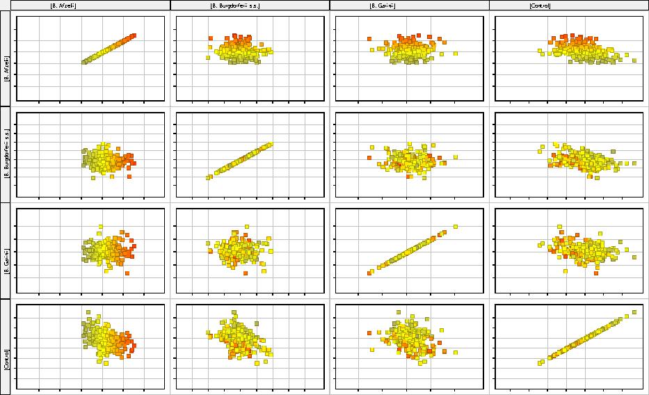
245

246 246

247

248
249
250 Asbrink E, Hovmark A Early and late cutaneous manifestations in Ixodes-borne borreliosis (Erythema migransborreliosis, Lyme borreliosis). Ann NY AcadSci; 539: Burgdorfer W., Barbour A. G., Benach J. L., Grunwaldt E., Davis J. P Lyme disease A tick-borne spirochetosis? Science vol Choudhry Z, Rikani AA, Choudhry AM,Tariq S, Zakaria F, Asghar MW, Sarfraz MK, Haider K, Shafiq A, Mobassarah NJ. Sonic hedgehog signalling pathway: a complex network. Ann Neurosci Jan;21(1): Coller HA, Sang L, Roberts JM. A new description of cellular quiescence. PLoS Biol Mar;4(3):e83. Epub 2006 Mar Darby IA, Laverdet B, Bonté F, Desmoulière A. Fibroblasts and myofibroblasts in wound healing. Clin Cosmet Investig Dermatol Nov 6;7: Dignass AU, Podolsky DK. Cytokine modulation of intestinal epithelial cell restitution: central role of transforming growth factor beta. Gastroenterology Nov;105(5): Gatza CE, Oh SY, Blobe GC. Roles for the type III TGF-beta receptor in human cancer. Cell Signal Aug;22(8): Gern L Borrelia burgdorferi sensu lato, the agent of Lyme borreliosis: life in the 250
251 wild. Parasite 15: Gurney A, Axelrod F, Bond CJ, Cain J, Chartier C, Donigan L, Fischer M, Chaudhari A, Ji M, Kapoun AM, Lam A, Lazetic S, Ma S, Mitra S, Park IK, Pickell K, Sato A, Satyal S, Stroud M, Tran H, Yen WC, Lewicki J, Hoey T. Wnt pathway inhibition via the targeting of Frizzled receptors results in decreased growth and tumorigenicity of human tumors. Proc Natl Acad Sci U S A Jul 17;109(29): Hu X, Chung AY, Wu I, Foldi J, Chen J, Ji JD, Tateya T, Kang YJ, Han J, Gessler M, Kageyama R, Ivashkiv LB. Integrated regulation of Toll-like receptor responses by Notch and interferon-gamma pathways. Immunity Nov 14;29(5): Lin J. C, Duell K, Konopka J. A Microdomain Formed by the Extracellular Ends of the Transmembrane Domains Promotes Activation of the G Protein-Coupled α-factor Receptor Mol Cell Biol Mar; 24(5): Endothelial cells dynamically compete for the tip cell position during angiogenic sprouting Jurgens HA, Johnson RW. Environmental enrichment attenuates hippocampal neuroinflammation and improves cognitive function during influenza infection. Brain Behav Immun Aug;26(6): Kang P, Wan M, Huang P, Li C, Wang Z, Zhong X, Hu Z, Tai S, Cui Y. The Wnt antagonist sfrp1 as a favorable prognosticator in human biliary tract carcinoma. PLoS One Mar 3;9(3):e Kopan R, Ilagan MX. The canonical Notch signaling pathway: unfolding the activation mechanism. Cell Apr 17;137(2): Krakauer T. PI3K/Akt/mTOR, a pathway less recognized for staphylococcal superantigen-induced toxicity. Toxins (Basel) Nov 15;4(11): Larabee JL, Shakir SM, Barua S, Ballard JD. Increased camp in monocytes augments Notch signaling mechanisms by elevating RBP-J and transducin-like enhancer of Split (TLE). J Biol Chem Jul 26;288(30): Martin P. Wound healing--aiming for perfect skin regeneration. Science.1997 Apr 4;276(5309): Murray-Zmijewski F, Slee EA, Lu X. A complex barcode underlies the heterogeneous response of p53 to stress. Nat Rev Mol Cell Biol Sep;9(9): Nardelli D.T., Callister M. S., Schell F. R Lyme Arthritis: Current Concepts and a Change in Paradigm. CLINICAL AND VACCINE IMMUNOLOGY, p Vol Nowak-Chmura M., Siuda K Ticks of Poland. Review of contemporary issues and latest research. Ann Parasitol.; 58(3): Pollina EA, Legesse-Miller A, Haley EM, Goodpaster T, Randolph-Habecker J, Coller 251
252 HA. Regulating the angiogenic balance in tissues. Cell Cycle Jul 1;7(13): Richard S., Oppliger A. Zoonotic occupational diseases in forestry workers Lyme borreliosis, tularemia and leptospirosis in Europe. Ann Agric Environ Med 2015; 22(1); Shapiro ED, Gerber MA. Lyme disease. Clin Infect Dis Aug;31(2): Epub 2000 Sep Simmons GE, Pandey S, Nedeljkovic-Kurepa A, Saxena M, Wang A, Pruitt K. Frizzled 7 expression is positively regulated by SIRT1 and β-catenin in breast cancer cells. PLoS One Jun 4;9(6):e Straubinger RK, Summers BA, Chang JF, Appel MJG Persistence of Borreliaburgdorferi in experimentally infected dogs after antibiotic treatment. J ClinMicrobiol; 35: Takahashi K, Tanabe K, Ohnuki M, Narita M, Ichisaka T, Tomoda K, Yamanaka S. Induction of pluripotent stem cells from adult human fibroblasts by defined factors. Cell.2007 Nov 30;131(5): Tijsse-Klasen E, Sprong H, Pandak N. Co-infection of Borrelia burgdorferi sensu lato and Rickettsia species in ticks and in an erythema migrans patient. Parasit Vectors Dec 10;6: Uren A, Reichsman F, Anest V, Taylor WG, Muraiso K, Bottaro DP, Cumberledge S, Rubin JS. Secreted frizzled-related protein-1 binds directly to Wingless and is a biphasic modulator of Wnt signaling. J Biol Chem Feb 11;275(6): Welc-Falęciak R., Hildebrandt A., Siński E Co-infection with Borrelia species and other tick-borne pathogens in humans: Two cases from Poland. Ann Agric Environ Med 2010, 17, Wong TY, Solis MA, Chen YH, Huang LL. Molecular mechanism of extrinsic factors affecting anti-aging of stem cells. World J Stem Cells Mar 26;7(2): Wu N, Gidrol X. The wind rose of human keratinocyte cell fate. Cell Mol Life Sci Dec;71(24): Wu SY, de Borsetti NH, Bain EJ, Bulow CR, Gamse JT. Mediator subunit 12 coordinates intrinsic and extrinsic control of epithalamic development. Dev Biol Jan 1;385(1): Yamanaka, S. (2007). Strategies and new developments in the generation of patientspecific pluripotent stem cells. Cell Stem Cell 1,
253 Interleukina 8 (CXCL8) i jej receptory w fibroblastach skóry zakażonych krętkami Borrelia burgdorferii Interleukin-8 (CXCL8) and its receptors in skin fibroblast infected spirochete Borrelia burgdorferii Muc -Wierzgoń M. 1, Rajs A 2., Nowakowska Zajdel E. 1, Kokot T 1, Wierzgoń A 2, Gola J. 2, Kruszniewska Rajs C. 2, Dudek S. 3, Jasik K. 4, Kozieł P. 1, Walkiewicz K. 1,Benjamin Grabarek 2, Mazurek U. 2 1 School of Public Health in Bytom, Medical University of Silesia, Katowice, ul. Poniatowskiego 15, Department of Internal Diseases. 2,3,4 School of Pharmacy with the Division of Laboratory Medicine in Sosnowiec, Medical University of Silesia, Katowice, ul. Poniatowskiego 15, 2 Department of Molecular Biology, 3 Department of Pharmacognosy and Phytochemistry, 4 Institute of Structural Skin Research. autor korespondencyjny: ckruszniewska@sum.edu.pl Abstract Introduction: Lyme borreliosis can be caused by at least three species of spirochetes Borrelia: B. burgdorferi sensu stricto, B. garinii and B. afzelii. The mechanisms responsible for the induction of lesions due to infection spirochete are not fully understood. IL8 is a member of the CXC chemokine family, one of the main mediators of the inflammatory response. IL8 also has cytotoxic activity and is involved in many pathological processes, for example: in the etiology of skin diseases or induce rheumatoid arthritis. In this study the profile of expression level of genes encoding interleukin 8 (IL8, CXCL8) and its two receptors was analyzed. Additionally, the expression of genes involved in signaling pathways activated by IL8 was evaluated. Material and methods: Profile of gene expression was analyzed in skin fibroblasts cells (NHDF) infected spirochete Borrelia burgdorferi sensu stricto, B. garinii, and B. afzelii. Profile of mrna expression levels were determined by microarray technology (Affymetrix, HG- U133A). Analysis of the results was conducted using the PL-Grid Infrastructure ( Results: There was no changes in expression profile of genes encoding interleukin 8 and its two receptors ( CXCR1 and CXCR2). Among 612 transcripts for genes involved in IL8 signaling pathway 41 was significantly changed. Wstęp Borelioza (choroba z Lyme) jest chorobą odkleszczową, która przenoszona jest poprzez kleszcze z rodziny Ixodes. Bakteriami wywołującymi tą chorobę są Gram ujemne krętki z rodziny Spirochetaceae - Borrelia burgdorferi sensu lato. Krętki te, z racji różnic genetycznych podzielone zostały na 18 subtypów, z których przede wszystkim trzy: Borrelia 253
254 burgdorferi sensu stricto, Borrelia garinii oraz Borrelia afzelii są patogenne dla człowieka. Zakażenie krętkami Borellia może doprowadzić do ogólnoustrojowej choroby wpływając między innymi na działanie serca, stawów, układu nerwowego i mózgu oraz wywołania zmian skórnych (Biesiada i wsp. 2010). Borrelia burgdorferi sensu stricto, która głównie występuje na obszarach Stanów Zjednoczonych i Europy, charakteryzuje się wywoływaniem głównie objawów stawowych u zakażonych osób. Zakażenia pozostałymi dwoma subtypami, które głównie występują w Europie i Azji, charakteryzuje się przede wszystkim objawami neurologicznymi, szczególnie w zakażeniu Borrelia garinii, gdyż ten patogen wykazuje szczególne powinowactwo do układu nerwowego (Żarowski i wsp. 2007, Zajkowska i Pancewicz 2008). Do zakażenia krętkami dochodzi po ukąszeniu kleszcza, jednak nie od razu, ale po upływie czasu. Czas od ukąszenia do zakażenia pozostaje wciąż obiektem badań. W 2015 roku Cook w swojej pracy analizowała szereg doniesień traktujących właśnie o tym, jaki czas jest konieczny do zakażenia organizmu gospodarza. Wynik jego analizy nie dał jednak konkretnej odpowiedzi na pytanie, a czas od ukąszenia do zakażenia może różnić się w zależności od gatunku kleszcza, jak również od subtypu bakterii i wahać się od kilku godzin do nawet kilku dni. Ryzyko zakażenia Borrelia wzrasta jednak niezaprzeczalnie z czasem, zatem konieczne jest jak najszybsze usunięcie kleszcza (Cook 2015). Skóra jest kluczowym elementem w zakażeniu Borrelia. W pierwszej kolejności jest celem krętków, a manifestacja zakażenia następuje poprzez wystąpienie rumienia wędrującego, który pojawić się może już po kilku dniach od ukąszenia. Rumień wędrujący pojawia się u ok.50-80% pacjentów. Do objawów skórnych pojawiających się w wyniku zakażenia zaliczamy również limfocytomę, która pojawia się rzadko, bo zaledwie u około 1% zakażonych (Żarowski i wsp. 2007). W przewlekłej, nieleczonej boreliozie może dojść również do zanikowego zapalenie skóry, a proces ten obejmuje głównie obszary kończyn. Ta postać zapalenia skóry występuje u chorych zakażonych subtypem B. afzelii, pojawia zazwyczaj wiele lat po ukąszenie i występuje raczej u dorosłych osób (Biesiada i wsp. 2010, Schramm i wsp. 2012). Głównymi komórkami skóry są fibroblasty. Są to komórki, które produkują substancje macierzy łącznotkankowej, czyli substancji włóknistej i podstawowej, a jedną z najważniejszych funkcji fibroblastów jest wytwarzanie kolagenów (Zegarska i Woźniak 2007 ). Prócz swojej podstawowej funkcji związanej z macierzą zewnątrzkomórkową fibroblasty mają zdolność komunikowania się z innymi komórkami tj, komórkami dendrycznymi skóry, komórkami tucznymi, makrofagami i keratynocytami. Ponadto uczestniczyć mogą w procesie homeostazy tkankowej, rekrutacji leukocytów oraz regulacji procesu zapalnego. Szeroki i wyspecjalizowany panel działania fibroblastów w warunkowaniu odpowiedniego środowiska komórek i układu cytokin/chemokin może wskazywać na ich udział również w systemie odpornościowym (Schramm i wsp. 2012). Cytokiny są białkami wydzielanymi przez różne komórki, mające zdolność modulowania wielu zjawisk biologicznych w organizmie. Wiele cytokin bierze udział w regulacji wrodzonej oraz nabytej odpowiedzi immunologicznej (Kontny 2011). Wśród cytokin możemy wyodrębnić grupę chemokin chemotaktycznych cytokin. Chemokiny są białkami wytwarzanymi konstytutywnie lub ich produkcja zostaje indukowana przez odpowiednie czynniki. Główną rolą chemokin jest rekrutacji różnych populacji leukocytów w celu prawidłowego działania układu odpornościowego, jak również w walce z patogenami. Ponadto chemokiny mogą również pełnić funkcje w embriogenezie, organogenezie czy angiogenezie (Waśniowska 2004, Sekuła i Majka 2011). Po zakażeniu krętkami w organiźmie gospodarza dochodzi do indukcji cytokin. Zmiany w profilu cytokin, zarówno na poziomie mrna, jak i białka, obserwowane są przy różnych formach boreliozy tzn. skórnej, stawowej 254
255 czy neuroboreliozy (Grygorczuk i wsp. 2004, Shin i wsp. 2007, Rupprecht i wsp 2008, Ramesh i wsp 2013). Celem niniejszej pracy była ocena zmiany profilu ekspresji genów w komórkach ludzkich fibroblastów (NHDF ang. Normal Human Dermal Fibroblasts) zakażonych trzema subtypami krętków Borrelia tj.: B. burgdorferi sensu stricto, B. garinii oraz B. afzelii względem kontrolnej linii komórkowej NHDF. Analizie zostały poddane transkrypty genów kodujących chemokinę 8 - CXCL8 (interleukinę 8 IL8) oraz receptory, dla których stanowi ona ligand CXCR1 oraz CXCR2. Ponadto analizie został poddany panel genów kodujących białka związane z ścieżkami sygnałowymi, które powiązane są z CXCL8. Materiały i Metody Hodowla komórkowa. Kultury bakterii prowadzone były w pożywce BSK-H (Barbour'a, Stoenner'a, Kelly'ego; Sigma-Aldrich, St. Louis, MO, USA), w mikroaerofilnych warunkach oraz temperaturze 37 o C. Komórki linii NHDF hodowano w dedykowanej pożywce FBM dla fibroblastów (ang. Fibroblast Basal Medium; Lonza, Basel, Szwajcaria) wzbogaconej zasadowym czynnikiem wzrostu fibroblastów, insuliną oraz gentamycyną (FGM TM SingleQuots TM ; Lonza, Basel, Szwajcaria). Hodowla prowadzona była w naczyniach hodowlanych z powierzchnią Nunclon (pow. 25cm 2 ) wyposażonych w filtry bakteriologiczne (Nunc, Wiesbaden, Niemcy). Po osiągnięciu konfluencji dodano do hodowli linii NHDF krętki trzech genogatunków Borrelia: B. burgdorferi sensu stricto, B. garinii oraz B. afzelii ( stosunek ilościowy bakterii do fibroblastów 10:1). Hodowlę komórek linii NHDF w obecności krętków Borrelia oraz hodowle kontrolne NHDF prowadzono przez okres 24 godzin. Po zakończonej inkubacji komórki zostały zebrane i przechowywane do czasy ekstrakcji RNA. Ponadto, po określonym czasie inkubacji fibroblastów w obecności bakterii wykonany została test proliferacji TOX6 (Sigma- Aldrich, St. Louis, MO, USA). Ekstrakcja kwasu rybonukleinowego. Całkowity RNA został wyizolowany z komórek linii NHDF z użyciem odczynnika TRIZOL (Invitrogen, Carlsbad, CA, USA) zgodnie z dołączona instrukcją producenta. Otrzymane ekstrakty całkowitego RNA oczyszczone zostały z wykorzystaniem trawieniea DNazą I (deoksyrybonukleazą I) na kolumnach zestawu RNeasy Mini Kit (Qiagen, Hilden, Niemcy). Otrzymany RNA oceniany został ilościowo z wykorzystaniem spektrofotometru GeneQuant II firmy Pharmacia Biotech (Uppsala, Szwecja) oraz jakościowo techniką elektroforezy w 0,9% żelu agarozowym barwionym bromkiem etydyny. Mikromacierze oligonukleotydowe. Transkryptom mrna został wyznaczony techniką mikromacierzy oligonukleotydowych HG-U133A ( Affymetrix, Santa Clara, CA, USA). Całkowity RNA (8ug) stanowił matrycę do syntezy dwuniciowego cdna (SuperScript Choince System; Invitrogen Life Technologies, Kalifornia, USA). Kolejnym etapem była synteza biotynylowanego crna (BioArray HighYield RNA Transcript Labeling Kit; Enzo Life Science, New York, USA), który następnie pofragmentowano i poddano hybrydyzacji z sondami na płytce HG-U133A. Po 16-to godzinnej hybrydyzacji płytki poddano płukaniu i znakowaniu kompleksem streptawidyna fikoerytryna, zgodnie z protokołem EukGEWS2v4 zalecanym przez producenta. Pomiar intensywności fluorescencji został wykonany przy użyciu skanera GeneArray Scanner G2500A. Analizę otrzymanych wyników przeprowadzono z wykorzystaniem Infrastruktury PL-Grid ( i programu GeneSpring
256 Wyniki Analiza wyników opiera się na porówaniu poziomu ekspresji genów w poszczególnych grupach badanych. W przeprowadzonym doświadczeniu zastosowano 4 grupy badane tj: grupę kontrolną K linia komórkowa NHDF hodowana bez obecności krętków Borrelia oraz trzy grupy badane, czyli linie komórkowe NHDF hodowane w obecności poszczególnych subtypów krętków Borrelia i nazwane od nazwy bakterii tj. : B. sensu stricto, B. afzelii oraz B. garinii W pierwszym etapie analizy ocenie poddano profil ekspresji mrna kodujących CXCL8 oraz jej dwa receptory CXCR1 i CXCR2. Po przeprowadzeniu jednoczynnikowego testu Anova nie zaobserwowano statystycznie znamiennych różnic w poziomie ekspresji analizowanych genów w poszczególnych grupach badanych. Ryc.1 Wynik grupowania hierarchicznego profili znormalizowanych sygnałów fluorescencji 612 mrna w transkryptomach wyznaczonych metodą mikromacierzy oligonukleotydowy HGU 133A. Kolejny etap analizy obejmował panel 612 ID mrna kodujących geny związane z aktywowanymi przez CXCL8 ścieżkami sygnalizacyjnymi. W pierwszej kolejności przeprowadzono klasteryzację hierarchiczną, której wynik wskazał na zróżnicowanie w poziomie ekspresji pomiędzy kontrolna grupą NHDF a fibroblastami zakażonymi krętkami Borrelia. Ponadto zaobserwowano podobieństwo w profilu ekspresji genów w komórkach NHDF po zakażeniu w porównaniu do kontroli. Wynik przedstawiono na Ryc.1. Następny etap analizy polegał na wyznaczeniu genów różnicujących poszczególne grupy badane. W tym celu przeprowadzono jednoczynnikowy test Anova z testem post hoc Tukeya. Wynik testu Anova wskazał, że spośród 612 transkryptów 41 uległo statystycznie istotnej zmianie w poziomie ekspresji (Tab.I) Tab. I Wynik analizy wariancji przeprowadzonej jednoczynnikowym testem ANOVA, wskazujący liczbę ID mrna związanych z aktywnością biologiczną CXCL8, różnicujących grupy transkryptomów komórek fibroblastów skóry zakażonych krętkami Borrelia i grupy kontrolnej. Wartość p Wszystkie ID mrna p < 0,05 p < 0,02 p < 0,01 p < 0,005 p < 0,001 Liczba
257 ID mrna Tab. II. Wynik testu wielokrotnych porównań post-hoc test Tukeya, wskazującego liczbę reprezentatywnych ID mrna różnicujących grupy transkryptomów zakażonych krętkami Borrelia Kolorem czarnym zaznaczono liczbę 41 ID mrna wyznaczonych testem jednoczynnikowej ANOVA, jako różnicujące badane grupy transkryptomów. Kolorem czerwonym zaznaczono liczbę ID mrna, reprezentatywnych dla różnicowania porównywanych grup transkryptomów przy założeniu że wartość p < 0,05. Kolorem zielonym liczbę ID mrna, niereprezentatywnych w różnicowaniu porównywanych grup Grupa transkryptomów B. sensu stricto B. garinii B. afzelii K B. sensu stricto B garinii B. afzelii K Przeprowadzony test Tukeya pozwolił dodatkowo na wskazanie liczby IDmRNA, które różnicują pomiędzy sobą konkretne grupy. Wyniki testu przedstawia Tab.II. Otrzymane listy IDmRNA, które różnicowały badane grupy naniesione zostały na diagram Venn ( Ryc.2), który pozwolił na przedstawienie graficzne zbiorów genów różnicujących wspólnych oraz specyficznych dla przeprowadzonych porównań. Zaobserwowano, że tylko 3 IDmRNA są wspólne dla różnicowania komórek NHDF zakażonych poszczególnymi subtypami bakterii od kontroli. Są to transkrypty kodujące receptor naskórkowego czynnika wzrostu- EGFR (ang. epidermal growth factor receptor), czynnik wzrostu fibroblastów 13 FGF13 ( ang. fibroblast growth factor 13) oraz zależną od wapnia i kalmoduliny kinazę typu 2 - CAMKK2 (calcium/calmodulin-dependent protein kinase kinase 2). EGFR oraz CAMKK2 ulegają wyciszeniu, natomiast FGF13 ulega nadekspresji w porównaniu wszystkich 3 grup badanych względem kontroli. 257

258 Ryc. 2 Diagram Venn grupujący mrna wytypowane testem post-hoc Tukeya po teście jednoczynnikowym ANOVA (Tab.II) w zależności od specyficzności różnicowania grup transkryptomów. Tab.III. Wykaz transkryptów różnicujących badane grupy względem kontroli specyficznych dla poszczególnych porównań oraz kierunek zmiany ekspresji w komórkach NHDF zakażonych krętkami Borrelia. B. afzelii vs K B.garinii vs K B.sensu stricto vs K Transkrypty ulegające wyciszeniu Transkrypty ulegające nadekspresji MAPK12, BNIP2 MAPKAPK2, MAP3K8, FOS, TGFB3, ELK1, PXN, SHC1 RGS4, RGS4, MAP2K3, DUSP6, HGF, NF1, HRAS, CSPG4, KRAS, TAOK3 MAP3K11, TAOK2, MAP3K12 FPR1, CD24, GRM1, CXCR4, MET Dyskusja Zakażenia bakteryjne wpływają na zmianę profilu ekspresji i produkcji białek biorących udział w odpowiedzi immunologicznej przeciwko patogenowi. W trakcie zakażenie dochodzi do uwolnienia szeregu cytokin, między innymi chemokiny CXCL8 znaną również pod nazwą interleukina-8 (IL8), która jest silną cytokiną prozapalną. CXCL8 działa głównie jako chemoatraktant dla neutrofili i limfocytów, powodując ich migrację do miejsca zakażenia. Ponadto, CXCL8 pobudza aktywacje prozapalnych proteaz- metaloproteinazy 2 i 9 czy propaptycznego białka BIM (Ramesh i wsp. 2013). Poprzez aktywacje receptorów CXCL-1 i/lub CXCL-2 dla których jest ona ligandem dochodzi do uruchomienia kaskady sygnałowej i aktywacji szlaków związanych z obroną organizmu. Rola układu CXCL8 i receptorów CXCL-1 i CXCL-2 opisywana w odniesieniu do centralnego układu nerwowego, gdzie aktywność wzrasta podczas procesów patologicznych (Semple i wsp. 2010), może to sugerować również 258
259 o udziale tej chemokiny w zaburzeniach neurolgicznych pojawiających się przy neuroboreliozie. Ocena aktywności CXCL8 w zakażeniu Borrelia została podjęta przez różne zespoły badawcze. W większości przypadków badania dowodziły o znacznym wzroście aktywności tej chemokiny podczas zapalenia. W dużej mierze wyniki badań opierają się na ocenia poziomu ekspresji białka CXCL8, zarówno w hodowlach komórkowych, jak i próbkach pobranych od chorych. Grygorczuk i wsp, w swoim doświadczeniu oceniali poziom CXCL8 u chorych z różnymi odmianami boreliozy (tj. z występującym rumieniem wędrującym, stawową postacią boreliozy oraz z neuroboreliozą). Poziom chemokiny oceniali w surowicy oraz w płynie mózgowo-rdzeniowym u chorych na neuroboreliozę. We wszystkich przypadkach odnotowali wzrost poziomu białka CXCL8 w odniesieniu do grupy kontrolnej (Grygorczuk i wsp. 2004). Podobne wyniki otrzymali również inni Moniuszko i wsp. u chorych z neuroboreliozą (Moniuszko i wsp. 2014). Ponadto, niezależne badania Grygorczuk i wsp. oraz Shin i wsp. pokazują, że poziom tej chemokiny spada po zastosowaniu terapii antybiotykowej u chorych, którzy odpowiadają na leczenie (Grygorczuk i wsp. 2004, Shin i wsp. 2007). Poziom CXCL8 badany był również w hodowlach komórkowych, które infekowano krętkami Borrelia. Takie doświadczenie przeprowadził w 2013 między innymi zespół Ramesh i wsp. badając między innymi wpływ krętków na wydzielanie cytokin w komórkach zwoju korzenia grzbietowego małp oraz w ludzkich komórkach Schwanna, obserwując wzrost CXCL8 w obu przypadkach (Ramesh i wsp. 2013). W przedstawionej pracy ocenie poddano profil ekspresji genów kodujących CXCL8 raz jej dwa receptory. Po 24 godzinnej inkubacji komórek NHDF z trzema subtypami Borrelia nie odnotowano istotnych zmian na poziomie ekspresji mrna badanych genów. Sugerować to może, że po tym czasie być może CXCL8 nie jest aktywna na poziomie mrna, ale jako funkcjonujące białko. Dopełnieniem więc przeprowadzonych badań byłaby ocena poziomu białka po przeprowadzonej inkubacji, bądź też analiza ekspresji genów w krótszym czasie od zainfekowania. Potwierdzeniem tego mogą być badania Sandholm i wsp. którzy wykazali, wzrost aktywności CXCL8 oraz innych cytokin na poziomie białka już po 4-ro godzinnej inkubacji krwi z krętkami Borrelia, co wskazuję na szybką odpowiedzi immunologicznej po zakażeniu (Sandholm i wsp. 2014). W przeprowadzonym przez autorów eksperymencie analizie poddano również panel 612 IDmRNA genów związanych z aktywnością CXCL8. Mimo, iż w przypadku samej chemokiny i jej receptorów nie odnotowano różnic w poziomie ekspresji, to zmiana w profilu genów z nią związanych jest zauważalna. W pierwszej kolejności przeprowadzona została klasteryzacja hierarchicza ( ryc.1), która pozwoliła na wykazanie podobieństwa pomiędzy badanymi grupami transkryptomów. Po przeprowadzonej analizie widoczny jest podział na dwie grupy, gdzie w jednej została umieszczona tylko kontrola, natomiast w drugiej wszystkie 3 grupy badane. Sugeruje to, że w przypadku zakażenia wszystkimi trzema subtypami Borrelia dochodzi do zmiany w profilu genów związanych z CXCL8. Spośród 612 transkryptów 41 charakteryzuje się zmienionym poziomem ekspresji. Próba wytypowania genów różnicujących wskazała po przeprowadzonym teście Anova, że 41 transkryptów wykazuje statystyczne różnice w poziomie ekspresji, a test post-hoc Tukeya pozwolił na sprecyzowanie genów charakterystycznych dla różnicowani poszczególnych grup. Wśród genów różnicujących 3 transkrypty były wspólne dla przeprowadzonych porównań. Wskazywać to może, że aktywność tych trzech genów ulega zmianie po zakażeniu krętkami Borrelia, niezależnie od subtypu patogenu. Ponadto warto tu zauważyć, że wszystkie trzy analizowane subtypy wywołały ten sam kierunek zmian ekspresji. Większość spośród genów różnicujących komórki NHDF zakażone odpowiednimi subtypami bakterii względem kontroli 259
260 jest specyficznych dla poszczególnych porównań. Taki wynik może być skutkiem różnic w mechanizmie działania poszczególnych subtypów bakterii. Strle i wsp. wykazali, że różnice w poziomie aktywności cytokin i chemokin po zakażeniu mogą być wynikiem obecności innego subtypu patogenu w organizmie. Badania ich pokazały, że krętki Borrelia burgdorferi w znaczniejszym stopniu indukują produkcję cytokin i chemokin niż porównywane Borrelia garinii i Borrelia afzelii (Strle i wsp. 2009). Różnice te mogą być może są również przyczyną występowania różnych objawów boreliozy. Różnorodność genów różnicujących w poszczególnych porównaniach grup badanych może również wynikać z faktu, że subtypy Borrelia charakteryzuje inny czas zakażania organizmu gospodarza, o czym w swojej pracy przeglądowej napisał Cook (Cook 2015). Zatem po czasie 24 godzin po zainfekowaniu komórek różnymi subtypami komórki linii NHDF mogły być na różnym etapie zakażenia. Otrzymane wyniki nie dały konkretnego obrazu zmian w profilu ekspresji genu CXCL8 w odpowiedzi na zakażenie krętkami Borrelia. Mimo że nie wykazano różnic na poziomie mrna dla chemokiny i jej receptorów, to jednak widoczne są zmiany w aktywności genów związanych ze ścieżkami, na które wpływa CXCL8. Sugeruje to zatem, że chemokina ta bierze udział w mechanizmach obronnych po zakażeniu krętkami, co potwierdzają liczne badania wskazujące na jej istotny wzrost na poziomie białka. Warto to jednak wspomnieć, że cytokiny/chemokiny i ich receptory wykazują się redundancją, czyli wywoływaniem takiego samego efektu w komórce przez różne cytokiny, dlatego że dla jednego receptora może być kilka różnych ligandów. Przedstawiony w pracy eksperyment i otrzymane wyniki wymagają zatem dalszej analizy i dopełnienia szerszym panelem badań. Spis literatury 1. Biesiada G., Czepiel J., Leśniak M., Garlicki A., Mach T Lyme disease: review. Arch Med Sci. 20; 8(6): Cook M Lyme borreliosis: a review of data on transmission time after tick attachment. Int J Gen Med. 8: GrygorczukS., Pancewicz S., Zajkowska J., Kondrusik M., Świerzbińska R., Hermanowska- Szpakowicz T Concentrations of Macrophage Inflammatory Proteins MIP-1α and MIP- 1 and Interleukin 8 (Il-8) in Lyme Borreliosis. Infection. 32: Kontny E Patogeneza reumatoidalnego zapalenia stawów. Część III cytokiny i procesy destrukcyjne. Reumatologia. 49(3): Moniuszko A., Czupryna P., Pancewicz S., Rutkowski K., Zajkowska O., Swierzbińska R., Grygorczuk S., Kondrusik M., Owłasiuk P., Zajkowska J Evaluation of CXCL8, CXCL10, CXCL11, CXCL12 and CXCL13 in serum and cerebrospinal fluid of patients with neuroborreliosis. Immunol Lett. 157(1-2): Ramesh G., Santana-Gould L., Inglis F., England J., Philipp T. The Lyme disease spirochete Borrelia burgdorferi induces inflammation and apoptosis in cells from dorsal root ganglia J Neuroinflammation. 10: Rupprecht T., Koedel U., Fingerle V., Pfister HW The Pathogenesis of Lyme Neuroborreliosis: From Infection to Inflammation. Mol Med. 14(3-4): Sandholm K., Henningsson A., Säve S., Bergström S., Forsberg P., Jonsson N., Ernerudh J., Ekdahl K arly cytokine release in response to live Borrelia burgdorferi Sensu Lato Spirochetes is largely complement independent. PLoS One. 9(9):e Schramm F., Kern A., Barthel C., Nadaud S., Meyer N., Jaulhac B., Boulanger N Microarray analyses of inflammation response of human dermal fibroblasts to different strains of Borrelia burgdorferi sensu stricto. PLoS One. 7(6):e
261 10. Sekuła M., Majka M Udział chemokin w wybranych procesach komórkowych. Journal of Laboratory Diagnostics. 47 (3): Semple B., Kossmann T., Morganti-Kossmann M Role of chemokines in CNS health and pathology: a focus on the CCL2/CCR2 and CXCL8/CXCR2 networks. J Cereb Blood Flow Metab. 30(3): Shin J., Glickstein L., Steere A High Levels of Inflammatory Chemokines and Cytokines in Joint Fluid and Synovial Tissue Throughout the Course of Antibiotic-Refractory Lyme Arthritis. ARTHRITIS & RHEUMATISM. 56 (4): Strle K., Drouin E., Shen S., El Khoury., McHugh G., Ruzic-Sabljic E., Strle F., Steere A Borrelia burgdorferi stimulates macrophages to secrete higher levels of cytokines and chemokines than Borrelia afzelii or Borrelia garinii. J Infect Dis. 200(12): Waśniowska K Chemokiny - perspektywy zastosowania związków blokujących ich działanie w terapii. Postepy Hig Med Dosw. 58: Zajkowska J., Pancewicz A Wybrane aspekty patogenezy i diagnostyki neuroboreliozy. Polski Przegląd Neurologiczny.3(2): Zegarska B., Woźniak M Wpływ estrogenu na zmiany zachodzące w skórze. Przegląd Menopauzalny. 4: Żarowski M., Justyna Młodzikowska-Albrecht J., Steinborn B Symptomatologia boreliozy u dzieci i młodzieży leczonych w Klinice Neurologii Wieku Rozwojowego AM w Poznaniu. Neurologia Dziecięca. 15(30):
262 262
263 Analiza histologiczna wątroby szczura pod wpływem terapii przeciwpierwotniakowej azytromycyną, atowakwonem i prokwanilem Jan Słodki 1, Hubert Okła 1,2, Krzysztof P. Jasik 1, Aleksandra Słodki 1, Aniela Grajoszek 3, Beata Rozwadowska 1,4, Marta Albertyńska 1,4 1 Zakład Badań Strukturalnych Skóry, Katedra Kosmetologii Wydział Farmaceutyczny z Oddziałem Medycyny Laboratoryjnej w Sosnowcu Śląski Uniwersytet Medyczny w Katowicach, Sosnowiec, ul. Kasztanowa 3, , jan.slodki@gmail.com 2 Instytut Nauki o Materiałach, Wydział Informatyki i Nauki o Materiałach, Uniwersytet Śląski w Katowicach, ul. 75 Pułku Piechoty 1A, Chorzów 3 Centrum Medycyny Doświadczalnej Śląski Uniwersytet Medyczny w Katowicach, Katowice, ul. Medyków 4, Wojewódzka Stacja Sanitarno-Epidemiologiczna w Katowicach, Katowice, ul. Raciborska 39, Histological analysis of rat s liver changed by antiprotozoal therapy consisted of azithromycin, atovaquone and proquanil. Abstract Treatment of pregnant organisms is always a challenge for medicine. Our previous studies have shown that drug therapy consisting of azithromycin, atovaquone and proguanil used in pregnant rats infected with B. microti is hepatotoxic. It is not known what was the most aggravating factor babesiosis, pregnancy or pharmacotherapy. Therefore, we decided to check and compare if pregnancy itself may be the most aggravating factor. For this purpose we have done an experience, which has proved how azithromycin, atovaquone and proguanil affects the liver of uninfected nonpregnant rats. Materials and methods: The used material from 20 females. The first group consisted of five livers collected during the section from non-infected, non-pregnant female rats treated for seven days with azithromycin, atovaquone and proguanil. The material was analyzed by light microscopy prepared by standard histological procedure, stained by Masson's tricoloured method. The second control group consisted of five uninfected female rats, pregnant, untreated, and administered only saline for seven days. The third control group consisted of liver obtained from infected pregnant rats treated by this same pattern as the study group in this experiment. The fourth control group consisted of five livers derived from infected, 263
264 untreated pregnant female rats. Results: In our study have not observed significant changes in the morphological structure of the liver. There were neither major necrotic nor vacuolic changes. Structure of the blood vessels showed proper construction. The only significant change observed in the material was different degree in color saturation of hepatocytes. This condition may be associated with changes in their metabolism as a result of the applied pharmacotherapy. Conclusion: Treatment with azithromycin, atovaquone and proguanil does not cause major changes in the structure and functioning of the liver. Key words: Babesia microti, pregnancy, azithromycin, atovaquone, proquanil, liver and histology. 1.Wstęp Wątroba jako centrum metaboliczne jest jednym z najważniejszych narządów w organizmie ludzkim i zwierzęcym. Odpowiada za produkcje białek niezbędnych do życia oraz związków istotnych dla trawienia. Pełni również ważną funkcję w metabolizmie, sprzęganiu i usuwaniu farmaceutyków z organizmu m. in za pomocą sprzęgania cząsteczek chemicznych z kwasem glukuronowym. Przy udziale cytochromu P 450 w wątrobie odbywają się pierwsze etapy aktywacji dużej ilości leków. W leczeniu babeszjozy używane są substancje, które poprzez swój mechanizm działania mogą bezpośrednio negatywnie oddziaływać na wątrobę leczonego organizmu. Do takich należą atowakwon, prokwanil i azytromycyna. Atowakwon to substancja chemiczna należąca do grupy naftochinonów, pochodnych naftalenu. Jest on syntetycznym analogiem koenzymu Q, czyli białkiem mitochondrialnym biorącym udział w transporcie elektronów (Pudney 1999). Wykazuje on szerokie spektrum m. in. przeciwko Plasmodium spp., Pneumocystis carinii, Babesia spp., Toxoplasma gondii. Mechanizm działania w największym stopniu został poznany na modelu Plasmodium spp..wiadomo, że przez swoje podobieństwo do ubichinonu zaburza on transport elektronów w łańcuchu oddechowym. Skutkuje to zniszczeniem potencjału na błonie mitochondrialnej (Srivastava 1997). W efekcie ograniczona zostaje synteza wielu enzymów niezbędnych do życia pierwotniakom np. DHOD dehydrogenaza dihydroorotanowa biorącą udział w syntezie pirymidyn. Enzym ten jest niezbędny do życia Plasmodium więc jego zaburzenie skutkuje śmiercią patogenu (Hammond 1985). Z różnicą potencjałów elektrycznych na błonie związana jest też synteza ATP. Synteza ta również może być zakłócona przez atowakwon (Hudson 1991). Odziaływanie na Babesia spp. nie jest dokładnie poznane ale podejrzewa się, że ten sam mechanizm tj. zniszczenie transportu elektronów w mitochondrium odpowiada za właściwości przeciewpierwotniakowe wobec innych patogenów. Biorąc pod uwagę fakt, że wątroba zawiera dużą liczbę mitochondriów z racji pełnienia swoich funkcji fizjologicznych pada podejrzenie, że atowakwon może negatywnie wpływać na jej funkcjonowanie. Prokwanil jest prolekiem, którego mechanizm działania został poznany na podstawie obserwacji na Plasmodium spp.. Prokwanil ma niewielką aktywność przeciwpierwotniakową. W postaci niezmetabolizowanej w znacznym stopniu obniża efektywną dawkę atowakwonu. Po transformacji w wątrobie przy udziale cytochromu P450 3A i 2C do aktywnej formy cykloguanilu staje się aktywnym inhibitorem reduktazy dihydrofolianowej. Enzym ten uczestniczy w przemianach kwasu foliowego, co jest niezbędne w procesie reprodukcyjnym pasożyta. W efekcie pasożyt nie może się namnażać w krwinkach 264
265 czerwonych. Ze względu na nieliczne doniesienia o jego wpływie na wątrobę, mechanizm hepatotoksyczności nie został do tej pory opisany. Azytromycyna to lek, który w badaniach na chomikach wykazał, że jego dodatek do kuracji atowakwonem zmniejszył liczbę nawrotów babeszjozy spowodowanej inwazją B. microti (Wittner 1996). Krause w badaniu porównującym skuteczność azytromycyny z atowakwonem w stosunku do klindamycyny z chininą, wykazał porównywalną skuteczność eliminowania patogenu. Azytromycyna z atowakwonem wywołała zdecydowanie mniejszą liczbę działań niepożądanych (Chandrupatla 2002). Mechanizm bójczy azytromycyny w stosunku do Babesia spp. pozostaje nieznany. W literaturze fachowej pojawiają się hipotezy, że długi okres półtrwania oraz zdolność do penetracji tkanek może odgrywać tu znaczącą rolę. Babeszjoza ciężarnych szczurów wywołana inokulacją Babesia microti, zastosowana farmakoterapia, jak również sama ciąża wpływają hepatotoksycznie na wątroby samic szczura (Słodki i wsp. 2014). Nie wiadomo która zmienna wpływa najbardziej niekorzystnie na ten narząd. Z tego powodu przeprowadzono analizę wpływu leczenia azytromycyną, atowakwonem i prokwanilem na wątrobę samic szczurów nieciężarnych i niezarażonych B. microti. 2. Materiały i metody. W doświadczeniu użyto 20 szczurów szczepu Wistar (tab. 1). Zwierzęta przechowywano w metalowych klatkach i hodowanych w standardowych warunkach laboratoryjnych. Aby zrealizować cel badawczy, zwierzęta podzielono na 4 grupy, tzn. grupę badawczą i 3 kontrolne. Samice niezarażone, nieciężarne, leczone Pięć samic poddano farmakoterapii składającej się z azytromycyny w dawce 150mg/kg/dzień (Zetamax, Pfizer Europe, Sandwich, Wielka Brytania), atowakwonu 100mg/kg/dzień oraz prokwanilu w dawce 40mg/kg/dzień Malarone, Glaxo Wellcome S.A., Aranda de Duero, Burgos, Hiszpania). Atowakwon i prokwanil występują na rynku polskim wyłącznie w postaci tabletki pod handlowa nazwą Malarone. Preparat ten zostały roztarty w moździerzu aptecznym w celu uzyskania leku w postaci sypkiej. Następnie preparat odważono proporcjonalnie do wagi zwierzęcia. Azytromycyna pod nazwą handlową Zetamax występuje w postaci granulatu, dlatego lek ten został odważony bez konieczności zmiany formulacji. Leki rozpuszczono w soli fizjologicznej i podawano sondą dożołądkową o stałej porze dnia, tj. o szóstej rano. Po siedmiu dniach terapii samice poddano eutanazji poprzez inhalację izofluranem a następnie pobrano narządy do analizy. Wątroby przeznaczone do analizy histologicznej utrwalono w roztworze Bouina w temperaturze pokojowej. Stosując rutynową technikę histologiczną materiał zatopiono w parafinie, a następnie przy użyciu mikrotomu saneczkowego uzyskano skrawki grubości 6 um. Po wybarwieniu metodą Massona, skrawki odwodniono i zamknięto w żywicy DPX (POCh, Gliwice, Polska). Preparaty analizowano w mikroskopie świetlnym Olympus BX60. Samice zarażone, ciężarne, leczone 265
266 Pierwszą grupę kontrolną stanowiły samice które zarażono poprzez inokulację dootrzewnową (ip) 0.5 ml krwi pochodzącej od szczura zarażonego szczepem wzorcowym B. microti (ATCC ). Poziom parazytemii kontrolowano za pomocą analizy cienkiego rozmazu z krwi obwodowej barwionej metodą May-Grünwalda-Giemsy (POCh, Gliwice, Polska). W trzecim tygodniu parazytemii samice zostały pokryte metodą haremową. Moment zapłodnienia kontrolowano wymazami cytologicznymi. W czternastym dniu ciąży wprowadzono leki wg tego samego reżimu co w grupie badanej. Po siedmiu dniach leczenia, czyli w dwudziestym pierwszym dniu ciąży dokonano eutanazji, oraz sekcji, podczas której pobrano narządy do analizy. Samice zarażone, ciężarne, nieleczone Drugą grupę kontrolną stanowiło pięć zarażonych, nieleczonych samic, którym podawano roztwór soli fizjologicznej sondą dożołądkową od 14 dnia ciąży. Zarażenie i reszta procedur była taka sama jak w pierwszej grupie kontrolnej. Materiał również pobrano podczas sekcji w 21 dniu ciąży. Samice niezarażone, ciężarne, nieleczone Trzecią grupę kontrolną stanowiły samice niezarażone, ciężarne, nieleczone od których pobrano materiał podczas sekcji w 21 dniu ciąży. Ilość szczurów Zarażenie Ciąża Leczenie Grupa badana 5 Nie Nie Tak Pierwsza grupa kontrolna 5 Tak Tak Tak Druga grupa kontrolna Trzecia grupa kontrolna 5 Tak Tak Nie 5 Nie Tak Nie Tab. 1. Charakterystyka grup szczurów wykorzystanych w doświadczeniu. 3. Wyniki Obserwacje makroskopowe. Wątroby zwierząt niezarażonych, nieciężarnych, leczonych. W trakcie sekcji nie zaobserwowano zmian anatomicznych pobieranych narządów. Były one prawidłowo rozwinięte bez obrzęków, przebarwień ani krwawych wybroczyn. Wątroby szczurów zarażonych, ciężarnych, leczonych. Wątroby zarażonych, leczonych samic charakteryzowały się licznymi przekrwieniami oraz niejednorodną barwą. Ponadto były powiększone, obrzmiałe z żółtobrunatnymi naciekami (Ryc. 1) 266
. Torebka łącznotkankowa nie była pogrubiona (Ryc. 3).")
267 Obserwacje w mikroskopie świetlnym wątroby szczurów niezarażonych, nieciężarnych, leczonych Analiza nie ujawniła poważnych zmian wątroby na poziomie histologii ogólnej i szczegółowej. Nie odnotowano ani zmian wakuolizacyjnych ani nekrotycznych (Ryc. 2). Torebka łącznotkankowa nie była pogrubiona (Ryc. 3). Nie odnotowano zwiększonej ilości kolagenu ani jego wnikania w głębsze warstwy wątroby. W trakcie analizy zaobserwowano różnicę barwliwości jąder oraz cytoplazmy hepatocytów (Ryc. 4). Hepatocyty wykazywały prawidłowe kształty i rozmiary we wszystkich trzech strefach gronek wątrobowych. Również budowa naczyń krwionośnych była prawidłowa (Ryc. 5). Ryc. 1 Żółtobrunatne nacieki na obrzmiałej, powiększonej wątrobie samic zarażonych, ciężarnych, leczonych Ryc. 2. Prawidłowa struktura hepatocytów. Brak zmian wakuolizacyjnych i nekrotycznych. Wątroba niezarażonej, nieciężarnej leczonej samicy. Ryc. 3. Niepogrubiona torebka łącznotkankowa otaczająca wątrobę. Brak zwiększonej ilości kolagenu w strukturze narządu. Wątroba niezarażonej, nieciężarnej leczonej samicy. 267
268 Ryc. 4. Różnobarwliwość hepatocytów. Komórki wykazują prawidłowy kształt. Wątroba niezarażonej, nieciężarnej leczonej samicy. Ryc. 5. Prawidłowa struktura naczynia krwionośnego. Różna barwliwość hepatocytów w bliskim otoczeniu naczynia krwionośnego. Wątroba niezarażonej, nieciężarnej leczonej samicy. Ryc. 6. Różnobarwliwość hepatocytów. Zmiany wakuolizacyjne i nekrotyczne w strukturze wątroby. Wątroba zarażonej, ciężarnej leczonej samicy. Ryc. 7. Niewielkie zmiany wakuolizacyjne oraz pasma tkanki łącznej w strukturze wątroby zarażonych, ciężarnych, nieleczonych samic. Wątroba niezarażonej, ciężarnej, nieleczonej samicy. Obserwacje w mikroskopie świetlnym wątroby zwierząt zarażonych, ciężarnych, leczonych Poszczególne hepatocyty w wątrobach zarażonych, leczonych samic szczurów wykazywały różnobarwliwość względem siebie. Jednocześnie ich cytoplazma była homogenna, jednolicie wybarwiona w porównaniu z heterogenna, niejednolicie wybarwioną cytoplazmą grupy kontrolnej. W bezpośredniej okolicy naczyń krwionośnych oraz przestrzeni Dissego zaobserwowano liczne komórki Kupffera. W naczyniach żylnych wątroby widoczne były monocyty, limfocyty oraz neutrofile w większej ilości niż w porównywalnych miejscach w preparatach otrzymanych z grupy zwierząt kontrolnych. W preparatach widoczna jest duża ilość zmian wakuolizacyjnych oraz wykrzepin krwi (Ryc. 6). Odnotowano również pasma tkanki łącznej w bezpośredniej okolicy komórek Ito. Obserwacje w mikroskopie świetlnym wątroby zwierząt zarażonych, ciężarnych, nieleczonych. 268
269 W obserwowanym materiale zaobserwowano podobne zmiany jak w przypadku preparatów pochodzących od zarażonych leczonych szczurów ale w znacznie mniejszym stopniu. Widoczne są zmiany wakuolizacyjne, wykrzepiny krwi oraz pasma tkanki łącznej w okolicy komórek Ito ale są one mniejsze i rzadziej spotykane w porównaniu z grupą zarażoną, ciężarną, leczoną (Ryc. 7.). Dodatkowym powtarzalnym obrazem są liczne metorozity B. microti obserwowane w przekrojach przez erytrocyty w świetle naczyń krwionośnych. Wątroby zwierząt niezarażonych, ciężarnych, nieleczonych (dane niepublikowane). W materiale uzyskanym do zarażonych, ciężarnych, nieleczonych samic obserwowano prawidłową strukturę wątroby. Nie odnotowano zmian nekrotycznych ani wakuolizacyjnych. Hepatocyty wykazywały prawidłową, jednorodną barwliwość. W naczyniach krwionośnych w okolicy przestrzeni Dissego nie obecne były komórki układu odpornościowego. Torebka otaczająca wątrobę nie była pogrubiona. Pomiędzy hepatocytami nie dostrzeżono włókien kolagenowych. 4. Dyskusja Wpływ azytromycyny na wątrobę Azytromycyna jest uznawana za bezpieczny lek wykorzystywany w leczeniu infekcji górnych dróg oddechowych oraz zapalenia ucha środkowego. Istnieje niewielka ilość doniesień o jej negatywnym wpływie na wątrobę. Na hepatotoksyczność azytromycyny ma wpływ jej długi czas półtrwania oraz osiąganie stężenia 50 razy większego w tkankach niż w surowicy (Das 2011). Istnieją doniesienia opisujące przypadki pacjentów którzy w wyniku reakcji nadwrażliwości na azytromycynę doświadczyli cholestatycznego zapalenia wątroby, co wymagało jej transplantacji. W biopsji obserwowano cholestazę wewnątrzwątrobową oraz nacieki limfocytów i eozynofili w okolicy żyły wrotnej. Syndromy te ustąpiły po odstawieniu leku. (Chandrupatla 2002, Irvine 2011).W analizie przypadku 26 - letniej kobiety, u której pojawiło się cholestatyczne zapalenie wątroby w wyniku zastosowania azytromycyny biopsja wykazała zmniejszenie prześwitu dróg żółciowych oraz złogi włóknika w okolicy triad wątrobowych. Odnotowano zmniejszone światło przewodów żółciowych oraz przewlekły, umiarkowany stan zapalny z charakterystycznym naciekiem licznych komórek plazmatycznych, neutrofili i eozynofili. Badanie szczegółowe wykazało złogi żółci, makrofagi o budowie piankowej ze względu na duża ilość fagosomów, niewielkie ilości ziarniniaków, zmiany wakuolizacyjne i pleomorfizm hepatocytów, zgrupowania glikogenu, ciała Councilmana w niewielkich ilościach, oraz martwiczo-zapalne ogniska w miąższu wątrobowym (Maggioli 2012). Obserwacje te są częściowo zgodne z wynikami naszej poprzedniej pracy (Słodki i wsp. 2014). Zastosowana przez nas kuracja składająca się z azytromycyny, prokwanilu i atowakwonu u zarażonych, ciężarnych zwierząt spowodowała zmiany o charakterze hepatotoksycznym. Wiele komórek wątrobowych wykazywało zmiany nekrotyczne lub/i apoptotyczne oraz zawierało krople lipoproteidowe. W analizowanych preparatach obserwowano komórki Kupffera w bezpośredniej okolicy przestrzeni Dissego. W naczyniach żylnych wątroby widoczne były monocyty, limfocyty oraz neutrofile w większej ilości niż w preparatach uzyskanych od zwierząt niezarażonych, ciężarnych, nieleczonych, do których 269
270 były porównywane. Zmian tych nie zaobserwowano również w aktualnym doświadczeniu u nieciężarnych, niezarażonych, leczonych samic. Chandrupatla (2002) przedstawili przypadek pacjenta, u którego przyjmowanie azytromycyny przyczyniło się do wystąpienia cholestazy wewnątrzzrazikowej oraz wewnątrz kanalików żółciowych a nagromadzenie z tego powodu mediatorów stanu zapalnego doprowadziło do powstania stanu zapalnego w tkankach wokół żyły centralnej i wrotnej. Mimo zwiększonej ilości eozynofilii nie udowodniono stanu zapalnego przewodów żółciowych, okołoprzewodowych obrzęków, zwłóknień ani uszkodzeń wątroby. Wpływ i bezpieczeństwo stosowania azytromycyny na wątrobę zwierząt ciężarnych Azytromycyna jest substancją, która może być użyta w zwalczaniu malarii w trakcie ciąży (Nosten i wsp. 2006). Er (2013) wykazał na modelu zwierzęcym, że azytromycyna może być użyteczna w przypadkach zapobiegania utraty ciąży w wyniku zakażeń. Badanie przeprowadzone na zwierzętach przez Kemp i wsp. (2014) również sugeruje, że jest ona bezpieczna w ciąży. Nasze aktualne doświadczenie na nieciężarnych, niezarażonych leczonych szczurach nie wykazało istotnych zmian w strukturze wątroby. Sugeruje to, że sama farmakoterapia nie jest hepatotoksyczna. Z drugiej strony taka sama farmakoterapia w połączeniu z zarażeniem B. microti oraz ciążą w poprzednim doświadczeniu wywołała efekt hepatotoksyczny oraz prawdopodobnie przyczyniła się do przedwczesnej śmierci części badanych zwierząt. Wpływ i bezpieczeństwo stosowania Atowakwonu / Proquanilu (Malarone) na wątrobę Malarone jest wykorzystywany przede wszystkim w zapobieganiu malarii. Lek jest używany w lecznictwie od niedawna, dlatego istnieje skąpa ilość przypadków opisujących powikłania po jego stosowaniu. Dane na temat jego wpływu na wątrobę ograniczają się do przejściowego podwyższenia wartości enzymów wątrobowych (Hitani 2006). Nie istnieją informacje o wpływie preparatu Malarone na wątroby ciężarnych organizmów. Istnieje również mało informacji na temat wpływu atowakwonu / proquanilu na ciężarne organizmy (Björn 2011, AlKadi 2007). Na-Bangchang (2005) podawali przez 3 dni Malarone w 3 trymestrze kobietom ciężarnym. Ocenili, że jest on efektywny i bezpieczny. Pacjentki dobrze tolerowały kuracje atowakwonem/prokwanilem, nie zaobserwowano ani poważnych skutków ubocznych ani samoistnych poronień. W badaniu kohortowym Penelope i wsp. ocenili, że przyjmowanie m. in. proquanilu w pierwszym trymestrze nie miało znaczącego wpływu na nieprawidłowe urodzenia (Phillips-Howard 1998). Inne badanie, potwierdziło, że kombinacja atowakwonu i diclazuril jest bezpieczna dla ciąży (Phillips-Howard 1998). Bezpieczeństwo stosowania atowakwon/proquanil w ciąży potwierdziły też inne zespoły (Irvine 2011, Nosten 2006, McGready 2005, McGready 2003). W naszym poprzednim badaniu nie doszło do samoistnych poronień. LD 50 nie zostało przekroczone, jednak w pojedynczych przypadkach doszło do przedwczesnego zgonu badanych zwierząt. Trudno jest jednoznacznie stwierdzić co było tego przyczyną zgonu czy farmakoterapia czy inokulacji B. microti czy może ciąża. W innym badaniu na ciężarnych myszach udowodniono, że terapia atowakwonem znacząco ochroniła samice przed zapaleniem wątroby, powiększeniem śledziony, zapaleniem jelita grubego, zapaleniem mięśnia 270
271 sercowego, oraz bólu w wyniku zarażenia toksoplazmozą (Oz 2012 ). W naszym doświadczeniu śledziony nie były analizowane, jednak wątroby po zastosowanej kombinacji leków w poprzednim doświadczeniu były powiększone, nabrzmiałe i miały brunatnożółte nacieki. 5. Wnioski Na podstawie naszego aktualnego doświadczenia nasuwa się wniosek, że farmakoterapia azytromycyną, atowakwonem i prokwanilem nie powoduje poważnych zmian w histologicznej strukturze i funkcjonowaniu wątroby zwierząt niezarażonych, nieciężarnych. Zaobserwowane różnice barwliwości hepatocytów wynikają prawdopodobnie z uchwycenia różnych stanów metabolicznych komórek. Kolejnym niezbędnym etapem doświadczenia będzie analiza ultrastruktury pobranych narządów. Dostarczy ona bardziej szczegółowych danych na temat wpływu zastosowanej przez nasz farmakoterapii na poszczególne organelle komórkowe. Pomoże to precyzyjniej określić jaki wpływ na hepatocyty ma azytromycyna, prokwanil i atowakwon. 6. Podziękowania Praca powstała dzięki pomocy finansowej Śląskiego Uniwersytetu Medycznego w Katowicach numer KNW 2 006/D/4/K. 7. Spis literatury 1. AlKadi, H.O., Antimalarial drug toxicity: a review. Chemotherapy 53: Björn P, Anders H Atovaquone-proguanil use in early pregnancy and the risk of birth defects. Arch Intern Med 171: Das, B.K., Azithromycin induced hepatocellular toxicity and hepatic encephalopathy in asymptomatic dilated cardiomyopathy. Indian J Pharmacol 43: Er, A., Azithromycin prevents pregnancy loss: reducing the level of tumor necrosis factor-alpha and raising the level of interleukin-10 in rats. Mediators Inflamm. 2013: Government of Canada, H.C., ARCHIVED - Canadian Adverse Reaction Newsletter, Volume 17, Issue 1, January 2007 (report). 6. Hammond, D.J., Burchell, J.R., Pudney, M., Inhibition of pyrimidine biosynthesis de novo in Plasmodium falciparum by 2-(4-t-butylcyclohexyl)-3-hydroxy-1,4- naphthoquinone in vitro. Mol. Biochem. Parasitol. 14: Hitani, A., Nakamura, T., Ohtomo, H., Nawa, Y., Kimura, M., Efficacy and safety of atovaquone-proguanil compared with mefloquine in the treatment of nonimmune patients with uncomplicated P. falciparum malaria in Japan. J. Infect. Chemother. 12, Hudson, A.T., Dickins, M., Ginger, C.D., Gutteridge, W.E., Holdich, T., Hutchinson, D.B., Pudney, M., Randall, A.W., Latter, V.S., C80: a potent broad spectrum antiinfective agent with activity against malaria and opportunistic infections in AIDS patients. Drugs Exp Clin Res 17:
272 9. Chandrupatla, S., Demetris, A.J., Rabinovitz, M., Azithromycin-induced intrahepatic cholestasis. Dig. Dis. Sci. 47: Irvine, M.-H., Einarson, A., Bozzo, P., Prophylactic use of antimalarials during pregnancy. Can Fam Physician 57, Kemp, M.W., Miura, Y., Payne, M.S., Jobe, A.H., Kallapur, S.G., Saito, M., Stock, S.J., Spiller, O.B., Ireland, D.J., Yaegashi, N., Clarke, M., Hahne, D., Rodger, J., Keelan, J.A., Newnham, J.P., Maternal intravenous administration of azithromycin results in significant fetal uptake in a sheep model of second trimester pregnancy. Antimicrob. Agents Chemother. 58: Krause, P.J., Lepore, T., Sikand, V.K., Gadbaw, J., Burke, G., Telford, S.R., Brassard, P., Pearl, D., Azlanzadeh, J., Christianson, D., McGrath, D., Spielman, A., Atovaquone and azithromycin for the treatment of babesiosis. N. Engl. J. Med. 343, Maggioli, C., Santi, L., Zaccherini, G., Bevilacqua, V., Giunchi, F., Caraceni, P., A Case of Prolonged Cholestatic Hepatitis Induced by Azithromycin in a Young Woman. Case Reports in Hepatology 2011: e McGready, R., Ashley, E.A., Moo, E., Cho, T., Barends, M., Hutagalung, R., Looareesuwan, S., White, N.J., Nosten, F., A randomized comparison of artesunate-atovaquoneproguanil versus quinine in treatment for uncomplicated falciparum malaria during pregnancy. J. Infect. Dis. 192: McGready, R., Keo, N.K., Villegas, L., White, N.J., Looareesuwan, S., Nosten, F., Artesunate-atovaquone-proguanil rescue treatment of multidrug-resistant Plasmodium falciparum malaria in pregnancy: a preliminary report. Trans. R. Soc. Trop. Med. Hyg. 97: Na-Bangchang, K., Manyando, C., Ruengweerayut, R., Kioy, D., Mulenga, M., Miller, G.B., Konsil, J., The pharmacokinetics and pharmacodynamics of atovaquone and proguanil for the treatment of uncomplicated falciparum malaria in third-trimester pregnant women. Eur. J. Clin. Pharmacol. 61: Nosten, F., McGready, R., d Alessandro, U., Bonell, A., Verhoeff, F., Menendez, C., Mutabingwa, T., Brabin, B., Antimalarial drugs in pregnancy: a review. Curr Drug Saf 1: Oz, H.S., Tobin, T., Atovaquone ameliorate gastrointestinal toxoplasmosis complications in a pregnancy model. Med. Sci. Monit. 18: Phillips-Howard, P.A., Steffen, R., Kerr, L., Vanhauwere, B., Schildknecht, J., Fuchs, E., Edwards, R., Safety of mefloquine and other antimalarial agents in the first trimester of pregnancy. J Travel Med 5: Pudney, M., Gutteridge, W., Zeman, A., Dickins, M., Woolley, J.L., Atovaquone and proguanil hydrochloride: a review of nonclinical studies. J Travel Med 6 Suppl 1: S Shih, C.M., Wang, C.C., Ability of azithromycin in combination with quinine for the elimination of babesial infection in humans. Am. J. Trop. Med. Hyg. 59: Słodki J, Okła H, Jasik KP., Słodki A, Grajoszek A, Rozwadowska B Analiza histologiczna wątrób szczurów zmienionych pod wpływem farmaceuttyków wykorzystywanych w leczeniu inwazji Babesia microti. Stawonogi : zagrożenie zdrowia człowieka i zwierząt Srivastava, I.K., Vaidya, A.B., A mechanism for the synergistic antimalarial action of atovaquone and proguanil. Antimicrob. Agents Chemother. 43:
273 24. Wittner, M., Lederman, J., Tanowitz, H.B., Rosenbaum, G.S., Weiss, L.M., Atovaquone in the treatment of Babesia microti infections in hamsters. Am. J. Trop. Med. Hyg. 55:
274 274
275 Analiza histologiczna śledziony płodowej szczura zmienionej pod wpływem inwazji Babesia microti Aleksandra Słodki 1, Krzysztof P. Jasik 1, Jan Słodki 1, Hubert Okła 1,2, Aniela Grajoszek 3, Beata Rozwadowska 1,4, Marta Albertyńska 1,4 1 Zakład Badań Strukturalnych Skóry, Katedra Kosmetologii Wydział Farmaceutyczny z Oddziałem Medycyny Laboratoryjnej w Sosnowcu Śląski Uniwersytet Medyczny w Katowicach, Sosnowiec, ul. Kasztanowa 3, , slodki.aleksandra@gmail.com 2 Instytut Nauki o Materiałach, Wydział Informatyki i Nauki o Materiałach, Uniwersytet Śląski w Katowicach, ul. 75 Pułku Piechoty 1A, Chorzów 3 Centrum Medycyny Doświadczalnej Śląski Uniwersytet Medyczny w Katowicach, Katowice, ul. Medyków 4, Wojewódzka Stacja Sanitarno-Epidemiologiczna w Katowicach, Katowice, ul. Raciborska 39, Histological analysis of fetal spleen changed by Babesia microti invasion. Abstract The impact of Babesia microti infection in maternal organism on structure and function of the spleen in developing fetus is not known. In this paper we present the results of histological analysis of fetal spleen, collected in the twenty-first day of gestation. Materials and methods: The analyzed material are fetal spleens collected from rats inoculated with B. microti reference strain. (ATCC 30221). Parasitemia was monitored by peripheral blood smears stained by May-Grunwald-Giemsa method. In the twenty-first day pregnant females were euthanized by inhalation of isoflurane.. The material was fixed in Bouin's solution, then it is prepared according to standard histological procedures. The slides were cut by a microtome and then stained by tricolor Masson staining. Results: Fetal spleens collected from females infected with B. microti did not show major pathological changes. The structure of the spleen is correct, without inflammatory infiltrates. Neither necrotic nor vacuolic changes were seen. Conclusion: Babesiosis has little effect on the structure of fetal spleen. Key words: Babesia microti, pregnancy, fetal spleen and histology. 1.Wstęp 275
276 Babeszjoza to pasożytnicza choroba wywoływana przez pierwotniaki z rodzaju Babesia. Inwazja ta, u człowieka może nastąpić wskutek ukąszenia przez zarażonego kleszcza w Europie najczęściej jest nim Ixodes ricinus, albo w drodze transfuzji krwi pochodzącej od zainwadowanego donora (Mierzejewska 2014). Według danych literaturowych schorzenie to, w swoim przebiegu porównywane jest do malarii i często opisywane jest jako malaria północy (Watkins 1991). Babeszjoza w największym stopniu dotyczy zwierząt hodowlanych jak: konie, krowy, owce, psy. Odnotowuje się jednak coraz częstsze zakażenia człowieka piroplazmami B. divergens i B. microti (Meldrum 1992). W ciągu ostatniej dekady dostrzega się zwiększoną aktywność kleszczy, co ma ścisły związek ze zwiększoną zachorowalnością ludzi na choroby przenoszone przez te pajęczaki (TBD Tick-borne Diseases). Dodatkowymi czynnikami zwiększającymi prawdopodobieństwo zachorowalności na babeszjozę jest splenektomia, zaburzenia odporności oraz podeszły wiek. Symptomy ludzkiej babeszjozy są mało specyficzne. Najczęściej są to gorączka oraz anemia hemolityczna. Ponadto, chorobie zwykle towarzyszy, zmęczenie, bóle mięśni i ogólne złe samopoczucie. Objawy cięższe i mogące stanowić zagrożenie życia, występują u osób z immunosupresją (Meer-Scherrer 2004). Śledziona pełni bardzo ważne funkcje w organizmie. Jest on drugim, pod względem znaczenia, organem dla układu odpornościowego. Odpowiedzialny jest za inicjację obrony przed różnymi czynnikami patogennymi, oraz za przefiltrowywanie krwi z obcego materiału oraz starych, zużytych erytrocytów (Cesta 2006). Czynności te w śledzionie mają miejsce w dwóch jej obszarach białej i czerwonej miazdze (Nolte i Martjin 2002, Kurper i wsp. 2002). Szczególne funkcje narząd ten sprawuje w życiu płodowym, ponieważ odpowiada za hematopoezę. Proces ten zachodzi w miazdze czerwonej (Cesta 2006). W późniejszych etapach ontogenezy rolę tą przejmuje szpik kostny. W przypadku zaistnienia dysfunkcji hemopoetycznych szpiku kostnego śledziona może powrócić do czynności wykonywanych w trakcie życia płodowego. Obecność makrofagów i specyficzna budowa strefy brzeżnej śledziony implikuje fakt, że śledziona pełni istotną funkcję immunologiczną (Kraal 1992). Biorąc pod uwagę to, że B. microti przenika przez łożysko, a zatem potencjalnie może inwadować organizm od chwili implantacji, istotne jest poznanie jaki wpływ wywiera ten patogen na morfogenezę śledziony. Niniejsza praca ma na celu określenie jaki wpływ ma infekcja Babesia microti na budowę histologiczną śledziony płodowej szczurów. 2. Materiały i metody W doświadczeniu obserwowano śledziony pobrane z 20 zarażonych płodów pochodzących od samic szczurów rasy Wistar. Zarażenie ich odbyło się poprzez iniekcję dootrzewnową 0,5 ml krwi pochodzącej od samicy zarażonej szczepem wzorcowym B. microti (ATCC ). Kontrolę parazytemii przeprowadzono poprzez analizę rozmazu krwi obwodowej, zebranej z żyły grzbietowej ogona samic szczura, barwionej metodą May-Grunwalda-Giemsy. Zaobserwowanie merozoitów B. microti w erytrocytach stanowiło potwierdzenie infekcji. W 21 dniu choroby samice zostały pokryte metodą haremową. Pierwszy dzień ciąży liczony był od momentu zaobserwowania plemnika w wymazie cytologicznym. W 21 dniu ciąży samice poddano eutanazji za pomocą izofluranu. W trakcie sekcji z płodów pobrano śledziony, które utrwalono w roztworze Bouina (mieszanina kwasu pikrynowego, lodowego kwasu octowego oraz zbuforowanej formaliny w stosunku objętościowym (15:5:1). Po 7 dniach utrwalacz wypłukano 80% alkoholem etylowym, a materiał odwodniono w szeregu 276
277 alkoholowym (90%, 96%, 99,8%), prześwietlono w benzenie i zatopiono w mieszaninie benzenu z parafiną w stosunku (1:1) i ostatecznie w parafinie. Przy użyciu mikrotomu saneczkowego uzyskano skrawki grubości 6um naklejono je na szkiełkach podstawowych stosując albuminę z tymolem. Uzyskane preparaty wybarwiono trójbarwną metodą wg Massona (Chang 2008) i zamknięto szkiełkami nakrywkowymi przy użyciu żywicy DPX. Preparaty analizowano w mikroskopie świetlnym Olympus BX60. Grupę kontrolną stanowiły 3 niezarażone, nieleczone samice, z których śledziony płodowe zostały pozyskane i przygotowane do analiz w ten sam sposób co materiał badany. 3. Wyniki Struktura śledziony płodowej nie wykazuje zmian pod wpływem infekcji B. microti. Nie odnotowano zmian nekrotycznych ani wakuolizacyjnych (Ryc. 1). Miąższ barwi się prawidłowo i jednolicie. Naczynia zatokowe wykazują prawidłową, niezmienioną patologicznie strukturę. Torebka otaczająca narząd nie jest pogrubiona, nie zaobserwowano też zwiększonej ilości włókien kolagenowych zarówno w jej strukturze jak i w okolicy sznurów Billrotha (Ryc. 2). W śledzionach płodowych zaobserwowano prawidłową strukturę zrazikową(ryc. 3). W obrębie naczyń występują różnobarwliwe erytrocyty (Ryc. 4). Ryc. 1. Prawidłowo wykształcone struktury śledziony płodowej. H - hilus (wnęka), T - trabeculus (belka), A - tętnica centralna, P - PALS, F follicle (pęcherzyk), RP - czerwona miazga. Ryc. 2. Brzeżna część śledziony płodowej. Widoczna prawidłowa struktura miąższu oraz niepogrubiona torebka łącznotkankowa. 277

278 Ryc. 3. Struktura zrazikowa śledziony, prawidłowo rozwinięta czerwona miazga. Ryc. 4. Formującą się beleczka odchodzącą od torebki łącznotkankowej śledziony oraz różnobarwliwe erytrocyty. 4. Dyskusja Obserwacje w mikroskopie świetlnym Babeszjoza to choroba, która ma wpływ na funkcjonowanie wielu narządów. Szczególnie narażona na jej negatywny wpływ jest śledziona. Narząd ten pełni ważną funkcję w eliminowaniu patogenów występujących we krwi oraz w produkcji immunoglobulin. Z tego powodu jednym z charakterystycznych symptomów babeszjozy jest splenomegalia. W życiu płodowym narząd ten pełni funkcje hematopoetyczne. Nie istnieją dane literaturowe na temat wpływu babeszjozy na śledzionę płodową. Wyniki naszego badania możemy skonfrontować z wpływem innych przedstawicieli Apicomplexa, takich jak Plasmodium spp. czy Toxoplasma spp. na strukturę śledziony dorosłych zwierząt lub ludzi i śledziony płodowej. Corkill i wsp. (1989) w badaniu przyżyciowym za pomocą USG monitorował rozmiary śledzion noworodków urodzonych przez matki zamieszkałe w rejonach endemicznych dla malarii. W porównaniu do śledzion pochodzących z terenów wolnych od zachorowań na malarię, śledziony te były ponad dwukrotnie większe. Powiększenie śledzion noworodków było prawdopodobnie związane z narażeniem matek w okresie ciąży na antygeny malarii. Przenikały one przez barierę krew-łożysko narażając płody na ich bezpośrednie działanie. Podobne zjawisko zaobserwował u ludzi Bruce-Chwatt 278
279 i wsp. (1956). Natomiast Odoula i wsp. (1982) w doświadczeniu przeprowadzonym post mortem na myszach inokulowanych Plasmodium berghei zaobserwował powiększenie śledzion płodowych, mimo braku zarażenia płodu. Wiele innych zespołów badawczych zaobserwowało powiększenie śledziony noworodków po urodzeniu przez matki narażone na malarię (Metselaar 1956, Mackerras i Aberdeen 1946). Powiększenia śledziony płodowej można więc oczekiwać w m. in w chorobach, które prowadzą do anemii hemolitycznej. Śledziona płodowa biorąc udział w hematopoezie krwi, kompensuje ubytek erytrocytów w trakcie choroby. W naszym badaniu pomimo potwierdzenia parazytemii matki nie zaobserwowaliśmy powiększenia śledziony u płodu. Wiadomo, że śledziona musi być co najmniej dwukrotnie powiększona, aby była palpacyjnie rozpoznana jako zmieniona (Corkill 1989). Nie odnotowane przez nas powiększenie albo związane jest z niewielkimi rozmiarami śledzion płodowych i mikroskalą zjawiska albo z tym, że babeszjoza w niewielkim stopniu wpływa na ten narząd we wczesnych etapach ontogenezy. Zjawisko to zasługuje na uwagę, ponieważ słaby wpływ patogenu na rozwój płodów i zdolność rozrodczą inwadowanych samic gryzoni stanowi element przystosowania pasożyt-żywiciel. Dzięki temu rozprzestrzenianie B. microti jest efektywniejsze, a zagrożenie epidemiologiczne wyraźnie większe. Poza śledzionami płodowymi zwierząt i ludzi opisano również śledziony osobników dorosłych zwierząt (Metselaar 1956, Mierzejewska 2014, Oduola 1982, Watkins 1991) i ludzi (Fox i wsp. 2006, Krause i wsp. 2008, Shih i wsp. 1997, Mathewson 1984, Meldrumi wsp. 1992, Meer- Scherrer i wsp. 2004, Surra i Jesus 2015). W większości przypadków u chorych na babeszjozę odnotowywano powiększenie tego narządu. Dkhil (2009) analizował śledziony myszy inokulowanych Plasmodium chabaudi. W wyniku zarażenia nastąpiła proliferacja komórkowa w białej miazdze. Granica pomiędzy białą a czerwoną miazgą zaczęła zanikać a narząd powiększył się. Odnotowano zmiany wakuolizacyjne oraz ciemniejsze wybarwienie większości komórek śledziony. Przestrzenie sinusoid były powiększone. W miarę rozwoju parazytemii rozróżnienie białej od czerwonej miazgi było coraz mniej możliwe. Wynikało to z hiperplazji tkanki limfatycznej. Komórki w stanie apoptozy były liczne a śledziona zdecydowanie zwiększyła swój rozmiar w ostatnich dniach parazytemii (Kraal 1992, Kuper i wsp. 2002). Są to obserwacje odmienne od naszych. Mimo, że śledziony płodowe zostały pobrane w trzecim tygodniu parazytemii można było z łatwością odróżnić czerwoną miazgę i strukturę zrazikową. W preparatach nie obserwowano zmian wakuolizacyjnych ani nekrotycznych. Śledziona płodowa zachowała prawidłową strukturę. Ze względu na rozmiar pobieranego narządu masa nie była kontrolowana, jednak ani rozmiar ani obserwacje zewnętrzne nie wykazały istotnych różnic pomiędzy śledzionami płodowymi pochodzącymi od zarażonych babeszjozą samic od śledzion kontrolnych. 5. Wnioski Na podstawie naszego doświadczenia można stwierdzić, że babeszjoza w niewielkim stopniu wpływa na strukturę śledziony płodowej. Potrzebne są kolejne badania, sprawdzające wpływ dostępnych farmakoterapii przeciwpierwotniakowych na embriogenezę rozwijającego się płodu. Obserwacje nasze mogą pomóc w odpowiedzi na istotne pytanie czy bezpieczniejsza jest farmakoterapia rozwijającego się płodu, przeprowadzona jeszcze w trakcie ciąży, czy leczeniu powinno się poddać noworodka. 279
280 6. Spis literatury 1. Bruce-Chwatt, L.J., Biometric study of spleen- and liver-weights in Africans and Europeans, with special reference to endemic malaria. Bull. World Health Organ. 15: Cesta, M.F., Normal structure, function, and histology of the spleen. Toxicol Pathol 34: Corkill, J.A., Brabin, B.J., MacGregor, D.F., Alpers, M.P., Milner, R.D., Newborn splenic volumes vary under different malaria endemic conditions. Arch Dis Child 64: Dkhil, M.A.E., Apoptotic changes induced in mice splenic tissue due to malaria infection. J Microbiol Immunol Infect 42: Kuper F, C., de Heer, E., Van Loveren, H., Vos, J.G., Immune System, in: Wallig, W.M.H.G.R.A. (Ed.), Handbook of Toxicologic Pathology (Second Edition). Academic Press, San Diego, Kraal, G., Cells in the marginal zone of the spleen. Int. Rev. Cytol. 132: Mackerras, I.M., Aberdeen, J.E.C., A malaria survey at Wewak, New Guinea. Med. J. Aust. 2: Metselaar, D., Spleens and holoendemic malaria in West New Guinea. Bull World Health Organ 15: Mierzejewska E.J., Welc-Falęciak, R., Bednarska, M., Rodo, A., Bajer, A., The first evidence for vertical transmission of Babesia canis in a litter of Central Asian Shepherd dogs. Ann Agric Environ Med 21: Oduola, A.M., Holbrook, T.W., Galbraith, R.M., Bank, H., Spicer, S.S., Effects of malaria (Plasmodium berghei) on the maternal-fetal relationship in mice. J. Protozool. 29: Watkins, R.A., Moshier, S.E., O Dell, W.D., Pinter, A.J., Splenomegaly and reticulocytosis caused by Babesia microti infections in natural populations of the montane vole, Microtus montanus. J. Protozool. 38: Shih, C.M., Liu, L.P., Chung, W.C., et.al Human babesiosis in Taiwan: asymptomatic infection with a Babesia microti-like organism in a Taiwanese woman. J. Clin. Microbiol. 35(2): Meldrum, S.C., Birkhead, G.S., White, D.J., Benach, J.L., Morse, D.L., Human babesiosis in New York State: an epidemiological description of 136 cases. Clin. Infect. Dis. 15: Meer-Scherrer, L., Adelson, M., Mordechai, E., Lottaz, B., Tilton, R., Babesia microti infection in Europe. Curr. Microbiol. 48: Chang, J.Y.F., Kessler, H.P., Masson trichrome stain helps differentiate myofibroma from smooth muscle lesions in the head and neck region. J. Formos. Med. Assoc. 107: Fox, L.M., Wingerter, S., Ahmed, A., Arnold, A., Chou, J., Rhein, L., Levy, O., Neonatal babesiosis: case report and review of the literature. Pediatr. Infect. Dis. J. 25: Mathewson, H.O., Anderson, A.E., Hazard, G.W., Self-limited babesiosis in a splenectomized child. Pediatr Infect Dis 3: Krause, P.J., Gewurz, B.E., Hill, D., Marty, F.M., Vannier, E., Foppa, I.M., Furman, R.R., Neuhaus, E., Skowron, G., Gupta, S., McCalla, C., Pesanti, E.L., Young, M., Heiman, D., Hsue, G., Gelfand, J.A., Wormser, G.P., Dickason, J., Bia, F.J., Hartman, B., Telford, S.R., 280
281 Christianson, D., Dardick, K., Coleman, M., Girotto, J.E., Spielman, A., Persistent and relapsing babesiosis in immunocompromised patients. Clin. Infect. Dis. 46:
282 282
283 Trudności w diagnostyce boreliozy u dzieci Barbara Koleżyńska¹ ², Józef Kurek¹, Krzysztof Solarz², Marek Ślusarz ¹SP ZOZ Szpital Wielospecjalistyczny w Jaworznie, Chełmońskiego 28, Jaworzno. e- mail: bkolezynska@szpital.jaworzno.pl ²Zakład Parazytologii, Śląski Uniwersytet Medyczny w Katowicach, ul. Jedności 8, Sosnowiec Difficulty of diagnosis of Lyme disease in children Abstract The clinical picture of the Lyme disease is very mixed and depends not only on the spirochete species that enters the organism but also on the general health and individual susceptibility of the infected person. There is high susceptibility to the spirochete infection in human, however, being ill does not permanent immunity. Not only adults are exposed to ticks bite, the problem concerns children more. In the case of children the disease has noncharacteristic course which creates great difficulties in diagnosis and the additional problem is to take the medical history particularly in the field of tick bites. If immediately after the bite there is no erythema, considering the possibility of infection with Borrelia spirochete in the later stage is little, especially when the results obtained from ELISA test were negative. The result is that the disease can be detected in the late stage of disseminated infection. Both in children and adults there are no characteristic symptoms that might suggest infection with Borrelia spirochete. Thus, in the case of noticing changes in behaviour or the cognitive functions of the child of school age, that occur suddenly without clear reasons, the differential diagnostics towards the Lyme disease should be conducted. Astute observer: teacher, school psychologist, nurse may notice a decrease in the abilities of remembering, learning new things, difficulties in communicating their thoughts, expressing themselves, growing aversion to social interaction. Other symptoms that may appear are sensitivity to light and sound, blood pressure fluctuations, sleep disorders, headaches or wandering pains often considered as growing pains. There may also appear ADHA-type behaviour disorders, aggressive behaviour and dysfunctions such as dyslexia or dyscalculia. The paper describes the case of 8-year-old boy with positive medical history in terms of tick bite and positive result towards Borrelia in IgG by ELISA and Western-blot tests. The reason for the child s parents contacted the doctor was the appearance of longitudinal skin change behind the left ear at the border of the pinna, which was not accompanied by any other symptoms. 283
284 Wstęp Kleszcze są nieodzownymi pasożytami zewnętrznymi lądowych kręgowców. Wszystkie stadia rozwojowe odżywiają się krwią. Z uwagi na możliwość przenoszenia bakterii, wirusów i pierwotniaków jednym ze skutków ich pasożytowania u ludzi może być borelioza wywoływana przez krętki Borrelia. W zależności od gatunku krętka, który wniknie do organizmu tak będzie się przedstawiał obraz chorobowy. Borrelia garinii powoduje zaburzenia ze strony układu nerwowego, Borrelia burgdorferii s. str. - objawy reumatologiczne a Borrelia afzelii - przewlekłe zanikowe zapalenie skóry. Obraz kliniczny jest bardzo zróżnicowany i zależny nie tylko od fazy choroby ale również od ogólnego stanu zdrowia i wrażliwości osobniczej osoby zakażonej (Buczek A. 2010). U człowieka występuje duża wrażliwość na zakażenie krętkiem natomiast przechorowanie nie daje trwałej odporności. Nie tylko dorośli są narażeni na pokłucie przez kleszcze, w większym stopniu problem dotyczy dzieci, jednakże w literaturze można znaleźć nieliczne opisy przypadków. (Duszczyk i wsp. 2001; Talarek 2002; Willis et al. 2003; Shapiro 1999). Przebieg boreliozy u dzieci jest zazwyczaj mało charakterystyczny, rzadko występuje rumień wędrujący czyli typowy objaw pierwszej fazy zakażenia. Badania serologiczne wykonane zbyt wcześnie nie są pomocne w postawieniu rozpoznania; odpowiedź humoralna pojawia się po 2-4 tygodniach od momentu pokłucia przez kleszcza. Możliwość uzyskania wyników fałszywie dodatnich wynikających z podobieństwa antygenowego krętka do wielu patogenów nie pomaga w postawieniu właściwej diagnozy (Andrzejewski i wsp. 2011). W pracy opisano przypadek 8 letniego chłopca z dodatnim wywiadem w zakresie pokłucia przez kleszcza, ze zmianą skórną za lewym uchem. 112 gatunków kleszczy należących do12 rodzajów z liczby około 872 poznanych do tej pory gatunków jest odpowiedzialnych za występowanie zmian skórnych u ludzi. Obraz zmian skórnych jest uzależniony od czynników leżących zarówno po stronie żywiciela jaki i pasożyta. W pierwszym przypadku są to poziom odporności oraz stopień uszkodzenia tkanek podczas żerowania, w drugim gatunek i stadium rozwojowe kleszcza (postaci dojrzałe powodują większe zmiany w tkance żywiciela) oraz długość żerowania. Obraz makroskopowy może być bardzo zróżnicowany, począwszy od braku występowania jakichkolwiek zmian skórnych, poprzez zmiany zapalne, małe rany, zaczerwienienie, obrzęk, zapalenie węzłów chłonnych aż do wystąpienia rumienia obrączkowatego, owrzodzeń, zmian krostkowych, guzów skóry. Zmianom skórnym towarzyszy odpowiedź immunologiczna, w której biorą udział komórki prezentujące antygen, przeciwciała, układ dopełniacza, limfocyty T i B, bazofile, komórki tuczne, eozynofile i inne. Kontakt z antygenami kleszczy powoduje wyzwolenie zarówno odpowiedzi immunologicznej typu humoralnego, jak i komórkowego. Przy wniknięciu antygenu kleszczowego do organizmu człowieka w odpowiedzi pierwotnej uwalniane są immunoglobuliny klasy M. Przy ponownym kontakcie z antygenem ma miejsce odpowiedź wtórna, w tej reakcji przeciwciała mają większą swoistość, występują wyższe poziomy przeciwciał klasy IgG, IgA, IgE, a niższe IgM. Rozpoznanie boreliozy powinno opierać się na wywiadzie epidemiologicznym, obrazie klinicznym oraz dodatnich wynikach testów serologicznych (Buczek A. 2010) Materiały i metody W dniu r. 8-letni chłopiec A.S. zgłosił się wraz z Rodzicami do Izby Przyjęć Uniwersyteckiego Szpitala Dziecięcego w Krakowie z powodu utrzymującej się od 2 tygodni podłużnej zmiany skórnej za uchem lewym w granicy przyczepu małżowiny. Zmianie 284

285 towarzyszył niewielki obrzęk i odczyn zapalny - bez cech ropnia. Chłopiec nie gorączkował, przy przyjęciu temperatura ciała 36,7 C. Zmianę nakłuto, nie uzyskując wypływu treści ropnej. Rycina 1. Zmiana za uchem. Obraz z dnia Chłopiec został odesłany do domu z zaleceniem konsultacji i ewentualnego dalszego leczenia pediatrycznego i/lub dermatologicznego. Następnego dnia ( ) rodzice wykonali u chłopca podstawowe badania laboratoryjne, a także, zupełnie przypadkowo, badanie w kierunku boreliozy przy pomocy testu ELISA. Uzyskano następujące wyniki: krwinki czerwone: /ul [N: ], krwinki białe: 6 220/ul [N: ], OB 3/6 [N: <15], segmenty 35% [N: 40-55], kwasochłonne 5% [N: 0-4]. Borelioza IgG 0,66 [0-0,20 wynik ujemny, powyżej 0,20 wynik dodatni], borelioza IgM 0,17 [0-0,20 wynik ujemny, 0,20-0,32 wynik wątpliwy, powyżej 0,32 wynik dodatni]. W związku z dodatnim wynikiem w kierunku infekcji Borrelia uzyskanym w teście ELISA rodzice wykonali w dniu badanie testem Western-blot uzyskując negatywny wynik w klasie IgM oraz pozytywny w klasie IgG. Uzyskano dodatnią odpowiedź w zakresie VisE, p30, p25, OspC. W dniu u chłopca odbyła się konsultacja pediatryczna. W wywiadzie rodzice podali, że około 3 lata wcześniej u chłopca zauważono kleszcza w skórze, przypuszczalnie czas ekspozycji był krótki, kleszcz został usunięty w całości. Jednakże od tego czasu chłopiec sporadycznie uskarża się na bóle stawowe - kolanowe i łokciowe. Pojawiły się dwukrotnie bóle zamostkowe, chłopiec diagnozowany kardiologicznie, zmian nie stwierdzono. W badaniu podmiotowym nie stwierdzono odchyleń od stanu normalnego. Brak obrzęków okolicy dużych stawów. Węzły chłonne nie powiększone. Temperatura ciała w normie, w badaniu USG jamy brzusznej nie uwidoczniono zmian patologicznych. Wykonano powtórne badania laboratoryjne, w tym badanie w kierunku boreliozy, zmiana za uchem mogła odpowiadać naciekowi limfocytarnemu. Morfologia: krwinki białe: 5,5 K/uL [N: 6-15], krwinki czerwone: 4,82 M/uL [N: 3,8-5,6], monocyty 1% [N: 3-8], limfocyty 53% [N: 20-45]. Próby wątrobowe bez zmian. Test Western-blot w kierunku Borrelia ujemny w klasie IgM, dodatni 285

286 w klasie IgG. Dodatni wynik w zakresie VisE Borrelia afzelii, VisE Borrelia burgdorferi, p41 Flagellin, OspC. W dniu chłopiec został skonsultowany przez lekarza dermatologa. Na podstawie wywiadu chorobowego, obrazu klinicznego, zmiany skórnej odpowiadającej pseudochłoniakowi boreliozowemu lub chłoniakowi limfocytarnemu oraz dostarczonych wyników badań laboratoryjnych rozpoznano u chłopca boreliozę. Zastosowano antybiotykoterapię. Z uwagi na uczulenie na Penicylinę zastosowano Azytromycynę w dawce 250mg 1x dziennie przez 12 dni. Po upływie 10 dni zmiana za uchem widocznie się zmniejszyła. Rycina 2. Zmiana za uchem. Obraz z dnia Po zakończonej antybiotykoterapii, w dniu chłopiec konsultowany przez lekarza chorób wewnętrznych. Zalecono kontrolne badania laboratoryjne: morfologia, mocz, kreatynina, próby wątrobowe. Zaproponowano włączenie do leczenia ziół - czystek 2x dziennie. 286

![4500-13500], rozmaz w normie. W teście Western-blot w kierunku Borrelia w klasie IgM wynik negatywny, w klasie IgG wynik pozytywny VIsE (17), p30 (65), p25, OspC (42).](/docs-images/84/89343407/images/287-1.jpg "Podczas kolejnej wizyty lekarskiej zalecono utrzymanie ziół jako terapii podtrzymującej oraz zalecono wykonanie badania w kierunku Borrelia przy pomocy testu Western-blot.")
287 Rycina 3,4. Naciek limfocytarny za uchem. Obraz z dnia i W zaleconych badaniach laboratoryjnych wykonanych w dniu brak odchyleń; próby wątrobowe w normie, bilirubina w normie, w moczu nieliczne bakterie, morfologia: krwinki czerwone [N: ], krwinki białe [N: ], rozmaz w normie. W teście Western-blot w kierunku Borrelia w klasie IgM wynik negatywny, w klasie IgG wynik pozytywny VIsE (17), p30 (65), p25, OspC (42). Podczas kolejnej wizyty lekarskiej zalecono utrzymanie ziół jako terapii podtrzymującej oraz zalecono wykonanie badania w kierunku Borrelia przy pomocy testu Western-blot. Po ponad 5 miesiącach od pojawienia się zmiany obraz przedstawia się następująco: 287

.")
288 Rycina 5,6. Naciek limfocytarny za uchem. Obraz z dnia i Wyniki W przypadku dzieci choroba ma niecharakterystyczny przebieg przez co stwarza duże trudności diagnostyczne, a dodatkową trudnością jest zebranie wywiadu, szczególnie w zakresie pokłucia przez kleszcze (Shapiro 1999). Borrelia lymphocytoma - pseudochłoniak boreliozowy, który wystąpił u chłopca jako obiektywny objaw toczącej się infekcji jest łagodną zmianą umiejscowioną w skórze lub tkance podskórnej. Ma wygląd niebieskoczerwonawej lub sinawej grudki albo nacieku o średnicy 1-5cm. Najczęściej jest to zmiana pojedyncza, ale może przyjąć postać rozsianą. Znika dość szybko po antybiotykoterapii. Może wystąpić w każdym okresie boreliozy, ale najczęściej pojawia się w II okresie. Podobny obraz daje chłoniak limfocytarny skóry (LBC), może być umiejscowiony na płatkach usznych, brodawkach, otoczkach sutkowych lub mosznie. Jest łagodną zmianą, przybiera postać grudki lub guzka o sinoczerwonej barwie i wzmożonej spoistości. Powstaje kilka tygodni do 10 miesięcy od kontaktu, niekoniecznie w miejscu ukłucia. Nie leczony może się utrzymywać nawet kilka lat (6). 288
 Jeśli bezpośrednio po pokłuciu nie występuje rumień, wzięcie pod uwagę możliwości infekcji krętkiem Borrelia burgdorferi w późniejszym etapie jest nikłe, szczególnie przy ujemnych wynikach")
289 Ryc. 7. Umiejscowienie chłoniaka limfocytarnego na płatku usznym. (źródło: dostęp ) Jeśli bezpośrednio po pokłuciu nie występuje rumień, wzięcie pod uwagę możliwości infekcji krętkiem Borrelia burgdorferi w późniejszym etapie jest nikłe, szczególnie przy ujemnych wynikach uzyskiwanych w teście ELISA. Powoduje to, że choroba może zostać wykryta na późnym etapie, w stadium rozsianej infekcji. U dzieci, jak i u dorosłych, nie występują charakterystyczne objawy, które mogłyby sugerować infekcję krętkiem Borrelia. Zatem należałoby prowadzić diagnostykę różnicową w kierunku boreliozy w przypadku stwierdzenia u dziecka w wieku szkolnym zmian w zachowaniu oraz w funkcjach poznawczych, które pojawiają się nagle, bez jasnych przyczyn. Wnikliwy obserwator: nauczyciel, pedagog szkolny, pielęgniarka mogą zauważyć zmniejszenie możliwości zapamiętywania, uczenia się nowych rzeczy, trudności w komunikowaniu własnych myśli, wysławianiem się, niechęć do kontaktów społecznych. Może występować nadwrażliwość na światło i dźwięk, wahania ciśnienia tętniczego, zaburzenia snu, bóle głowy czy bóle wędrujące często uznawane jako bóle wzrostowe. Mogą również występować zaburzenia zachowania typu ADHD, zachowania agresywne, dysfunkcje jak dysleksja czy dyskalkulia (Gałęziowska 2010). Dyskusja W grupie polskich dzieci, u których rozpoznano boreliozę najczęściej występującymi objawami były: bóle głowy, zmęczenie i osłabienie, omdlenia, bóle mięśniowo-stawowe, zaburzenia pamięci i koncentracji, obniżenie nastroju, drażliwość, obwodowe porażenie nerwu twarzowego. Rzadziej występował rumień wędrujący (amerykańskie źródła podają, że występuje jedynie w około 10% przypadków), dolegliwości stawowe, kołatanie serca, nadwrażliwość na światło i dźwięk (Żarowski i wsp. 2006). Wraz z czasem trwania choroby zauważa się pogorszenie wyników w nauce, nadwrażliwość emocjonalną, wycofanie z pełnienia ról społecznych w grupie rówieśników, obniżenie ilorazu inteligencji i dalsze upośledzenie funkcji poznawczych. Zajęcie układu nerwowego i mięśniowego znajdzie odzwierciedlenie w zaburzeniach widzenia, dysfunkcji układu moczowego np. częstsze korzystanie z toalety, a także drętwienia rąk, nóg, ust. Dzieci często korzystają z pomocy pielęgniarki szkolnej, w boreliozie może dochodzić do omdleń, uporczywego bólu głowy, dolegliwości gastrycznych, przez co częstotliwość wizyt w gabinecie zabiegowym może ulec zwiększeniu (Gałęziowska 2010). 289
290 Na podstawie obserwacji 28 dzieci z rozpoznaną boreliozą wynika, że u dzieci rzadko występuje rumień wędrujący jako pierwszy charakterystyczny obraz zakażenia a także rzadziej niż u dorosłych występują zmiany stawowe. Często jest to przyczyną rozpoznania choroby dopiero w jej późniejszej fazie (Andrzejewski 2010). Jeśli jednak zmiany skórne występują to na podstawie obserwacji klinicznych zwraca się uwagę na ich umiejscowienie w obrębie głowy i szyi co może wynikać z mniejszego wzrostu dziecka i większej możliwości penetracji tych części ciała przez kleszcze (Hengge et al. 2004; Steere 2001; Mullergger et al. 2008). W przypadku gdy u dzieci objawy są niecharakterystyczne bardzo trudno o postawienie właściwej diagnozy, szczególnie w najmłodszej grupie pacjentów. Rodzice najczęściej podają: stany podgorączkowe i gorączki, ponadto dzieci uskarżają się na bóle w obrębie całego ciała, bóle głowy, bóle kostno-stawowe i mięśniowe (objawy pseudogrypowe) (Andrzejewski i wsp. 2010). Duże różnice w manifestacji objawów pomiędzy grupą dorosłych a dzieci dotyczą układu kostno-stawowego. Dzieci rzadziej zgłaszają dolegliwości ze strony pojedynczych dużych stawów, a w USG rzadko stwierdza się cechy przewlekłego stanu zapalnego (Shapiro 1999; Andrzejewski i wsp. 2010). Jednakże zwraca się uwagę, że objawy można już zaobserwować u coraz młodszych pacjentów np. zapalenie stawu kolanowego u 1,5 rocznego dziecka (Willis et al. 2003; Saulsbury 2005). Najczęściej wyniki badań laboratoryjnych a w tym wskaźniki stanu zapalnego nie wykazują odchyleń od normy i niestety nie są pomocne w rozpoznawaniu boreliozy (Andrzejewski i wsp. 2010). W przypadku objawów ze strony układu kostno-stawowego i nerwowego przy braku informacji w wywiadzie, że doszło do pokłucia przez kleszcze bez towarzyszących charakterystycznych zmian skórnych w diagnostyce bierze się czasami pod uwagę zakażenie bakteriami Mycoplasma pneumoniae i Chlamydophila (Andrzejewski i wsp.2011). Wykonanie testu w kierunku tych bakterii bez równoczesnego uwzględnienia dwuetapowych badań w kierunku infekcji Borreliaburgdorferi może wydłużyć czas postawienia właściwego rozpoznania a co za tym idzie wdrożenia odpowiedniego leczenia. Należy zaznaczyć fakt, że jedynym sposobem zapobiegania chorobom odkleszczowym u dzieci i dorosłych jest unikanie pokłucia przez kleszcze. Nie dysponujemy szczepionką chroniącą przed boreliozą. Aby zapobiec inwazji należy ograniczyć przebywanie w wilgotnych lasach będących siedliskami kleszczy, unikanie siadania na trawie (np. podczas wycieczek szkolnych), stosowanie właściwego ubioru oraz staranne oglądanie całego ciała po powrocie ze spaceru. Ubranie ściśle przylegające do skóry, z długim rękawem i długimi nogawkami uniemożliwiają przytwierdzenie się kleszcza do skóry, a jasne kolory ubrań ułatwiają zauważenie intruza zanim dojdzie do inwazji. Na skórę oraz ubranie należy zastosować repelenty odstraszające kleszcze, chociaż w przypadku dzieci szczególnie należy mieć na uwadze ich toksyczne działanie i ograniczyć ich stosowanie do niezbędnego minimum. W przypadku pokłucia należy jak najszybciej usunąć kleszcza ze skóry. Czas żerowania odgrywa istotną rolę w występowaniu zakażenia. Kleszcza należy uchwycić tuż przy skórze pincetą i jednym ruchem wyciągnąć ku górze. Wówczas jest najmniejsze ryzyko uszkodzenia kleszcza i przedostania się na skórę lub do uszkodzonych tkanek zawartości hemocelu z chorobotwórczymi drobnoustrojami. Ranę należy zdezynfekować (Buczek A. 2010), osoba 290
291 usuwająca kleszcza powinna starannie umyć ręce, a najlepiej jeśli zabieg będzie wykonywany w rękawiczkach aby zapobiec wtarciu drobnoustrojów z uszkodzonego kleszcza w skórę wykonującego zabieg. Wnioski: 1. Zakażenie krętkiem Borrelia burgdorferi u dzieci charakteryzuje się dużym zróżnicowaniem obrazu klinicznego. 2. Brak kontaktu z kleszczem w wywiadzie, nie wyklucza zakażenia krętkiem Borrelia burgdorferi. 3. Zmiany skórne o typie chłoniaka limfocytarnego powinny być poddane diagnostyce różnicowej w zakresie boreliozy. 4. Nagłe zmiany w dotychczasowym zachowaniu dzieci w wieku szkolnym, a także obniżenie zdolności poznawczych powinny być wskazaniem do rozpoczęcia diagnostyki w kierunku infekcji. 5. Informacja o objawach towarzyszących chorobom odkleszczowym powinna być kierowana nie tylko do lekarzy, ale również do pedagogów, nauczycieli, pielęgniarek szkolnych oraz rodziców. 6. Wczesne rozpoznanie i wdrożenie odpowiedniego leczenia prowadzą do poprawy stanu klinicznego i ustąpienia objawów. 7. Jedynym sposobem zapobiegania boreliozie jest unikanie pokłucia poprzez unikanie wilgotnych lasów będących siedliskami kleszczy, unikanie siadania na trawie, stosowanie odpowiedniego ubioru podczas wycieczek do lasu, a po powrocie dokładne sprawdzenie całego ciała pod kątem inwazji przez kleszcze. Spis literatury 1. Andrzejewski A. Woźniakowska-Gęsicka T. Kwilman E Analiza przebiegu klinicznego zakażenia krętkiem Borrelia Burgdorferi u dzieci. Przegl. Pediatr. 40(2) s Andrzejewski A. Woźniakowska-Gęsicka T Trudności w interpretacji wyników testów serologicznych w kierunku zakażenia krętkiem Borrelia Burgdorferi u dzieci - opis przypadków. Przegl. Pediatr. 41(4) s Buczek A Choroby pasożytnicze. Epidemiologia, diagnostyka, objawy. Koliber. Lublin. 4. Duszczyk E. Kowalik-Mikołajewska B Zakażenia Borrelia burgdorferii u dzieci - doświadczenia własne. Przegl. Epidemiol. 55: Gałęziowska E. Borelioza - choroba groźna, lecz mało znana Hengge U.R. Tannapfel A. Tyring S.K. Erbel R. Arendt G. Ruzicka T Lyme boreliosis. Lancet Infect. Dis. 4: Mullergger R. R. Glatz M Skin manifestationns of Lyme boreliosis: diagnosis and management. Am. J. Clin. Dermatol. 9: Saulsbury F.T Lyme arthritis in 20 children residing in a non -endemic area. Clin. Pediatr. 44: Shapiro E.D Choroba z Lyme. Pediatr. Dypl. 3:
292 11. Steere A.C Lyme disease. N. Engl. J. Med. 12: Talarek E Przebieg kliniczny i wyniki leczenia boreliozy u dzieci. Ped. Pol. 77: Willis A. A. Widman R. F. Flyn J. M Lyme arthritis presenting as acute septic arthritis in children. J. Pediatr. Orthop. 23: Żarowski M. Młodzikowska-Albert J. Steinborn B Symptomatologia boreliozy u dzieci i młodzieży leczonych w Klinice Neurologii Wieku Rozwojowego AM w Poznaniu. Neurol. Dziec. 15 (30):
293 Ocena występowania zakażenia krętkami Borrelia u pracowników Nadleśnictwa Kobiór w latach Marta Albertyńska 1,2, Beata Rozwadowska 1,2, Grzegorz Hudzik 1, Hubert Okła 2,3, Aleksandra Słodki 2, Jan Słodki 2, Justyna Jakubas-Zawalska 4, Krzysztof P. Jasik 2, Andrzej S. Swinarew 3 1 Wojewódzka Stacja Sanitarno-Epidemiologiczna w Katowicach, Katowice, ul. Raciborska 39, , malbertynska@yahoo.pl 2 Zakład Badań Strukturalnych Skóry, Katedra Kosmetologii, Wydział Farmaceutyczny z Oddziałem Medycyny Laboratoryjnej w Sosnowcu, Śląski Uniwersytet Medyczny w Katowicach, Sosnowiec, ul. Kasztanowa 3, Instytut Nauki o Materiałach, Wydział Informatyki i Nauki o Materiałach, Uniwersytet Śląski w Katowicach, Chorzów, ul. 75 Pułku Piechoty 1A, Zakład Parazytologii, Wydział Farmaceutyczny z Oddziałem Medycyny Laboratoryjnej w Sosnowcu, Śląski Uniwersytet Medyczny w Katowicach, Sosnowiec, ul. Jedności 8, Assessment of the occurrence Borrelia infections among forestry workers in forest inspectorate in Kobiór in the years Abstract Lyme disease is caused by spirochetes of Borrelia burgdorferi sensu lato complex. This zoonosis is transmitted to humans by infected Ixodes ticks. The diversity of B. burgdorferi s.l. strains and polymorphism of their antigens cause many diagnostic problems. The aim of this study was to determine antiborrelia antibodies in blood serum samples collected from forestry workers in Voivodeship Sanitary-Epidemiological Station in Katowice during years. Materials and methods: In order to verify the presence of IgM and IgG antibodies against B. burgdorferi s.l. 309 serum samples were examined. Analyses were conducted using Elisa and Western blot method. The presence of IgM and IgG antibodies was determined with using commercial kits Borrelia IgM Elisa Recombinant Antigen, Borrelia IgG Elisa Recombinant Antigen (Biomedica, Austria), recomline Borrelia IgG and recomline Borrelia IgM (Mikrogen, Germany). Results: Among the patients Elisa tested there was detected at least fraction of IgM and IgG antibodies. IgM group represented 75 samples (24,3%), whereas IgG 54 (17,5%). 75 samples have been tested by Western-blot analysis in the IgM group and 54 in IgG group. Positive results have been reached for 17 IgM class samples and 23 for IgG. Among all positive results 293
294 in Elisa analysis there were 22,7% of results confirmed for IgM class and 42,6 % for IgG class. The whole tested sample (309) showed 5,5% of positive results for IgM class and 7,4 % for IgG. Key words: Borrelia burgdorferi sensu lato, Elisa, Western blot, IgM, IgG. Wstęp Borelioza z Lyme (krętkowica odkleszczowa) jest wieloukładową chorobą wywoływaną przez krętki Borrelia burgdorferi sensu lato. Są to Gram ujemne bakterie z rodziny Spirochaetaceae. Krętki kompleksu Borrelia burgdorferi sensu lato po raz pierwszy zostały wyizolowane z kleszczy i opisane przez Willy Burgdorfera w 1982 roku (Burgdorfer i wsp. 1982). Dotychczasowe badania molekularne izolatów B. burgdorferi s.l. pochodzących ze zwierząt rezerwuarowych i kleszczy wykazały duże zróżnicowanie genetyczne krętków. Potwierdzono i opisano 19 genogatunków krętków kompleksu B. burgdorferi s.l. z czego 9 z nich to genogatunki patogenne dla człowieka tj.: B. garinii, B. afzelii, B. burgdorferi sensu stricto, B. spielmanii, B. bavariensis, B. lusitaniae, B. bissettii, B. valaisiana oraz B. finlandensis. Spośród nich, do najczęściej odnotowywanych czynników etiologicznych boreliozy należą B. burgdorferi s.s., B. afzelii i B. garinii (Mannelli i wsp. 2012, Michalik i Zajkowska 2013). Borelioza z Lyme jest chorobą odzwierzęcą. Krętki Borrelia przenoszone są na człowieka przez kleszcze z rodzaju Ixodes (Anquita i wsp. 2003, Mączka i Tylewska- Wierzbanowska 2010). Głównym wektorem boreliozy w Europie jest kleszcz pospolity Ixodes ricinus (Szlendak 2013, Wasiluk i wsp. 2011). Kleszcze żyją w wilgotnych lasach, zagajnikach, żerując na gryzoniach, ptakach, zwierzętach wolnożyjących jak i domowych. U człowieka w przebiegu boreliozy wyróżnia się trzy stadia chorobowe: wczesna infekcja miejscowa (rumień wędrujący - erythema migrans, objawy grypopodobne), wczesna infekcja rozsiana (zakażenie wielu układów i narządów - nawracające dolegliwości stawowe, rumień wędrujący mnogi, objawy podrażnienia lub zapalenia opon mózgowo-rdzeniowych, zapalenie nerwu twarzowego lub innych nerwów czaszkowych, zapalenie mięśnia sercowego), późna infekcja (niedowład, porażenie nerwów obwodowych, zapalenie mózgu lub opon mózgowo-rdzeniowych, zapalenie nerwów czaszkowych, zaburzenia czucia, zaburzenia psychiczne, przewlekłe zanikowe zapalenie skóry, bóle mięśniowo-stawowe, przewlekłe zmęczenie) (Stanek i wsp. 2011, Grygorczuk i Pancewicz 2011). Obraz kliniczny chorych - zakażonych poszczególnymi genogatunkami krętków jest odmienny ze względu na różnice ich tropizmu narządowego. Ma to istotne znaczenie przy wdrażaniu odpowiedniej terapii lekowej u pacjentów z objawami boreliozy (Michalik i Zajkowska 2013). W badaniach potwierdzono powodowanie przez B. afzelii wszystkich skórnych postaci boreliozy (rumień wędrujący, chłoniak limfatyczny skóry, przewlekłe zanikowe zapalenie skóry kończyn) (Shapiro i Gerber 2000, Zajkowska i wsp. 2008, Strle i Stanek 2009, Stanek i wsp. 2011). Objawy boreliozy ze strony układu kostno-stawowego determinowane są głównie zakażeniem krętkami B. burgdorferi s.s. (Grygorczuk i wsp. 2008). Natomiast w przypadku neuroboreliozy dominującym genogatunkiem jest neurotropowa B. garinii (Hildebrandt i wsp. 2003, Strle i wsp. 2006). Celem pracy była ocena częstości występowania przeciwciał przeciwko B. burgdorferi s.l. w surowicy krwi pracowników Nadleśnictwa Kobiór (woj. śląskie) w latach Materiały i metody 294
295 Materiał do badań stanowiły próbki surowicy krwi żylnej pracowników Nadleśnictwa Kobiór, dostarczone do Wojewódzkiej Stacji Sanitarno-Epidemiologicznej w Katowicach w latach Przebadano 309 próbek surowicy pacjentów pod kątem wykrycia przeciwciał w klasie IgM i IgG przeciwko B. burgdorferi s.l. metodą Elisa (ang. Enzyme-Linked Immunosorbent Assay). Próbki reaktywne w badaniu metodą Elisa (wyniki dodatnie i wątpliwe) przebadano następnie testem Western blot, w celu potwierdzenia uzyskanego wstępnie wyniku. Diagnostyka metodą Elisa Obecność przeciwciał w klasie IgM i IgG przeciwko Borrelia burgdorferi s.l. określono z wykorzystaniem komercyjnych zestawów testowych Borrelia IgM Elisa Recombinant Antigen, Borrelia IgG Elisa Recombinant Antigen (Biomedica, Austria). Badanie zostało przeprowadzone zgodnie z zaleceniami producenta zestawów. W zastosowanym zestawie dołki mikropłytki opłaszczono antygenami rekombinowanymi w celu poprawy specyficzności diagnostycznej testu (Tabela 1). Tabela 1. Antygeny wykorzystane w teście Elisa. IgM p21 = OspC (zewnętrzne białko powierzchniowe C B. afzelii, szczep PKo) p21 = ospc (zewnętrzne białko powierzchniowe C B. garinii, szczep 20047) IgG p21 = OspC (zewnętrzne białko powierzchniowe C B. burgdorferi sensu stricto, szczep B31) p21 = ospc (zewnętrzne białko powierzchniowe C B. garinii, szczep 20047) Antygeny p41/1 (wewnętrzny fragment flageliny B. garinii, szczep PBi) VlsE (białko fuzyjne różnych genogatunków) p18 (B. afzelii, szczep PKo) p100 (B. afzelii, szczep PKo) VlsE (białko fuzyjne różnych genogatunków) Wyniki przedstawiono w jednostkach BBU/ml (Biomedica Borrelia Units) (Tabela 2). Tabela 2. Kalkulacja ilościowa dla surowicy. BBU/ml Interpretacja 9 Wynik ujemny 9-11 Wynik wątpliwy Wynik nisko dodatni Wynik wysoko dodatni > 30 Wynik bardzo wysoko dodatni 295
296 Diagnostyka metodą Western blot Potwierdzenie obecności przeciwciał w klasie IgM i IgG przeciwko antygenom B. burgdorferi s.l. (Tabela 3) wykonano przy pomocy testów komercyjnych recomline Borrelia IgG, recomline Borrelia IgM (Mikrogen, Niemcy). Badanie zostało przeprowadzone zgodnie z zaleceniami producenta zestawów. Wyniki zostały określone na podstawie sumy punktów odpowiadających poszczególnym antygenom: dla wartości punktowej 5 wynik negatywny, 6 punktów wątpliwy, 7 punktów pozytywny (Tabela 4). Tabela 3. Antygeny wykorzystane w teście Western blot. Antygen Funkcja Genogatunek p100 Białko ściany komórkowej B. afzelii VlsE p58 Lipoproteina zewnętrznej błony komórkowej Niescharakteryzowany antygen specyficzny Borrelia Różne genogatunki B. garinii p41 Białko flageliny B. burgdorferi s.s. p39 Białko A błony Borrelia B. afzelii OspA Białko zewnętrznej błony komórkowej A B. afzelii OspC p18 Białko zewnętrznej błony komórkowej C Białko zewnętrznej błony komórkowej B. burgdorferi s.s., B. afzelii, B. garinii, B. spielmanii. B. burgdorferi s.s., B. afzelii, B. garinii, B. bavariensis, B. spielmanii. Tabela 4 Ocena punktowa antygenów. Antygen p100 VlsE p58 p41 p39 OspA OspC p18 Punkty IgG Punkty IgM Wyniki Łącznie przeanalizowano 309 próbek surowicy w kierunku wykrycia obecności przeciwciał w klasie IgM i IgG. Wszystkie próbki zostały przebadane testem Elisa, a następnie próbki z dodatnimi i wątpliwymi wynikami poddano analizie metodą Western blot. Testem Elisa w badanym materiale uzyskano wyniki pozytywne w klasie IgM dla 75 próbek, co stanowiło 24,3 % wszystkich przebadanych surowic, natomiast w klasie IgG uzyskano 54 wyniki pozytywne, co stanowiło 17,5 % przebadanych próbek. Za próbki pozytywne uznano zarówno próbki dodatnie, jak i wątpliwe (Tabela 5). 296
297 Testem Western blot przebadano 75 próbek surowicy w klasie IgM oraz 54 próbki w klasie IgG. Wyniki dodatnie uzyskano dla 17 próbek w klasie IgM oraz 23 w klasie IgG. Spośród wszystkich wyników pozytywnych uzyskanych metodą Elisa, testem Western blot potwierdzono 22,7 % wyników w klasie IgM oraz 42,6 % w klasie IgG. Z całej puli próbek poddanych badaniom (309) dodatnie potwierdzone wyniki (Elisa i Western blot) stanowiły 5,5 % w klasie IgM i 7,4 % w klasie IgG (Tabela 5). Dyskusja Rutynowa diagnostyka boreliozy według międzynarodowych wytycznych oraz Rekomendacji Grupy Roboczej (Krajowa Izba Diagnostów Laboratoryjnych, Narodowy Instytut Zdrowia Publicznego-Państwowy Zakład Higieny, Konsultant Krajowy w dziedzinie chorób zakaźnych, Klinika Chorób Zakaźnych i Neuroinfekcji Uniwersytet Medyczny w Białymstoku, Polskie Towarzystwo Wirusologiczne) powinna być dwustopniowa i oparta na zastosowaniu testów serologicznych Elisa i Western blot. W pierwszym etapie próbki bada się testem Elisa, który jest testem przesiewowym, a następnie uzyskane wyniki pozytywne i wątpliwe powinny być zweryfikowane testem Western blot (Chmielewski i wsp. 2014, Chmielewski i Tylewska-Wierzbanowska 2007, Zajkowska i wsp. 2008). Oznaczanie poziomu przeciwciał w teście przesiewowym należy wykonywać półilościowymi testami Elisa o wysokiej czułości. Następnie próbki, w których uzyskano wynik dodatni lub wątpliwy należy zbadać testem Western blot. Jest to test jakościowy o wysokiej swoistości, nie mniejszej niż 95%. Ze względu na zastosowanie w nim rozdzielonych rekombinowanych antygenów, pozwala on na prawidłową identyfikację specyficznych przeciwciał klasy IgM i IgG (Chmielewski i Tylewska-Wierzbanowska 2007). Rekomenduje się na terenie Europy wykorzystanie testów Western blot opartych na antygenach B. afzelii (szczep PKo), ponieważ charakteryzują się największą czułością (Chmielewski i wsp. 2014). Celem pracy była ocena częstotliwości występowania zakażeń wywoływanych przez B. burgdorferi s.l. u pracowników leśnictwa. Rodzaj i miejsce wykonywanej pracy związane są ze zwiększonym ryzykiem ukąszenia przez kleszcza, a co za tym idzie ze zwiększonym narażeniem na zakażenie Borrelia. Próbki surowicy krwi leśników przebadano w latach Procentowy udział wyników dodatnich w badaniach metodą Elisa wynosił dla klasy IgM od 9,3 % do 35,2 %, natomiast w klasie IgG było to min. 4,7 % oraz maks. 28,8 % (Tabela 5). Uzyskane wyniki potwierdzają, iż grupa ta jest narażona na zakażenie krętkami Borrelia. Podobne wnioski wysnuli Cisak i wsp. (2012) oraz Buczek i wsp. (2009). Dalsza analiza i badania metodą Western blot wykazały jednak, że spośród dodatnich wyników uzyskanych metodą Elisa, tylko 17 w klasie IgM i 23 w klasie IgG zostało potwierdzonych. Stanowi to odpowiednio 5,5 % wszystkich próbek dla klasy IgM oraz 7,4 % dla klasy IgG. Do podobnych wnisoków w swoich badaniach doszli Pańczuk i wsp. (2014), którzy wykryli obecność przeciwciał antyborrelia u osób ugryzionych przez kleszcze na poziomie 25% metoda Elisa, ale już potwierdzonych pozytywnych wyników było na poziomie 5,5%. Odbiega to od wyników Chmielewska-Badora i wsp. (2012), którzy wykazali dużo większą liczbę wyników pozytywnych (47,9%) wśród grupy leśników, jednakże opierali się tylko na teście Elisa. Wykazano zatem konieczność przeprowadzenia badania potwierdzającego metodą Western blot w celu zweryfikowania wyników i prawidłowej diagnostyki zakażenia Borrelia. Brak stosowania diagnostyki dwustopniowej i wykazywanie wysokiego stopnia zakażenia wśród 297
298 populacji tylko na podstawie nadczułego testu Elisa może bardzo zawyżać i zniekształcać rzeczywisty obraz ryzyka zagrożenia ze strony krętków B. burgdorferi s.l. Tabela 5. Wyniki badań uzyskane metodą Elisa. Rok Ogółem Przebadane próbki Stwierdzone przeciwciała IgM (%) Stwierdzone przeciwciała IgG (%) 23 (31,5) 21 (28,8) 12 (22,2) 5 (13,2) 9 (16,7) 4 (10,5) 19 (35,2) 13 (24,1) 4 (9,3) 12 (25,5) 2 (4,7) 5 (10,7) 75 (24,3) 54 (17,5) Wnioski 1. Na podstawie przeprowadzonych badań potwierdzono występowanie zakażenia krętkami kompleksu B. burgdorferi s.l. u pracowników Nadleśnictwa Kobiór, co potwierdza fakt, iż jest to grupa obarczona dużym ryzykiem ukąszenia przez kleszcze. 2. Częstotliwość występowania zakażeń krętkami B. burgdorferi s.l. wśród grupy leśników jest wysoka. 3. Potwierdzono konieczność stosowania dwustopniowej diagnostyki prowadzonej w kierunku zakażeń krętkami B. burgdorferi s.l. Podziękowania Badania były finansowane przez Śląski Uniwersytet Medyczny w Katowicach z umowy numer KNW-2-001/D/4/N. Spis literatury 1. Burgdorfer W. Barbour A.G. Hayes S.F. Benach J.L. Grunwaldt E. Davies J.P Lyme disease: a tick born spirochetosis? Science. 216: Mannelli A. Bertolotti L. Gern L. Gray J Ecology of Borrelia burgdorferi sensu lato in Europe: transmission dynamics in multi-host systems, influence of molecular processes and effects of climate change. FEMS Microbiol Rev. 36(4): Michalik J. Zajkowska J Ekologiczne uwarunkowania najczęstszych postaci klinicznych w Polsce i Europie. [W:] Buczek A. Błaszak C. red. Stawonogi. Aspekty medyczne i weterynaryjne. Lublin: Koliber. str , Anguita J. Hedrick M.N. Fikrig E Adaptation of Borrelia burgdorferi in the tick and the mammalian host. FEMS Microbiology Reviews. 27: Mączka I. Tylewska-Wierzbanowska S Cykl krążenia krętków Borrelia burgdorferi w środowisku. Post. Mikrobiol. 49(1):
299 6. Szlendak E Kleszcze (Acari: Ixodida) występujące w Polsce jako wektory chorób. [W:] Buczek A. Błaszak C. red. Stawonogi. Aspekty medyczne i weterynaryjne. Lublin: Koliber. str Wasiluk A. Zalewska-Szajda B. Waszkiewicz N. Kępka A. Szajda D.S. Wojewódzka-Żeleźniakowicz M. Ładny J.R. Pancewicz S. Zwierz Z.W. Zwierz K Lyme disease etiology, clinical courses diagnostics treatment. Prog Health Sci 1(2): Stanek G. Wormser G.P. Gray J. Strle F Lyme borreliosis. ( ). 9. Grygorczuk S. Pancewicz S.A Czy istnieje przewlekła borelioza z Lyme? [W:] Neuroinfekcje, streszczenia. VII Ogólnopolska Konferencja Naukowa, października 2011 r. Białystok. Klinika Chorób Zakaźnych i Neuroinfekcji Uniwersytetu Medycznego w Białymstoku. Białystok 2011: Shapiro E.D. Gerber M.A Lyme disease. Clin Infect Dis. 31: Zajkowska J. Pancewicz S.A. Grygorczuk S. Kondrusik M. Moniuszko A. Lakwa K Neuroborelioza wybrane aspekty patogenezy, diagnostyki i leczenia. Pol Merk Lek 24 (143): Strle F. Stanek G Clinical manifestations and diagnosis of Lyme borreliosis. Curr Prob Dermatol. 37: Stanek G. Fingerle V. Hunfeld K.P. Jaulhac B. Kaiser R. Krause A. Kristoferitsch W. O Connell S. Ornstein K. Strle F. Gray J Lyme borreliosis: Clinical case definitions for diagnosis and management in Europe. Clinical Microbiology and Infection. 17(1): Grygorczuk S. Zajkowska J. Kondrusik M. Moniuszko A. Pancewicz S. Pawlak-Zalewska W Niepowodzenia antybiotykoterapii w boreliozie układu kostno-stawowego. Przegl Epidemiol. 62: Hildebrandt A. Schmidt K.H. Wilske B. Dorn W. Straube E. Fingerle V Prevalence of four species of Borrelia burgdorferi sensu lato and coinfection with Anaplasma phagocytophila in Ixodes ricinus ticks in central Germany. Eur J Clin Microbiol Infect Dis. 22(6): Strle F. Ruzic-Sabljic E. Cimperman J. Lotric-Furlan S. Maraspin V Comparison of findings for patients with Borrelia garinii and Borrelia afzelii izolated from cerebrospinal fluid. Clin Infect Dis. 43(6): Chmielewski T. Dunaj J. Gołąb E. Gut W. Horban A. Pancewicz S. Puacz E. Szelenbaum-Cielecka D. Tylewska-Wierzbanowska S [W:] Diagnostyka laboratoryjna chorób odkleszczowych. Warszawa. str Chmielewski T. Tylewska-Wierzbanowska S Borelioza z Lyme, laboratoryjne metody rozpoznania zakażenia. Diagnosta laboratoryjny. 2(14): Cisak E. Zając V. Wójcik-Fatla A. Dutkiewicz J Risk of tick-borne diseases in various categories of employment among forestry workers in eastern Poland. Ann Agric Environ Med. 19(3): Buczek A. Rudek A. Bartosik K. Szymańska J. Wójcik-Fatla A Seroepidemiological study of Lyme borreliosis among forestry workers in southern Poland. Ann Agric Environ Med. 16:
300 21. Pańczuk A. Kozioł-Montewka M. Tokarska-Rodak M Exposure to ticks and seroprevalence of Borrelia burgdorferi among a healthy young population living in the area of southern Podlasie, Poland. Ann Agric Environ Med. 21(3): Chmielewska-Badora J. Moniuszko A. Żukiewicz-Sobczak W. Zwoliński J. Piątek J. Pancewicz S Serological survey in persons occupationally exposed to tickborne pathogens in cases of co-infections with Borrelia burgdorferi, Anaplasma phagocytophilum, Bartonella spp. and Babesia microti. Ann Agric Environ Med 19(2):
301 Urban foci of Ixodes ticks borreliosis or Lyme disease on the example of Kyiv, Ukraine Igor Nebogatkin The I.I. Schmalhausen Institute of Zoology of National Academy of Sciences of Ukraine, Departments of Acarology, Kyiv, Vul. B. Khmelnytskogo, 15, 01601, Ukraine, Summary In Europe, numbers of humans and house pets with Ixodes ticks borreliosis (Lyme disease) are increasing, necessitating studying foci of this infection. The aim of this research was to find out peculiarities of Lyme disease prevalence in Kyiv (Ukraine), in Out of 4900 sampled ixodids, 1800 ticks belonging to 9 species of 3 genera were studied using dark-field microscopy. It was revealed that peaks of extensiveness of ixodid infection rates occur every 4 years. Studying alive ticks slides from Kyiv, Borrelia burgdorferi s. l. were found in 458 ticks (28,38%) of I. ricinus, in 15 (71,43%) of I. apronophorus and in 7 specimens (70%) of I. trianguliceps. The extensiveness index of infection rates of ixodid ticks by borrelia are 3-20%, but in 1997 all 66 specimens of I. ricinus were infected. In urban foci circulation of borrelias includes ixodid ticks such as I. ricinus, I. apronophorus, I. lividus and I. trianguliceps, small mammals A. agrarius and S. sylvaticus, S. tauricus (flavicollis), Myodes (=Clethrionomys) glareolus, M. musculus, hedgehogs, hares and birds of shrubbery and litter. The results reveal high probability of human contact with I. ricinus which is the main disease vector, thus there is high risk of ticks-borne infections in urban foci. Introduction One of the main factors supporting ixodid ticks populations in urban environment is thought to be their transmission of tick-borne infections to humans and house pets. These infections include Ixodes tick borreliosis (ITB), also known as Lyme disease (Akimov, Nebogatkin, 1999, 2002; Pejchalová et al., 2007 and others). In Ukraine, number of disease cases is constantly increasing (Biletska et al., 2011). In 2013, ITB illness rate in Kyiv was 15,3 cases per citizens or 431 cases in total. Monitoring of ixodid ticks infected by ITB pathogens continues since 1989 (Akimov, Nebogatkin, 1997, 1999, 2001, 2002; Nebogatkin, 1999). This study is intended to observe peculiarities of urban loci of ITB on data from 26 years monitoring of Kyiv city. Material and methods The study was conducted at I. I. Schmalhausen Institute of Zoology of NAS of Ukraine and Central Sanitary Epidemiological Station of the Ministry of Health of Ukraine (CSES). The research took place in all over the city, in the surrounding forestries and in satellite settlements that belong in Kyiv agglomeration or are to be given in the city s regulation before The mites were collected on dragged flags and by combing small mammals, sampled from birds, cattle and small ruminants, hedgehogs, dogs, cats following 301
. Ticks were tested for borrelias while still alive after their idiosomas were dissected by thin needles in drops of saline solution, according to method proposed by Yu.")
302 standard methods (Tularemia, 1954). Presence of ITB pathogen was tested for by dark-field microscopy (DFM), a not complicated though reliable method (Kovalevsky et al., 1991; Junttila et al., 1999 and others). Ticks were tested for borrelias while still alive after their idiosomas were dissected by thin needles in drops of saline solution, according to method proposed by Yu. Kovalevsky et al. (1991) in 200 fields of view. For large samples, microscopy of stained fixed mounted slides was used. Bacteriological research was conducted at laboratories of CSES according to relevant departmental recommendations. The following quantitative measurements were used in the analysis of the material: abundance index (AI) and occurrence index (OI) (Tularemia, 1954). Raw data was analyzed by standard methods of biological statistics in Microsoft Excel 2010, and software packages SPSS v15, v and STATISTICA 6.1. Results Ixodid ticks were caught by cloth-dragging (over 500 km in total), combing of 1500 small mammals (from around Gero traps), taken from 400 dogs, cats, cows, hedgehogs and goats. Also shores of 24 city lakes at 8 city districts and 18 islands in the city were inspected (Fig. 1). number of points average % of infected ticks 100% ticks infected in 1997 Fig. 1 Number of points of ixodid collections and the percentage of infected tick s B. burgdorferi s.l. in districts of the city of Kyiv. We analyzed 1614 I. ricinus specimens, 21 I. apronophorus, 10 I. trianguliceps, 16 I. kaiseri, 1 I. arboricola, 1 I. lividus, 16 H. punctata, 22 H. concinna and 36 D. reticulatus in and Abundance indexes are as follows: 2.29 for I. ricinus, 0.03 for I. 302
303 apronophorus, 0.02 for I. kaiseri, 0.01 for I. trianguliceps, 0.03 for H. concinna, 0.02 for H. punctata, 2.99 for D. reticulatus. Ixodid numbers per 100 trap-days are: 5.35 I. ricinus, 0.4 I. apronophorus, 0.32 I. kaiseri, 0.28 I. trianguliceps. Borrelia burgdorferi sensu lato is found after in vivo examination in 458 specimens (28.38%) of I. ricinus, 15 (71.43%) I. apronophorus and 7 (70%) I. trianguliceps ticks. Infection rates range from 2.65% to 100% and mean infection rate is 22.17±1.65 % (Fig. 2). It is shown that extensiveness index of borrelias infection for female ixodids is 2.5 times higher (23.1%) than for males while the number of studied females was higher by a third. In 2012, 24 I. ricinus ticks were examined and borrelias were found in 20.83% of specimens. In 2013 in 16 ticks of this species, B. burgdorferi s.l. complex was found in 18.75%. In 2014, spirochetes were found in 39.31% of 26 I. ricinus. For the first time the borrelia infection of I. ricinus was found at the A.V. Fomin Botanical Garden and Ol gin island. The spirochetes were found in 77.8% cases (6 female and 3 male ticks were analyzed). The study leaves out analysis of infrequently caught D. reticulatus (36 specimens of 2103 in total) because Mátlová L. et. al. (1996) proved insignificance of this tick in circulation of borrelias in foci and transmission to humans. Additionally, 1090 ixodid ticks of two abundant species I. ricinus and D. reticulatus were tested in high-containment biological laboratory of CSES. Extensiveness of infection (МEI=22.92±5.86) и abundance indexes of ticks (МIA=3.2±0.94) for 26 years of monitoring are given on Fig frequency extensive indicator Fig. 2. Extensiveness index of ticks in Kyiv. Examination 513 animals (dogs, cats and hedgehogs) allowed us to collect 36 unfed ticks. Most of the dogs and cats were homeless (86.4%). Dogs make up more than 65% hosts on which nymphs, male and female ixodids feed. In , 11 species of small mammals were studied of 7 genera (Table 1). 303
304 Examination of avian species resulted in 32 ixodid ticks of three species. Of four I. ricinus nymphs collected from Parus major (2 birds) and Motacilla alba (also 2 birds), two ticks contained borrelias. Results Since 2000, only two ixodid species I. ricinus and D. reticulatus are abundant in Kyiv, supporting thesis of decreasing ixodid species diversity in urban landscapes. Species M.sub * M arv Table 1. Small mammals with borreliemia M oec * M(=C) gl Years ,00 33,33 50,00 33,33 16,67 5, not 2001 cau. 32,63 50,00 27,27 32,26 38,25 35,68 32,63 8,33 S sy Note: M. sub - M. (P.) subterraneus; M(=C) gl - Myodes (=Clethrionomys) glareolus; M arv - M. arvalis; M oec - M. oeconomus; S sy - S. sylvaticus; S (=fl)ta - S. tauricus (flavicollis); A agr - A. agrarius; M mus - M. musculus; M min - Micromys minutus (Pallas, 1771); S arn - S. araneus; S min - S. minutus; borreliemia was not found; * - single specimens were caught. Borrelias are found in I. ricinus in all of Kyiv districts (Fig. 1). For the first time in Ukraine, spirochetes are also found in I. apronophorus. Two females of this species were collected from M. (P.) subterraneus (new host species), from M. oeconomus (one nymph and four females) and S. (A). tauricus (flavicollis) (one male tick). Other I. apronophorus ticks were collected by cloth-dragging. These ixodid ticks were found on shores of three lakes and two islands. S (=fl)ta A agr M mus M min S arn S min 304
305 Fig. 3 Extensiveness index of borrelia infection and abundance index of ticks in Kyiv. In Kyiv, borrelias also are found in seven of ten I. trianguliceps ticks. One female tick was collected from European pine vole that was also infested with a female I. apronophorus and two female H. concinna ticks (Akimov, Nebogatkin, 2001). Previously, borrelias were detected in I. trianguliceps ticks from 30-km exclusion zone around Chernobyl nuclear power plant (Akimov, Nebogatkin, 1997). Histogram of extensiveness index of infection rates (Fig. 2) shows that the peaks lay in the 0-20% range while the 40-60% range is empty. This supports the findings that natural infection rates of ixodidae with borrelias is 2-20% and the high rates over 60% are rare. Abnormally high infection rates are observed in (Fig. 3). In В 1997, all of 66 I. ricinus specimens that were studied contained borrelias and two of them were still unfed and taken from human hosts. Similar results were produced by other researches (e.g. Burgdorfer W., Gage K. (1987) with I. dammini). In 1997 we revealed the four-year dynamics cycle of extensiveness of borrelias infection in Ukrainian ticks and found it to be similar in Russian Federation and Belarus (Akimov, Nebogatkin, 1997). We used autocorrelation analysis on 24 years of observations in the Kyiv city (Fig. 4) and confirmed previous findings. There are clear 4- and 6-year cycles of infection rates in ticks. The rates peaked in 1993, 1997, 2001, 2005 and

306 Fig. 4 The autocorrelation function index of degree of borrelia infection in ixodids in Kyiv, Ukraine. We measured the degree of borrealia infection in separate ticks and compared it to sickness rates per people. Three degrees of tick infection were chosen: weak (less than 10 spirochetes); mean (10 to 60 borrelias); high (more than 60 borrelias). The results are plotted at Fig. 5, data spanning years (the Lyme disease officially started to be registered in 2000; the registrations were continuous and representative). We found direct relationship between the mean degree of borrelia infection in ticks and the extensiveness of infection rates (r= 0,73), and inversely proportional relationship between weak degree of borrelia infection and extensiveness of infection rates (r= - 0,65). We also found inversely proportional relationship between the mean and high degrees of borrelia infection in ticks (r= - 0,8). Maximal degrees of borrelia infection are observed in Dnieprovsky, Darnitsky, Sviatoshinsky districts in forest parks, islands and suburbs that belong in city s area (Fig. 1). 306

307 Fig. 5. The degree of borrelia infection in individual ticks and extensiveness index of borrelia infection rates for humans in Kyiv In this study we confirm the previous findings that in cities the ticks are mainly feeding and being distributed by dogs (Akimov, Nebogatkin, 1999). The major role in feeding of immature ixodidae in nature belongs to sylvan mice («S. sylvaticus», S. tauricus (flavicollis) complex), and Myodes (=Clethrionomys) glareolus, AI were 21,2, 28,6 and 18,4 respectively. AI were maximal (34,5) in voles (infected ticks were taken from voles) while house mice had the least AI value (2,5). The voles inhabit almost all urban biotopes because they are among the most adapted to fragmented environment mammals. They are simultaneously the competent borrelia hosts in cities. Analysis of our results for small mammals and data of H. Biletska et al. (2011) led us to conclude that in urban landscapes the small rodents are borrelia s main hosts: both sylvan mice, sylvan voles and house mice; the common and root voles along with common and pygmy shrews are hosts of minor importance. The borrelia s circulation is supported by birds of shrubbery and litter (great tit, blackbird etc.), the hosts of immature ixodidae (Nebogatkin, 2014). The disjunction of ixodid ticks according to their ability to carry borrelias (Akimov, Nebogatkin, 1997) needed revision due to new data. The main vector and possibly the only species to infect humans is still I. ricinus The I. apronophorus, I. trianguliceps, and I. lividus (for the last species, the borrelia infection was confirmed by А. Movila et al., (2008)) also support the urban borrelia loci. The nidicole ixodid species from nests and burrows support circulation and persistence of borrelias in natural, anthropurgic and urban loci but are insignificant in Lyme disease epidemiology because they do not prey on humans. Other ixodid species only accidently have contact with borrelias as obligate hematophages. It should be noted that the nidicole species both support the epizootic process and lower the rates of contacts between humans and pathogens, thus assisting reducing the Lyme disease infection rates in humans (confirming the hypothesis «dilution effect»). 307
308 Conclusions In Kyiv, the extensiveness index of borrelia infection rates lies in the range of 3-20%, in some years reaching 100%. It is shown that the peaks in extensiveness of infection in ixodid ticks happen every four years. In the urban loci the borrelia s circulation includes: ixodid ticks I. ricinus, I. apronophorus, I. lividus and I. trianguliceps, small mammals A. agrarius, S. sylvaticus, S. tauricus (flavicollis), Myodes (=Clethrionomys) glareolus, M. musculus, hedghogs, hares and birds of shrubbery and litter, and stray dogs or foxes. Our results show the high possibility in cities of human contact with I. ricinus, the main borrelia s vector, and therefore, the significant risk of tick-borne infections in urban loci. References 1 Akimov I., Nebogatkin I Some results of the study of ticks in the foci of borreliosis in Ukraine. Vestnik zoologii. 4: [In Russian]. 2 Akimov I., Nebogatkin I Monitoring of paths of circulation of the activator of Lyme disease in urban natural refuges. Proceedings, VIII International Conference on Lyme Boreliosis and other Emerging Tick-Borne Diseases, June 1999: Akimov I., Nebogatkin I Borrelia and Borreliemia on Territory of Kiyv and in its Vicinity. Vestnik zoologii. 3: С Akimov I., Nebogatkin I Ticks of Kyiv Urbozoological and Epyzootological Aspects. Vestnik zoologii. 1: [In Russian]. 5 Biletska H., Lozynskiy I., Drul O., Semenyshyn O., Ben I., Shulgan A., Fedoruk V Natural Focal Transmissible Infections with Neurological Manifestations in Ukraine. In book: Flavivirus Encephalitis Ed. Dr. Daniel Ruzek.: Burgdorfer W., Gage K. L Susceptibility of the blacklegget tick, Ixodes scapularis, to the Lyme disease spirochete, Borrelia burgdorferi. Zbl. Bakt., Hyg. A. 263: Junttila T, Peltomaa M, Soini H, Marjamäki M, Viljanen M Prevalence of Borrelia burgdorferi in Ixodes ricinus ticks in urban recreational areas of Helsinki. J Clin Microbiol. 37: Kovalevsky J., Korenberg E., Nikitin I Optimization method for evaluating the degree of infestation and the individual ticks Borrelia infection. Med. parazitol. 3: [In Russian]. 9 Mátlová L., Halouzka J., Juricova Z., Hubalek Z Comparative experimental infection of Ixodes ricinus and Dermacentor reticulatus (Acari: Ixodidae) with Borrelia burgdorferi sensu lato. Folia Parasitologica 2: Movila A., Gatewood A., Toderas I., Duca M., Papero M., Uspenskaia I., Conovalov J., Fish D Prevalence of Borrelia burgdorferi sensu lato in Ixodes ricinus and I. lividus ticks collected from wild birds in the Republic of Moldova. Int. J. of Med. Micr. 298 S1: Nebogatkin I Birds as the feeders of ticks (Acari, Ixodida) in megalopolis of Kyiv. Vestnik zoologii 5: Nebogatkin I The problem of dissemination of ticks and tick-borne borreliosis in Kiev. The health of your pets. 5: [In Russian]. 308
309 13 Pejchalová K, Zákovská A, Mejzlíková M, Halouzka J, Dendis M Isolation, cultivation and identification of Borrelia burgdorferi genospecies from Ixodes ricinus ticks from the city of Brno, Czech Republic. Ann Agric Environ Med. 1: Tularemia (organizational and teaching materials). M. : Medgiz, 1954: [In Russian]. 309
310 310
311 The tick-borne diseases occurring among dogs and cats of Wysokie Mazowieckie county and Siemiatycze county Adam Kamil Trzeszczkowski, Bożena Kiziewicz Department of General Biology, Medical University of Białystok, Mickiewicza 2C, Białystok, Poland Abstract Paper presents research carried out in Wysokie Mazowieckie county and Siemiatycze county. The research consisted of conducting interviews and surveys among veterinarians. On the basis of the data gathered and interviews conducted, a short elaboration has been developed. Its main aim was to display the incidence and intensity of tick borne diseases in dogs and cats as well as various cause-effect relationships between a ticks, an animal, and a human. Surveys show that the most common tick-borne disease is babesiosis. This disease occurs only among dogs. In 2013, the veterinary clinics had about 583 cases of dogs contracting the disease and a year later about 635 individual cases. These data may differ slightly from the actual data due to lack of obligation to register the aforementioned diseases. In 2014 in Wysokie Mazowieckie county there was an observable increase in babesiosis of 1,61% compared to While in Siemiatycze county, there was an observable increase in incidence of babesiosis by 7,66%. There has been no registered cases of babesiosis among cats. Other diseases such as Lyme disease, tick-borne encephalitis and anaplasmosis, were found sporadically. Of the three, only the last one (among dogs) has been confirmed by laboratory tests. The other less common diseases were diagnosed based primarily on the characteristic symptoms without labwork. Introduction Characteristics of ticks People's awareness of ticks is seasonal due to ticks uneven activity throughout the year. Recently, there has been a lot of interest among people who want to know more about these parasites. One of the main reasons as for why ticks are infamous is their propensity to be vectors of many dangerous disease among humans as well as animals (Kempf et al. 2011). There are 900 species of ticks known worldwide. 70 of those species are found in Europe i.e. 8% of the total world population. Poland is home to 19 species that are 311
312 constantly present in fauna (Siuda 2013). That number makes up more than 2% total number of species. In spite of low diversity of the parasites in our country, they pose a serious problem for both medicine and economy (Nowak-Chmura 2013). Group of ticks of the most significance in medicine and veterinary includes 6 species: Argas polonicus, Argas reflexus, Ixodes ricinus, I. hexagonus, I. trianguliceps and Dermacentor reticulatus (Siuda 2013). Ticks are found throughout the entire area of Poland, but their distribution is not even. There are regions (e.g. northwest part of Poland) where the number of ticks is very high (Dobrzyński 2013). Moreover, as time goes by they become more prevalent in human habitats: in parks, on the squares, and lawns. This may be caused by an increased amount of homeless dogs and cats and large number of synanthropic animals that live in urban areas (Karbowiak and Siuda 2001, Rajković and Jurišić 2005). Human mobility also contributes to this new trend (Zygner and Wiśniewski 2006). Tick s life cycle consists of four primary stages: egg, larvae, nymph and adult (imago). A female can lay up to 5000 eggs, which develop into six-legged larvaes. Next, the larvae molt to eight-legged nymphs. The nymphs transforms into the adult form (imago) either male or female (Dobrzyński 2013). The fact that all transformations take place outside the host organism is significant. To achieve each stage blood feeding is required (Dobrzyński 2013, Siuda 2013). Considering the number upon which ticks feed scientists distinguish ticks: multi-host feeding, three-host feeding, two-host feeding and one-host feeding (Boczek and Błaszak 2005). The activity of many species of tick depends on seasonal variations of the climate. In Poland, activity of ticks starts in March and finishes in November (Siuda 2013). During this period there are observable additional peaks of feeding activity, both seasonal and diurnal. Spring seasonal peak appears in March and April while autumn season peak in September and October. The daily activity of these parasites is closely correlated with the activity of their hosts (Buczek 2010). Hard ticks spend most of their time on the ground. The vertical distribution of ticks is closely related to stages of development of this parasite. The larvae resides on short grass, whereas the nymph prefer medium-height plants and bush (less than one meter high). Adult ticks on the other hand, prefer trees and shrubs to the height of 1.5 meters. One may observe that this is connected with different hosts which they hunt for (Dobrzyński 2013). For example Dermacentor reticulatus feeds on small rodents and insectivorous mammals, therefore their vertical distribution do not need to achieve any substantial altitude. Adult form of the tick usually attacks big animals: European bison, elk, red deer and roe deer. Therefore, its distribution in the level of the forest is also not coincidental, but correlated with the host (Buczek 2005, Bogdaszewska et al. 2006). Horizontal distribution is various. Ticks are not equally distributed throughout all environments. They appear more often than not at the edge of forests rather than in the middle. They tend to prefer deciduous trees, grass, meadows, pastures and fields where their potential victim can be easily found (Dobrzyński 2013). Ticks detect host movement and other cues using Haller s organ (Klompen and Oliver 1993). They use specialized claws to snag onto the host s body (covered usually by fur or hair). When on the host, the ticks instinctively move upward to find a protected spot where they settle down to attach and feed. They begin feeding by cutting host s skin using the chelicerae. The hypostome is inserted into the small incision. The salivary glands of hard ticks secret a special cement-like substance. All ticks also secret many enzymes into the host s blood to counteract the effect of clotting. Ticks during feeding may secret many 312
313 pathogens to their hosts, what may turn out to be very dangerous for human and pets (Buczek 2010, Houseman 2013). Tick-borne diseases Tick is a major transmitter of infectious diseases because its organism is a comfortable habitat for pathogens. Due to various features like: firm attachment, slow feeding, longevity, dispersal by host, high reproductive potential, transovarial and transstadial transmission and adaptability ticks prove time and again to be an almost perfect infectious parasite (Boczek and Błaszak 2005). As specific arthropods, ticks transmit a large numerous agents of dangerous diseases endangering human and pets health. They are vectors of many pathogens which cause serious disease such as: Lyme disease, babesiosis, ehrlichiosis, anaplasmosis and tick-borne encephalitis (Zygner and Wiśniewski 2006). Lyme disease Lyme disease is an infectious, tick-borne disease first recognized in The factor which cause the Lyme disease is spirochete (bacteria) species of the Borrelia burgdorferi group (Mizak 2004). The species is classified to consist of subspecies such as: B. burgdorferi sensu stricte, B. afzelii and B. garinii. In Europe, they are treated as a single pathogen and called B. burgdorferi sensu lato (Lewandowski et al. 2013). Most cases of Lyme disease among humans and animals are reported in the United States of America where it is present in all states. Lower disease incidence takes place in China and Japan. In Europe, Lyme disease is recorded to occur in most countries including Poland (Mizak 2004). Here, the main vector of the disease is Ixodes ricinus (Zygner 2008). This species can parasitize over 300 different vertebrates, but an important role as a reservoir of pathogens is plays by only a few of them. They may be small rodents, rabbits, deers and roe deers (Mizak 2004). What matters in the spread of Lyme disease is different ways of bacteria transfer. This can be done in two ways: vertical and horizontal. In the vertical plane infected female can transmit pathogens to its offspring (transovarial transmission) whereas in transstadial transmission spirochetes move from one developmental stage to another. The horizontal pathogen s circulation is closely linked with tick s host, which is also a reservoir of pathogens. Ticks become infected by taking blood from an infected host (Wegner and Stańczak 1995). On the other hand, animal infections occur as well directly during feeding the tick (Dzięrzecka and Barszcz 2010). Blood obtained from the host stimulates proliferation of spirochetes in the intestine of the parasite which, after a certain time, contributes to a generalization of infection and the accuracy of bacteria in the arachnid salivary glands. Therefore, it can be assumed that the longer the tick feeds on the skin of the host the higher the probability of infection (Piekarska et al. 2014). There was also an observed association between the occurrence of the disease among domestic animals and humans (Dzięrzecka and Barszcz 2010). The symptoms of Lyme disease in pets differ from those with human, and usually occur little later after the tick s bite. Clinical illness among dogs occurs 2 to 5 months after the bite of an infected tick. Cats are also prone to catching Lyme disease, but it occurs rarely, even in endemic areas. The most common symptoms on pets are: lameness, swelling in the joints, reluctance to move, swollen lymph nodes, lethargy or depression, and loss of appetite. Occurrence of neurological symptoms such as nerve paralysis, seizures, including a change in behavior is also an option (Zygner 2008). 313
314 Babesiosis Babesiosis is a very dangerous disease for humans and animals. It was first described in the Biblical book of Exodus, in which one can read about many cases of cattle dying most likely as a result of the development of babesiosis (Skotarczak 2008). Among dogs, the main factor responsible for the induction is protozoa: Babesia canis and B. gibsoni. In Poland there is a species of B. canis represented by three subspecies: B. canis canis, B. canis vogel and B. canice rossi (Skotarczak 2007). Epidemiologically important is B. canis canis. It is a large (2,4µm-5µm) pear-shape organism. It is transmitted by Dermacentor reticulatus (Taboada and Lobetti 2006, Adaszek and Winiarczyk 2008). This tick lives mostly in the middle and western part of Poland. Therefore, these are areas with the most common occurrence of babesiosis in the country (Zygner and Grójska - Zygner 2012). Babesia canis after injection gets into the bloodstream with a tick s saliva and begins the attack on the dog's red blood cells. After entering the red blood cells, Babesia initiates its division after which it then leaves the erythrocytes causing cell lysis eventually while it attacks new cells (Taboada and Lobetti 2006). Continual presence of the protozoan in blood cells and plasma affects directly the incidence of the major symptoms of the disease (Madnay and Wiśniewska 2005). Among dogs, the development of the disease mainly depends on the type of protozoa and the immune status of the animal (Adaszek and Winiarczyk 2012, Zygner and Sobków 2012). The most common symptoms include: fever that comes and goes, lack of energy and appetite, pale gums, enlarged abdomen hematuria, yellow or orange skin, weight loss, anemia, vomiting, abnormal kidney and liver malfunction (Skotarczak 2007, Abramowicz 2007). Ehrlichiosis The causative agent of the disease are rickettsiales - Ehrlichia canis, which attacks lymphocytes and monocytes. Its vector is Rhipicephalus sanguineus. The disease does not develop immediately. Its first symptoms appear after 1 to 4 months after the tick bite. The main symptoms may include anemia, weight loss, shortness of breath and nosebleeds (Grygorczyk et al. 2004, Adaszek and Winiarczyk 2007, Dobrzyński 2013). Anaplasmosis Pathogen causing the disease is Anaplasma phagocytophilum (whose vector is Ixodes ricinus) and Anaplasma platys. The latter s vector is Rhpicephalus sanguineus. Anaplasma phagocytophilum causes granulocytic anaplasmosis. The first symptoms of the disease appear up to 2 weeks after the bite. The main symptoms include apathy, loss of appetite, fever, increased breathing frequency, inflammation of the spleen, liver, lungs and kidneys. Anaplasma platys triggers thrombocytotropic anaplasmosis in which pathogenic bacteria attack platelets. The most common symptoms are bleeding from nose and gums, weight loss, anorexia, cyclic thrombocytopenia (Dobrzyński 2013, Adaszek et al. 2014). European tick-borne encephalitis Tick infestation among dogs is a well recognized world-wide problem. The causative agent is Flavovirus, Ixodes ricinus being the vector. The TBE manifests itself almost identically as in clinical human cases. The disease is characterized by high fever and neurological symptoms. The first phase of the disease is manifested by increased 314
315 temperature of up to 39.5 C, then weakness and restlessness. This period lasts from 6 up to 12 days. After the first phase, the symptoms momentarily mute, and after a few days, the second phase occurs. This time it manifests with extreme body temperatures of up to 41.5 C. Another symptom is a headache that occurs due to increased cerebrospinal fluid pressure. The senses are also affected with photophobia and hypersensitivity to sounds. After some time, paralysis appear (Pfeffer and Dobler 2011, Dobrzyński 2013). Materials and methods Siemiatycze county and Wysokie Mazowieckie county are adjacent to each other. They lay in the south of Podlasie voivodeship, surrounded by Lubelskie voivodeship from the south, Zambrów county from the north, Mazowieckie voivodeship from the west and Bialystok county from the east. Podlaskie voivodeship is in northeastern part of Poland and borders Belarus. The total land area of both counties is km 2 ( The research was conducted in a period of six months ranging from October 2014 to March of The research consisted of conducting surveys and short interviews with veterinarians to investigate the intensity of tick-borne diseases in dogs and cats population, and various compounds cause-and-effect relationships between tick - animal - human. The study was conducted in the counties of Siemiatycze and Wysokie Mazwieckie. According to the data from the register of the Polish Chamber of Veterinary Surgeons from 19/12/2014 in both administrative counties there are 53 registered medical institutions. Thirty-two in Wysokie Mazowieckie and twenty-one in Siemiatycze ( Interviews were conducted in 41 sites which percentage wise is 77.35% of clinics in the studied area. Treatment of tick-borne diseases in domestic animals was noticed in 51.21% of cases (include intermittent treatment). Results Surveys show that the most common tick-borne diseases is babesiosis. This disease occurs only among dogs. In 2013, the veterinary clinics had about 583 cases of dogs and a year later about 635 individual cases (Table 1). These data may differ slightly from the actual data due to lack of obligation to register the aforementioned diseases. In 2014 in Wysokie Mazowieckie county there was an observable increase in babesiosis of 1,61% compared to While in Siemiatycze county, there was an observable increase in incidence of babesiosis by 7,66%. There has been no registered cases of babesiosis among cats. Other diseases such as Lyme disease, tick-borne encephalitis and anaplasmosis, were found sporadically. Of the three, only the last one (among dogs) has been confirmed by laboratory tests. Table 1. Incidence distribution of tick-borne diseases in dogs and cats from 2013 to 2014 in Wysokie Mazowieckie county and Siemiatycze county. Legend: W Wysokie Mazowieckie county, S- Siemiatycze county, C- cats, D- dogs, *- disease not confirmed by laboratory. County Diseases anaplasmosi Lyme disease ehrlichiosis babesiosis TBE s D C D C D C D C D C D C D C D C D C D C 315
316 W 5* S Summar y 12 * 17 * * 3 * * 1 * * 1 * 0 0 The frequency of occurrence tick-borne diseases during the year. According to the survey results, throughout the year, there are two peaks of activity during which the intensity of tick-borne diseases in dogs and cats is significantly increased. Most cases in clinics occur between March and April and September and October (Fig. 1). It can be exactly observed that the occurrence of diseases among domestic animals is correlated with the annual cycle of ticks activity. It is worth noticing that the cause of such a large number of cases at the beginning of March may not be about mass emergence of ticks but late disease prevention attempt by the owners. After a relatively safe period of winter, some of dogs or cats owners forget that, along with the rise of the environment temperature they ought to ensure the safety of their animals. Some of surveyed veterinarians also noticed that operating months can be misleading, and therefore they used the phrase "early spring" as the season of the rising temperatures, which increase the activity of ticks or the period of "mushroom picking" as the time associated with hot and humid autumn period. Changes in the incidence of early spring or mushroom season associated with periods of humidity and high temperature can result in changes in the temporal distribution of the population of ticks. precentage months Fig. 1. Incidence of tick borne disease through a year on specific area. The annual activity of ticks. According to the surveys and interviews with veterinary surgeons, nowadays there are no months completely free of the occurrence of ticks or tick borne diseases (Fig. 2). Cases discussed of the incidence of the mites on dogs or cats is observed as early as January. 316
317 According to veterinary surgeons the reason for this may be milder winters or temporary spikes in temperature. precentage Fig. 2. Incidence of ticks observed on dogs and cats. Protection against ticks. months In time of rapid and visible expansion of the tick, the effective protection of human and animal becomes a priority. The analysis of the surveys has revealed that even using repellents is not always 100% effective in shielding a dog or cat from parasites. In accordance to the survey, there were a few cases of tick feeding on a body of the animal despite protective measures taken (Fig. 3). This could be due to, as already mentioned, lack of ability to use available repellents, the out of date products or by the chemicals no longer being an effective deterrent. One should also note that some of the active substances do not remove ticks but only prevent them from feeding on the dog or cat. In this situation, there is a real possibility of transmitting the parasite to the home. 50 precentage from 1 to 2 from 3 to 5 from 6 to 10 from 11 to above 20 amount of cases when a secure animal 20still had a tick dog cat Fig. 3. Incidence of ticks observed on dogs protected by repellents. Prevention of tick-borne diseases. The most common form of prevention used with pets are anti-tick collar and spot-on repellents (Fig. 4). Many doctors state that the main disadvantage of the use of such preparations is that owners often do not obey the proper time-limit protection of the substances contained in repellents. There are situations when the owners mistake 4-weeks term with one month. Then, when the month is 30 or 31 days long, there may occur two or even three days periods during which a dog or cat is not protected. 317
318 precentage vaccinations tick collars anti tick drops vet check up prevention methods no prevention no customers with dogs and cats Fig. 4. Common prevention methods undertaken by inhabitants of Wysokie Mazowieckie county and Siemiatycze county. Veterinarians are also aware that the products do not provide 100% effectiveness in animal protection. In practice, some of the respondents had experienced cases when finding ticks on a dog that had been subjected to some protective measures. In extreme situations, the tick remained plugged under the repellent tick-collar itself. It is not known whether the same active ingredient contained in the product had been ineffective or perhaps it had been a result of the improper use of the product by the owner. Only 4% of vet clinics were asked for vaccinations by pet owners. This may be to owners ignorance stemming from ignorance on the availability of the vaccine or being unsure about its effectiveness, or the fear of health complications. Most veterinarians claim that the owners (who come to the office) try to take preventative measures against tick-borne diseases. Unfortunately, they also notice the frequent problem of lack of prevention and ignorance of the dangers of ticks and ignorance of how these diseases display. Areas on the dog's body preferred by ticks. The interview and surveys show that ticks are found all over the dog s body. However, some preference for stabbing in specific areas on the body of the animal can be noticed. Particularly sensitive place is head (around ears, eyebrows), abdomen, tail, and groin (Fig. 5). Interestingly, the place where a tick was seen rarely or almost never was paws. According to the respondents this is related to the thickness of the skin on the limbs. The head, back and abdomen are particularly exposed areas for contact with low vegetation where the parasites can be met. 318
319 belly groin body areas paws back tail neck head and ears Fig. 5. Body areas most prone to a tick bite. precentage What are the most common species of ticks appearing on dogs and cats in Wysokie Mazowieckie couty and Siemiatycze county? Vets are under no obligation to identify specific species of ticks in veterinary surgeries but nevertheless, basing on data indicating to high rate of occurrence of babesiosis among dogs, one can conclude that in the researched region there is a relatively large population of Dermacentor reticulatus, which is a vector of this disease. What animals are the most commonly ill with tick-borne disease? According to veterinarians there is no clear statement or an opinion on the subject of suitability of ticks to breed or age of dogs or cats. Animals that most often appear in the clinics are usually individuals living outside the home. This means that their main activity and habitat focuses on the backyard. These animals do not tend to be at home, or their stay in is limited to the bedtime. The second group of individuals that often visit the vet for surgeries are house or flat animals but set loose while being outdoor with or without their owner. The respondents also notice the interaction between the frequency of tick-borne diseases and the thickness of the hair cover. It is without a doubt that more tick-borne diseases were found among dogs with short hair (e.g. the type of wolf and north breeds dogs), to facilitate penetration of the tick. Direct or indirect risk to human health. Analysis of the interviews did not reveal a direct threat to humans from domestic animals bitten by ticks. This is due to the different forms of pathogens that reside on a human, dog or cat. However, intermediary risk occurrence is claimed. As mentioned earlier, 319
320 there has been reported very rare cases transferring tick through dog, to home. Parasites were then found on carpets or on the bed (if a dog or cat was lying there as well). There has been one case in 2013 when a tick transmitted by animal infected human with Borrelia spirochetes. It must therefore be ascertained that there is a real risk and frequent combing dogs or cats as well as checking the cover of their hair is a necessity. As emphasized by the veterinarians, it is a serious mistake to get rid of ticks on one s own. Often the tick is not killed but only thrown away. This may lead to a dire result because there is a possibility of that tick to be an adult female and, as such, if it was sufficiently satiated, it might lay eggs from which many hungry larvaes could hatch. What are the recommendations of veterinarians when it comes to prevention of tickborne diseases? In some vet offices unprotected animals accounted for 100% of sick specimen. The veterinarians recommend protective actions securing dogs and cats from ticks attacks. Collars are recommended very frequently because of their high efficiency and long-time protection. Taking into consideration that there never is a 100% protection against a tick it is also important to not leave a dog on the loose during a walk, or do not walk around the grounds covered with lush vegetation. A very important aspect of prevention is the control of animal skin after walking. Noticing the tick by the owner is an important element in the further proceedings by the veterinarian in case a disease occurs. Most common symptoms of canine babesiosis? The most common symptoms of babesiosis among dogs are lethargy, lack of appetite, hematuria (although there were cases without this symptom), high temperature. According to surveys the most common is apathy (Fig. 6). This is a very important symptoms it is not characteristic. Therefore, sometimes it is bound to happen that doge that are not frequently in contact with the owner, remain with the symptom unnoticed. Such dogs as described are diagnosed too late. Babesiosis, as emphasized by the respondents, is a disease that requires a rapid response from both the veterinarian, and the owner. It is therefore important to take the dog to the doctor right away if there are troubling signs. In a situation when we notice a tick on the animal, the animal should be observed in order to notice changes in behavior. 320
321 precentage symptoms Fig. 6. Most common symptoms of babesiosis. Typical backyard dogs are usually taken to the doctor after hematuria appears, which is a more unusual symptom than the apathy or lack of appetite. However, this means that the prognosis for treatment is frequently not very positive. The changes observed over the years associated with babesiosis. The changes observed over the years associated with babesiosis. In interviews, many doctors do not indicate significant changes in terms of the presence of ticks and associated with it babesiosis. However, there are some symptoms they do notice: an increase in severe cases of the disease or more frequent cases babesiosis associated with renal dysfunction. Another factor that varies from year to year, is that the disease has less and less specific symptoms. Discussion Nowadays, ticks are prevalent throughout the whole area of Poland. However, there are regions affected more than others (Dobrzynski 2013). Tick-related diseases are becoming a serious problem for people and animals alike; with the range of mites steadily increasing (Nowak-Chmura 2013). A testament to that are numerous publications describing the biology and ecology of ticks. Many interesting researches concerning population, activity and environmental preferences, or even factors influencing their populace were conducted in Lubelskie voivodeship (Biaduń 2008, Bartosik et al. 2011, Buczek et al. 2014). Most intensively researched species were Ixodes ricinus and Dermacentor reticulatus. The former is a vector for many pathogens such as Borelia burgdorferii or Anaplasma phagocytophilum. This tick s population also gained a new, urban habitat (Wójcik-Fatla et al. 2009). The most common cases of tick-related diseases amongst people caused by Ixodes ricinus concern northeastern part of Poland (Buczek 2010). Second most prevalent tick in Poland is Dermacentor reticulatus. Its area of habitation seems to be east of Vistula River (eastern and northeastern part) and correlate with its environmental needs (Bogdaszewska et al. 2006, 321
322 Buczek 2010). Dermacentor reticulatus is a vector for Babesia canis protozoan, which gives it a lot of clout in medicine, especially in veterinary medicine. B. canis brings on babesiosis in dogs (Dobrzyński 2013, Buczek et al. 2014). Therefore, one could logically conclude that the area most affected by tick attacks and, in turn, tick-related diseases is eastern (Lubelskie voivodeship) and northeastern (Podlasie voivodeship) Poland. A steady increase of tick-related diseases incidence in Podlasie voivodeship is a clear conclusion of conducted research. This conclusion is backed by other researches conducted. It is now clear that the population of ticks is ever increasing, as is its mobility (Dobrzyński 2013, Nowak-Chmura 2013). In Siemiatycze county and Wysokie Mazowieckie county there are 53 veterinary facilities. However, the agricultural character of the counties indirectly force the greater development of veterinary practices to deal with large animals. Typical small animal petclinics are present in relatively small amounts. Most such facilities are located in cities. The agricultural character of the counties also means that a specific approach to the part of the population of dogs and cats is taken. Reflection of this are the results and conclusions of the interviews. Dogs and cats as non-utilitarian animals are often deprived of right care among some of the owners. Its absence is often not deliberate and it is a result of a very complex relationship between a human and a dog. However, these trends are slowly beginning to improve. There are many factors contributing to the fact that ticks started to inhabit urban areas and that their population is on the rise. This situation correlates to more cases of tickrelated diseases among both people and pet animals, which should, in turn, spawn more indepth analyses (medical, veterinary, ecological) as well as gain more insight into this problem. Conclusions Most common tick-borne disease among domestic animals is babesiosis. Siemiatycze county and Wysokie Mazowieckie county has seen an increase in the incidence of tick borne disease. Tick-borne diseases relate to dogs than to cats to a much greater extent. The risk of tick attack and the disease is related to the way of the animal care, a place of rest and activity of the pets, the appropriate response to the alarming symptoms by owners, quick veterinary consultation, dog breed, kind of hair cover and the correct diagnosis. Acknowledgements The Authors wish to thank all of the veterinarians from Podlasie voivodeship who participated in the survey and who were so generous and shared their knowledge during various interviews and conversations. References 1. Abramowicz B Zaburzenia funkcji nerek i wątroby w przebiegu babeszjozy psów [Kidney and liver disturbances in the course of babesiosis in dogs]. Annales Universitatis Mariae Curie-Skłodowska Polonia. Sectio DD LXII: (in Polish). 322
323 2. Adaszek Ł., Winiarczyk S Ehrlichioza u psów [Ehrlichiosis in dogs]. Życie Weterynaryjne. Inst. 82(12): (in Polish). 3. Adaszek Ł., Winiarczyk S Babeszjoza psów wciąż aktualny problem [Dogs babesiosis still actually problem]. Wiad. Parazytol. Inst. 54(2): (in Polish). 4. Adaszek Ł., Winiarczyk S Różnorodność pierwotniaków Babesia izolowanych od psów w aspekcie kliniki choroby [Diversity of Babesia isolates in the aspects of clinical disease in dogs]. Życie Weterynaryjne. Inst. 87(1): (in Polish). 5. Adaszek Ł., Bartnicki M., Carbonero A., Ziętek W., Winiarczyk S Anaplazmoza trombocytarna utajona choroba psów [Thrombocytotropic anaplasmosis the hidden disease of dogs]. Mag. Wet. Inst. 23(202): (in Polish). 6. Bartosik K., Wiśniowski Ł., Buczek A Abundance and seasonal activity of adult Dermacentor reticulatus (Acari: Amblyommidae) in eastern Poland in relation to meteorological conditions and the photoperiod. Ann. Agric. Environ. Med. Inst. 18(2): Biaduń W Preferencje siedliskowe kleszcza pospolitego Ixodes ricinus L. na Lubelszczyźnie [Habitat preferences of the common tick Ixodes ricinus L. in Lublin region]. Wiad. Parazytol. Inst. 54(2): (in Polish). 8. Boczek J., Błaszak Cz Roztocza (Acari). Znaczenie w życiu i gospodarce człowieka [Mites (Acari). Importance in life and economy of human]. Wydawnictwo SGGW, Warszawa, Inc.7: (in Polish). 9. Bogdaszewska Z., Karbowiak G., Siuda K Występowanie i biologia kleszcza łąkowego Dermacentor reticulatus (Fabricius, 1794) w północno-wschodniej Polsce [The occurrence and biology of the ornate dog tick Dermacentor reticulatus (Fabricius, 1794) in north-eastern Poland]. In: Buczek A., Błaszak Cz. (eds.), Arthropods. The epidemiological importance. Koliber, Lublin: (in Polish). 10. Buczek A Atlas pasożytów człowieka [Atlas of human parasites]. Koliber, Lublin, Inc. 5: (in Polish). 11. Buczek A Choroby pasożytnicze. Epidemiologia. Diagnostyka. Objawy [Parasitic diseaes. Epidemiology. Diagnostics. Symptoms]. Koliber, Lublin, Inc. 8: (in Polish). 12. Buczek A., Bartosik K., Zając Z Changes in the activity of adult stages of Dermacentor reticulatus (Ixodida: Amblyommidae) induced by weather factors in eastern Poland. Parasites and Vectors. Inst. 7: Dobrzyński A Najniebezpieczniejsze choroby psów transmitowane przez kleszcze [The most dangerous canine tick-borne disease]. Weterynaria w Praktyce. Inst. 3: (in Polish). 14. Dzięrzecka M., Barszcz K Borelioza z Lyme u ludzi oraz zwierząt domowych i dziko żyjących [Lyme borreliosis in humans and domestic animals and wildlife]. Kosmos. Inst. 59: (in Polish). 15. Grygorczuk S., Hermanowska-Szpakowicz T., Kondrusik M., Pancewicz S., Zajkowska J Ehrlichioza Choroba mało znana i rzadko rozpoznawana w Polsce [Ehrlichiosis a disease rarely recognized in Poland]. Wiad. Lek. Inst. 57: (7-8): (in Polish). 16. Houseman R.M Guide to ticks and tick-borne diseases. University of Missouri Extension, Columbia: Karbowiak G., Siuda K Występowanie kleszcza pospolitego Ixodes ricinus (Acari: Ixodida) na terenach rekreacyjnych dużych aglomeracji miejskich w Polsce i 323
324 jego znaczenie epidemiologiczne [The occurrence of tick Ixodes ricinus (Acari: Ixodida) on the recreation areas of great urban agglomerations in Poland and their epidemiological importance]. In: Indykiewicz P., Barczak T., Kaczorowski G. (eds.), Bioróżnorodność i ekologia populacji zwierzęcych w środowiskach zurbanizowanych [Biodiversity and ecology of animals populations in urban environment]. Wydawnictwo NICE, Bydgoszcz: (in Polish). 18. Kempf F., De Meeûs T., Vaumourin E., Noel V., Taragelo ová V., Plantard O., Haylen D. J., Eraud C., Chevilion C., McCoy K. D Host races in Ixodes ricinus, the European vector of Lyme borreliosis. Infection, Genetics and Evolution. Inst. 11: Klompen J.S.H., Oliver J.H Haller s organ in the tick family Argasidae ( Acari: parasitiformes: Ixodida). J. Parasitol. Inst. 79(4): Lewandowski D., Urbanowicz A., Figlerowicz M Molekularne podłoże oddziaływań pomiędzy Borrelia burgdorferi, kleszczem i kręgowcem [Molecular interactions between Borrelia burgdorferi ticks and mammals]. Post. Mikrobiol. Inst. 52(1): (in Polish). 21. Madnay J., Wiśniewska M Babeszjoza psów [Canine babesiosis]. Annales Universitatis Mariae Curie-Skłodowska - Polonia. Sectio DD LX, 4: (in Polish). 22. Mizak Z Borelioza u ludzi i psów [Lyme disease in humans and dogs]. Życie Weterynaryjne. Inst. 79(9): (in Polish). 23. Nowak-Chmura M Fauna kleszczy (Ixodida) Europy Środkowej [The review of the fauna ticks (Acari: Ixodida) in select countries of Central Europe]. Wydawnictwo Naukowe Uniwersytetu Pedagogicznego, Kraków: (in Polish). 24. Piekarska J., Galli J., Janaczyk B Babeszjoza i borelioza nawroty choroby [Babesiosis and borreliosis recurrent infections]. Weterynaria w Praktyce. Inst. 4: (in Polish). 25. Preffer M., Dobler G Tick-borne encephalitis virus in dogs in this an issue? Parasites and Vectors. Inst. 59: The Polish Chamber of Veterinary Surgeon Rajković D.V., Jurišić A.D Ixodes ricinus as vector reservoir of Borrelia burgdorferi in an urban environment. Arch. Biol. Sci. Inst. 57(3): 13P-14P. 28. Siuda K Kleszcze Ixodida [Ticks Ixodida]. In: Błaszak Cz. (ed.), Zoologia. Stawonogi. Tom II. Część pierwsza [Zoology. Arthropods. Volume II. Part one]. Wydawnictwo Naukowe PWN, Warszawa: (in Polish). 29. Skotarczak B Babeszjoza człowieka i psa domowego; etiologia, chorobotwórczość, diagnostyka [Babesiosis of human and domestic dog; etiology, pathogenesis, diagnostics]. Wiad. Parazytol. Inst. 53(4) (in Polish). 30. Skotarczak B Babesiosis as a disease of people and dogs. Molecular diagnostic: a review. Veterinarni Medicina. Inst. 53(5) Taboada J., Lobetti R Babesiosis. In: Greene C.E. (eds.), Infectious diseases of the dog and cat 3 rd edition. Saunders, St. Louis, Inc The Gate of Podlasie Wegner A., Stańczak J Rola kleszczy w epidemiologii borelozy z Lyme [The role of ticks in the epidemiology of Lyme borreliosis]. Przeg. Epid. Inst. 49(3): (in Polish). 34. Wójcik-Fatla A., Szymańska J., Buczek A Choroby przenoszone przez kleszcze. Część I. Ixodes ricinus jako rezerwuar i wektor patogenów [Tick-transmitted diseases. 324
325 Part I. Ixodes ricinus as a reservoir and vector for pathogens]. Zdr. Publ. Inst. 119(2): (in Polish). 35. Wójcik-Fatla A., Szymańska J., Buczek A Choroby przenoszone przez kleszcze. Część II. Patogeny Borrelia burgdorferi, Anaplasma phagocytophilum, Babesia microti [Tick-transmitted diseases. Part II. Pathogens Borrelia burgdorferi, Anaplasma phafocytophilum, Babesia microti]. Zdr. Publ. Inst. 119(2): (in Polish). 36. Zygner W., Wiśniewski M Choroby przenoszone przez kleszcze zagrażające psom w Polsce [Tick-transmitted diseases which may threaten health of dogs in Poland]. Wiad. Parazytol. 52(2): (in Polish). 37. Zygner W Canine borreliosis. Życie Weterynaryjne. Inst. 83(10): (in Polish). 38. Zygner W., Grójska-Zygner O Babeszjoza psów jako posocznica pierwotniacza [Canine babesiosis as a protozoan sepsis]. Życie Weterynaryjne. Inst. 87(7): (in Polish). 39. Zygner W., Sobków K Diagnostyka babeszjozy psów w praktyce [Diagnostic of canine babesiosis in practice]. Życie Weterynaryjne. Inst. 87(9): (in Polish). 325
326 326
327 The role of developmental stages of Plodia interpunctella (Arthropoda, Insecta) as reservoirs/vectors of pathogenic fungi distributed in the human environment Lidia Chomicz 1, Bohdan Starościak 2, Monika Dybicz 3, Agnieszka Chruścikowska 1, Wanda Baltaza 1, Ilona Pilich 1, Konrad Perkowski 4, Julita Nowakowska 5, Paweł Zawadzki 6, Marcin Padzik 1, Piotr Wroczyński 7 1 Department of Medical Biology, Medical University of Warsaw,73Nowogrodzka Str., Warsaw; lidia.chomicz@wum.edu.pl 2 Department of Pharmaceutical Microbiology, Medical University of Warsaw, 3Oczki Str., Warsaw, Poland; 3 Chair and Department of General Biology and Parasitology, Medical University of Warsaw, 5Chałubinskiego Str., Warsaw 4 Department of Orthodontics, Medical University of Warsaw, 4Lindley a Str., Warsaw 5 Laboratory of Electron and Confocal Microscopy,Faculty of Biology, University of Warsaw, 1Miecznikowa Str., Warsaw, 6 Clinic of Cranio-Maxillo-Facial and Oral Surgery and Implantology, 4Lindleya Str., Warsaw; 7 Department of Bioanalysis and Drug s Analysis, Faculty of Pharmacy with the Laboratory Medicine Division, Medical University of Warsaw, 1Banacha Street, Warsaw Abstract The insect pests, Plodia interpunctella, recently found around the world with increasing frequency, destroy food in different areas where the products are stored, thus the pyralids cause major economic losses in stored food supplies. In this paper, results of our further interdisciplinary and multicentre investigations on the role of developmental stages of P. interpunctella as reservoirs/vectors of fungi pathogenic for humans are presented. Microscopic examinations, standard microbiology in vitro techniques, scanning electron microscopy as well as molecular analysis were applied for evaluation of fungi found in/on the pests. Some strains of the detected species produce mycotoxins and cause serious health risk as the etiological agents of opportunistic diseases. The food products contaminated with the pyralids may constitute reservoirs /vectors of opportunistic fungi, mostly the Aspergillus molds, causing a serious human health threats, especially for persons with weakened immune system. Introduction The Plodia interpunctella pyralid moths occur in different spaces of food storage warehouses and shops; they are often found in households. The insects feed and reproduce 327
328 on the surfaces of stored products; they infest a wide range of dry foodstuff (Johnson et al. 1992, Rees 2004, Nansen and Phillips 2004, Beckemeyer and Shrik 2004, Mohandass et al. 2007, Siemińska 2010, Chomicz et al. 2011, 2012, 2013, Arthur et al. 2013, Chruścikowska et al. 2014). Larvae spin massive amounts of silk that accumulate impurities: remnants of damaged foods, insect feces, egg shells. The infestation of food storage areas by the pyralids cause losses in food industry in different regions of world, also in Poland. Despite the fact that insect pests are recently detected with increasing frequency in human environments, the role as can play Plodia interpunctella developmental forms in spread of microbiota - potential human health risk factors - is still poorly known and described. In the previous papers, we described the results of our research on prevalence these pests in different spaces of human households and on species diversity of microorganisms that appear on/in these moths. The purpose of the analysis is the assessment of developmental forms of Plodia interpunctella in terms of their role as reservoirs/vectors of pathogenic fungi that can be distributed by these pests in the human environment. Material and methods The material for this analysis - Plodia interpunctella adult moths and larvae - have been collected from 2012 till 2014 in the urban and suburban environment: in several food storage warehouses and shops, various human households and in health facilities. In all, 400 adult moths trapped on the fly and also taken from various non-toxic pheromone traps (Bros, Raid, Expel, Molan) and 40 larvae captured directly from food products were evaluated. Insect samples were individually prepared for light microscope, in vitro techniques as well as for the scanning electron microscopy (SEM) and molecular examinations. Light microscope examinations and in vitro techniques of Plodia interpunctella developmental stages Adult moths and larval stages were prepared for detailed analysis in terms of the presence of microorganisms. Preliminary identification of Gram-positive and Gram-negative bacteriae were performed by microscopic assessment of their morphological features in Gram-stained preparations. Standard microbiology in vitro techniques were applied to detect bacteriae and fungi. The bacteriological agar and agar with 5% defibrinated sheep blood, Chapman s plate growth medium for recovery and isolation of Staphylococci, and Mc Conkey s medium to detect of Enterobacteriaceae were applied. Sabouraud medium and Chromagar -Candida BBL plates to detect fungi were used. Scanning electron microscope (SEM) examination of the pyralid developmental forms Some adult moths and larvae were examined on the ultrastructural level. Specimens were fixed in 2.5% glutaraldehyde in cacodylic buffer and next washed in 0.1 M cacodylate buffer (ph 7.2). The material was postfixed in 1% osmium tetroxide OsO 4 in ddh 2 O for 3hs and then washed three times in ddh 2 O. After postfixation, specimens were dehydrated by transfer through a graded series of ethanol (50% - 10 min, 70% - 24 h, 90% - 10 min, 96% - 10 min,) and dried on the open air. Next, a dry samples were mounted on aluminum stubs in 328

.")
329 different position and coated with gold in sputter coater (POLARON SC7620) and were examined in LEO 1430VP scanning electron microscope (Zeiss). Molecular analysis of Plodia interpunctella imago and larval samples to identify of fungi Some randomly selected moths and larvae of the pests were subject to molecular analysis in order to test them for the presence of Aspergillus strains. DNA extraction Imago, larvae and washings of these forms served for DNA extraction. Commercial kit for DNA isolation from tissue was applied (NucleoSpin, Macherey-Nagel, Düren, Germany). PCR conditions In order to detect fungi from Aspergillus genus, specific primers, designed for ITS1-5.8S rrna-its2 region, were used. PCR was performed in a volume of 50 μl. Reaction mixture consisted of 1 µl DNA, 10 pm of each primer, 0.2 mm of each dntp, 2.5 mm MgCl 2 and 1 U Taq DNA polymerase (KAPABIOSYSTEMS, Boston, USA). PCR was carried out in PTC-200 thermal cycler (MJ Research, Waltham, USA) in the following conditions: initial denaturation for 5min at 94 C; 40 cycles of denaturation for 30s at 94 C, annealing for 30s at 50 C, extension for 1min at 72 C. The PCR products were observed under UV light in 2% agarose gel stained with ethidium bromide (Promega, Madison, USA). Results The analysis of results of microscopic examination, assessment of in vitro cultures, molecular and scanning electron microscope investigations revealed in Plodia interpunctella moths the presence of Gram-positive and Gram-negative bacteria strains, yeast-like and mold species. Microbiota isolated from pyralids in various investigated sites Microscopic examination and assessment of in vitro cultures indicated presence of bacteriae and fungi deriving from Plodia interpunctella developmental forms: Gram- positive cocci: Enterococcus faecalis, Staphylococcus epidermidis, Micrococcus luteus, Bacillus sp.; Gram- negative bacteria strains: Escherichia coli, Klebsiella oxytoca; Yeast-like fungi: different strains of Candida albicans group; mold fungi: Aspergillus sp., A. niger, A. versicolor, A. clavatus, Penicilium sp.. Scanning electron microscope (SEM) Scanning electron microscope examination revealed the presence of numerous fungal filaments and pellets on surface of Plodia interpunctella imago and larvae (Fig.1-4) Fig.1 Adult moth Fig.2 Fungi on surface of wing 329


330 Fig.3 Fungal pellets on surface of wing Fig.4 Filamentous fungal structures Results of molecular investigations Designed primers, complementary to the end of 18S rrna fragment and the beginning of 28S rrna, produce the amplification product of ITS1-5.8S rrna-its2 region. The size of product is characteristic for different species of Aspergillus genus. PCR products were obtained from the isolates of 5 adult moths, 1 larva and 2 washing samples from adults. The positive products from adults and washings concerned different isolates. The products size varied and showed about 600 and 610 bp. The fragment of about 600 bp was obtained from the isolates of 5 adults, 1 larva and 1 washing sample. This fragment size suits the size of the products characteristic for a few Aspergillus species: A. flavus, A. fumigatus and A. niger. In case of 1 washing sample, the fragment of about 610 bp, is characteristic for A. terreus. Discussion and conclusions Results of these studies revealed that occurrence of Plodia interpunctella in the areas where food is produced and/or stored: in warehouses and shops, various human dwellings and in health facilities may cause not only economical looses but, additionally, contaminate of the products with microorganisms potentially pathogenic for humans, transmitted by the moths. Gram-positive M. luteus may be causative agent of serious pneumonia, septic arthritis, endocarditis (Murray et al. 2007). Enterococcus faecalis, the most common Gram- positive bacteria colonizing the human intestine may act as an opportunistic agent of serious or life - threatening infections in elderly persons and immunocompromised patients, pneumonia, nosocomial urinary tract infections; some strains of the bacterial species were detected in the oral cavity patients requiring surgical treatment (Perkowski et al. 2012). The bacteriae are recognized with increasing frequency as cause of hospital-acquired infections (Sramowa et al. 1992, Murray et al. 2007, Mikulak et al. 2014). Some Klebsiella oxytoca strains are multi-resistant to antibiotics; transmitted with food, they may cause diarrhoeal gastrointestinal infections. S. epidermidis strains are detected in oral cavity with stomatitis, from dental plaque and saliva; it may cause periodontal deterioration. Among Escherichia coli rods, many pathogenic strains occur - enteropathogenic, entertoxic, enterohemorrhagic; they are causative agents of nosocomial infections and sepsis. Yeast-like fungi Candida, mostly C. albicans group, considered to be opportunistic species cause local and systemic candidosis, opportunistic infections, they are factor of antibiotic treatment complications particularly dangerous for persons of high risk groups (Grajewski and Twarużek 2004, Murray et al. 2007, Bulpa et al. 2007, Chomicz et al. 2009, Krzysiak et al. 2011). 330
331 Molecular examinations based on the polymerase chain reaction PCR are now frequently applied to characterize fungi species (Henry et al. 2000, Guan et al. 2007). Targets for the genus level detection of Aspergillus include the two internal transcribed spacer (ITS) regions that offer distinct advantages over other molecular targets including increased sensitivity. Performed amplification of Aspergillus ITS1-5.8S rrna-its2 region allows fungi detection; basing on the PCR products size we can imply one or a few species which product sizes are included in a particular size range. However, PCR product sequencing is required for precise Aspergillus species and strain identification. Aspergillus is a large genus of economic, ecological and medical importance. The genus is divided into several subgenera and sections on the basis, among other, conidiogenous structure consisting of conidiophore, vesicle and phialides (Wiszniewska et al. 2004, Murray et al. 2007, Verma et al. 2011) Aspergillus strains are the species producing spores and mycotoxins that may cause serious health risk for humans (Wiszniewska et al. 2004, Zawirska and Adamski 2004, Bulpa et al. 2007, Jarzynka et al. 2010, Krzysiak et al. 2011). The dry conidia of the fungi dispersed by the pyralid pests in different spaces of the human environment may be inhaled from air, dust and water droplets. The forms are recorded as the causative agents of asthma and serious pulmonary infections; liver failure, causing jaundice and breathing difficulties may also occur (Murray et al. 2007, Bulpa et al. 2007, Jarzynka et al. 2010). Particularly, in the high-risk persons with severely compromised host defenses: undergoing chemotherapy due to cancer, transplant recipients receiving immunosuppressive drugs, infected with HIV, invasive aspergillosis may develop. In these persons, such infections develop as opportunistic diseases. Occurrence of mold strains on/in adult pyralid pests and larvae promote distribution of the fungi in different spaces of human environments. Consequences of an influence of the etiological agents of local and general infections, particularly of the respiratory system, can be short-term and even reversible, however, in many cases they may be long-term, and their late impact on the human health can be difficult to estimate. The fungal opportunistic diseases are serious threat particularly for persons with weakened immune system. The observance of sanitary and controlling standards for avoidance of dispersion of pyralids in the human environment is of great importance because of a serious disease risk associated with a role of the moths in transmission of pathogens that should be taken into consideration as important problem of public- health. References 1. Arthur, F.H., Campbell, J.F. Toews, M. D Distribution, abundance, and seasonal patterns of Plodia interpunctella (Hübner) in a commercial food storage facility. J. of Stor. Prod. Res. 43: Beckemeyer E. F., Shirk P. D Development of the larval ovary in the moth, Plodia interpunctella. J Insect Physiol 50: Bulpa P., Dive A., Sibille Y Invasive pulmonary aspergillosis in patients with chronic obstructive pulmonary disease. Eur Respir J 30: Chomicz L., Perkowski K., Siemińska Piekarczyk B., Starościak B., Piekarczyk P., Graczyk Z., Szałwiński M., Olędzka G., Graczyk T.K., Zawadzki P Assessment of various components of oral cavity ontocenosis as potential factors for pre and post- 331
332 surgery complications. II. Opportunistic fungi and protozoans in the oral cavity of orthodontic patients. Chir. Czaszk. Szczęk. Twarz. Ortop. Szczęk. 4: Chomicz L., Starościak B., Chruścikowska A., Kryczka T., Iwańczyk B., Olędzka G Microorganisms transmitted by pests from genus Plodia (Insecta: Lepidoptera, Pyralidae) as a risk for human health. In: Buczek A., Błaszak C. (eds.), Arthropods. Human and animal parasites. Akapit, Lublin: Chomicz L., Starościak B., Chruścikowska A., Olędzka G., Iwańczyk B., Padzik M., Baltaza W Evaluation of drug susceptibility/resistance of microorganisms transmitted by pyralid moths (Arthropoda: Insecta) occurring in human household environment. In: Buczek A., Błaszak C. (eds.), Arthropods. The medical and economic importance. Akapit, Lublin: Chomicz L., Chruścikowsk, A., Starościak B., Padzik M., Olędzka G., Baltaza W., Olczak M Plodia interpunctella (Arthropoda: Insecta) as a risk factor for spread of opportunistic/ pathogenic microorganisms in different areas of human environment. In: Buczek A., Błaszak C. (eds.), Arthropods. The medical and veterinary aspects, Koliber, Lublin: Chruścikowska A., Chomicz L., Starościak B., Padzik M., Asman M, Baltaza W., Olczak M The infestation of selected household areas in Poland with pyralid pests, health risk factors as reservoirs/vectors for human pathogens. n: Buczek A., Błaszak C. (eds.), Arthropods. Threat to human and animal health, Koliber, Lublin: Grajewski J., Twarużek M Zdrowotne aspekty oddziaływania grzybów pleśniowych i mikotoksyn. Alergia, 3: Guan H., Yang L., Guo J., Ma X., Wang H., You S Morphological and molecular identification of Aspergillus versicolor D 1 with selective reduction ability. Asian Journal of Traditional Medicines, 2: Henry T., Iwen P. C., Hinrichs S.H Identification of Aspergillus species using internal transcribed spacer regions 1 and 2. J Clin Microbiol 38: Jarzynka S., Dąbkowska M., Netsvyetayev, I., Swoboda-Kopeć, E Mikotoksyny niebezpieczne metabolity grzybów pleśniowych. Med Rodzinna. 4: Johnson J. A., Wofford P. L., Whitehand L. C Effect of diet and temperature on development rates, survival and reproduction of the Indian meal moth (Lepidoptera: Pyralidae). J Econ Entomol 85: Krzysiak P., Skóra M., Macura A. B Atlas grzybów chorobotwórczych człowieka. Med Pharm Polska: Mikulak E., Gliniewicz A., Myślewicz J., Królasik A Microorganism carried by insects in hospital environment. In: Buczek A., Błaszak C. (eds.), Arthropods. Threat to human and animal health, Koliber, Lublin: Mohandass S., Arthur F. H., Zhu K.Y., Throne J. E Biology and management of Plodia interpunctella (Lepidoptera: Pyralidae) in stored products. J Stored Prod Res. 43: Murray, P. R., Baron, E. J., Jorgensen, J. H., Landry, M. L., Pfaller, M. A Manual of clinical microbiology. 9 th edition. ASM Press, Washington, USA 18. Nansen C., Phillips T.W Attractancy and toxicity of an attracticide for Indianmeal moth, Plodia interpunctella (Lepidoptera: Pyralidae). J. Econ Entomol. 97:
333 19. Perkowski K., Chomicz L., Starościak B., Zadurska M., Szałwiński M., Zawadzki P., Piekarczyk P., Olędzka G Occurrence of pathogenic bacteria in the oral cavity of the orthodontic patients requiring surgical treatment. Stomatol. Współcz. 19: Rees D Insects of stored products. CSIRO Publishing, Collingwood, Australia. 21. Sambaraju K.R., Philips T.W Responses of adult Plodia interpunctella (Hübner) (Lepidoptera: Pyralidae) to light and combinations of attractants and light. J. Insect Behav. 21: Siemińska E Szkodliwe motyle: Omacnica spichrzanka - Plodia interpunctella Hübner Sramowa H., Daniel M., Absolonova V., Dëdicová D., Jedlicková Z., Lhotová H., Petrás P., Subertová V Epidemiological role of arthropods detectable in health facilities. J Hosp Infect. 20: Verma N., Bansal M. C., Kumar V Scanning electron microscopic analysis of Aspergillus niger pellets and biofilms under various process conditions. International Journal of Microbiological Research 2 (1): White, T., T. Burns, S. Lee, and J. Taylor Amplification and direct sequencing of fungal ribosomal RNA genes for phylogenetics. In. Innis M. A, Gelfand D. H., SninskyJ. J., White T. J. (ed.), PCR protocols. A guide to methods and applications. Academic Press, Inc., San Diego, Calif.: Wiszniewska M., Walusiak J., Gutarowska B., Żakowska Z., Pałczyński C Grzyby pleśniowe w środowisku komunalnym i w miejscu pracy istotne zagrożenie zdrowotne. Med Pr. 55: Zawirska A., Adamski Z Systemic fungal infections a tip of the iceberg? Nowiny Lekarskie 73:
334 334
335 Prognosis of the incidence of infestation by the head louse Pediculus humanus (Anoplura: Pedicullidae) in children in the Lublin Province as indicated by own long-term investigations Alicja Buczek, Katarzyna Bartosik, Zbigniew Zając, Patrycja Tokarzewska Chair and Department of Biology and Parasitology, Medical University, Radziwiłłowska 11 St., Lublin, Poland Abstract Although pediculosis is a common problem in Poland, studies on its epidemiology are seldom undertaken. In this paper, we present the results of long-term investigations of the prevalence and factors promoting pediculosis spread among schoolchildren of rural and urban schools in the Lublin Province (eastern Poland) as well as our prognosis of the occurrence of the disease in the study area. Introduction The human louse Pediculus humanus is one of the most widely distributed permanent human ectoparasites worldwide (Meinking 1999). The traditional approach distinguishes two subspecies of human lice, i.e. Pediculus humanus capitis and Pediculus humanus corporis. However, as currently reported by researchers, pure forms of both subspecies are rare. Typically, their intermediate forms are found (Piotrowski 1963, Kadulski et al. 2003). The medical importance of the human louse is related to the immediate effects of its parasitism and transmission of pathogenic organisms, e.g. Rickettsia prowazekii, Rickettsia quintana, and Borrelia recurrentis, by this arthropod. Among these pathogens, Rickettsia prowazekii played the greatest role in history, as they cause an infectious disease called typhus. Likewise elsewhere in the world, the great epidemics of typhoid have also affected the history and structure of Polish society in different eras (Bartosik et al. 2012, Lachowska-Kotowska et al. 2012). The head louse causes a scalp disease called pediculosis capitis. It major symptom is itchiness of the scalp. Scratching can damage the continuity of the skin, thereby increasing the risk of infection with pathogenic bacteria (Malcolm and Bergman 2007). Additionally, scratched wounds discharge seropurulent exudate, which solidifies into scabs and glues the hair together, forming a plica. Lice infestation sometimes causes lymphadenopathy near the site of louse parasitism, conjunctivitis (Ko and Elston 2004, Scott et al. 2005, Haukelbach et al. 2005), and allergic reactions in the nasal cavity (Fernandez et al. 2006). Cases of asthma attack induced by louse faeces have been reported as well (Buczek 2010). Pediculosis is particularly common in children (Falagas et al. 2008); hence, over the last 16 years, its incidence in this age group among the inhabitants of the Lublin Province has been monitored. This paper summarises the results of investigations of the prevalence of pediculosis among children and adolescents in the Lublin Province conducted by the researchers of the Chair and Department of Biology and Parasitology. An analysis of environmental factors that play an important 335
336 role in the epidemiology of the diseases is also presented. The data were used for prognosis of the incidence of the head lice disease in the Lublin Province. Material and methods We collected the information about the number of pediculosis cases among children and teenagers in urban and rural primary schools as well as in childcare institutions in the Lublin Province. Data on the incidence of pediculosis in were taken from medical records compiled by full-time school hygienists (Buczek et al. 2003). The examinations in this group were performed in 13 urban and 54 rural schools. In turn, in , the data were obtained directly from school directors and administrative staff, as the school hygienist positions had been dissolved by the education reform. Our investigations were conducted in 5 urban and 25 rural schools (Bartosik et al. in press). All schools were chosen randomly for the research, and schools where we were granted the Head s consent were only included in the investigations. In many schools visited by us to collect the data, we did not have access to medical information or we were informed about absence of head lice infestations, even if children s parents reported pediculosis cases to us. In 2002, we also carried out examinations in 9 orphanages located in the Lublin Province (Kawa et al. 2003). In order to assess the socioeconomic status of the population living in the analysed regions, we analysed data provided by the Central Statistical Office. Results In , we diagnosed pediculosis in 1.59% of children from the rural schools and 0.48% in the urban schools. During that period, the proportion of Pediculus humanus capitis infestations in different rural and urban communities ranged from 0 to 7.75% and from 0 to 2.5%, respectively (Buczek et al. 2004). In turn, in , the prevalence of head lice among children from rural areas was 3.52% and in children living in cities 0.98% (Bartosik et al. in press). Depending on the year of the study, the incidence of pediculosis in the children from the rural schools ranged from 0 to as much as 20%. In the urban schools, the differences in pediculosis incidence were significantly lower and ranged from 0 to 2.96%. There was a high prevalence of Pediculus humanus capitis among children from the orphanages. Pediculosis affected as many as 10% of the 451 children living in those institutions. In both periods, the investigations conducted in the schools in the Lublin Province and in the orphanages in 2002 demonstrated that lice infestations were more frequently diagnosed in girls. In , the ratio of Pediculus humanus capitis infections between girls and boys was 2:1, in :4, and in the orphanages 2:1. The analysis of the data obtained from the Statistical Office showed that the unemployment rate among the inhabitants of the rural and urban areas in ranged from 10.2 to 14.0% (mean 11.8%) and from 7.2 to 9.1% (mean 8.56%), respectively. The income per capita at that time was from to dollars in the rural areas and from to dollars in the urban area (Buczek et al. 2004). In the unemployment rate in the study area ranged from 13.1 to 14.2%. Discussion As shown by our investigations, the incidence of pediculosis among children and adolescents in the Lublin Province doubled within 9 years. A similar two-fold increase in the number of infestation cases diagnosed in children was reported from both the rural and urban areas. A large disparity in the pediculosis incidence between the rural schools can be noted during the latter period of our research. Most pediculosis cases (8-10%) were diagnosed in schools attended by children from orphanages and single-parent homes. As shown by the statistical data, the income per capita increased during the study period. Thanks to the European Union funds, the living and schooling conditions in the Lublin Province 336
337 improved as well. The infrastructure of the region changed and the length of water supply networks in the rural area increased substantially. However, the socioeconomic status of the population is still unsatisfactory. The agricultural character of the region accompanied by a low dynamics of restructuration of this branch of economy does not offer prospects for new jobs in the nearest future and, hence, for significant reduction in the unemployment rate in the Lublin Province. The socioeconomic conditions are, however, not the only factor that may exert an impact on pediculosis incidence. The disease occurs both in developed countries and in countries with underdeveloped economy. We believe that dissemination of knowledge about the epidemiology of the disease among the inhabitants of the Lublin Province is an essential factor that might contribute to reduction of pediculosis incidence, as their awareness of the routes and environmental conditions of the spread of head lice would be increased. It is also important that the inhabitants should acquire a habit of application of prophylaxis principles. In this regard, education of children and adolescents in school seems to be a key factor. The absence of full-time hygienists and nurses in a majority of the investigated schools undoubtedly contributes to the increased incidence of diseases contracted through direct contact. Our recent research (Bartosik et al., in press) indicates a considerable role of nurses in the maintenance of hygienic standards in school. Without a change in the population s education and without an increase in the budget allocated to health care in schools and other educational institutions or without upgrading residents living conditions, especially in the rural areas, no improvement in the health status can be expected in the region. The maintenance of pediculosis epidemics in some habitats of the Lublin region is possible owing to the biological characteristics of lice, mainly the short developmental cycle, considerable fecundity of females, and the route of transmission from human to human. The development of this parasite lasts days (Buczek 2010). However, comparative studies have revealed differences in the length of development of lice from different populations (Takano-Lee et al. 2003). During their lifetime, females lay about 300 eggs called nits (Sonnberg et al. 2010), which are strongly attached to the hair root with a cement substance. Such attachment of nits impedes removal thereof from hair by daily hygienic activities or by physical factors such as a stream of water or wind. Nits are also resistant to many chemicals, thereby making pediculosis control difficult (Bouvresse et al. 2012). In turn, maintenance of nymphs and adult stages in hair is facilitated by the morphological structure of the louse body, primarily by the and legs ensuring strong attachment of the insect to the hair as well as the structure of the mouthparts and the manner of blood ingestion. These active stages may also exhibit resistance to chemical compounds (Lopatina et al. 2014). As reported by some authors, the spread of head lice is associated with the cultural habits of different population groups, their sociological background, and the biological features of different races. Lice spread easily among people through direct physical contact. Hence, pediculosis is frequently diagnosed in kindergarten- and schoolchildren as well as members of a family living in one flat. Infestations by lice can also proceed when combs or headgear belonging to a pediculosis-affected person are used by other individuals. In the Lublin Province, head lice usually infest children aged from 6 to 12 (Buczek et al. 2001, Kawa et al. 2003, Buczek et al. 2004, Bartosik et al. in press ). Such correlations were observed in both study periods. As indicated by other epidemiological studies, pediculosis is usually diagnosed in children aged from 3 to 12 (Wegner et al. 1994, Lee et al. 2000, Lonc and Okulewicz 2000, Yoon et al. 2003, Ko and Elson 2004, Buczek et al. 2004, Toloza et al. 2009, Manrique-Saide et al. 2011). Our investigations carried out among 451 children living in the orphanages in the Lublin Province demonstrated a 10% incidence of head lice, mostly in the group of the 8-14-year-olds. The elevated pediculosis rate noted in this age group can be related to the fact that the children do not live in families, where child s hygiene is generally a greater concern. This is confirmed by investigations conducted by Sim et al. (2011), who demonstrated a lower pediculosis incidence in full families with 337
338 both working parents than that in single-parent families. The head lice disease also occurs less frequently in families with fewer children (Wegner et al. 1994). In our study, regardless of the environment, head lice more often infested girls, e.g. 10-year-old infested girls dominated in the orphanages (Kawa et al. 200 ) and girls aged 6-11 in schools (Buczek et al. 2001, 2004). The higher incidence of pediculosis in girls was repeatedly reported from various regions of the world (e.g. Piotrowski 1982, Buczek et al. 2004, Wegner et al. 1994, Gliniewicz et al. 2009, Hogan et al. 1991, Speera and Buettner 1999, Catalá et al. 2005, Leong et al. 2005, Willems et al. 2005, Toloza et al. 2009, Oh et la. 2010, Bugayong et al. 2011). The incidence of head lice in girls is associated with the greater length of their hair, in which lice find favourable living conditions and are hardly discernible by the infested individuals (Buczek et al. 2004, Ko and Elson 2004, Bartosik et al. in press). Although pediculosis is a common disease worldwide, investigations of its epidemiology are scarcely undertaken. The prevalence of head lice is usually documented by papers originating from various countries of South America (e.g. Paredes et al. 1997, Borges-Maroni et al. 2011, Manrique-Saide et al. 2011, Haukelbach et al. 2005, Toloza et al. 2009) and Asia (e.g. Arm and Nusier Bugayong et al. 2011, Sim et al. 2011, Saddozai and Karsullemankhal 2008, Mondal et al. 2010, Yuoseti et al. 2012, Al-Shawa 2006, Akisu et al. 2005). In Europe, however, this type of research has been conducted very seldom and only in certain regions of the continent (e.g. Buczek et al. 2004, Durand et al. 2007, Willems et al. 2005, Manjrekar et al. 2000). In Poland, epidemiological studies have been carried out in several centres, e.g. in Gdansk (Piotrowski 1982, Wegner et al ), Wrocław (Chylak et al. 1967, Lonc and Okulewicz 2000), Warsaw (Gliniewicz et al. 2008, 2009) and Lublin (Buczek et al. 2001, 2004 spr. W internecie prace Czy też w innych, jak np. w Katowickim, szczecińskim ). For reduction of the Pediculus humanus capitis population, it is essential that measures aimed at control of the pediculosis spread among children and other individuals in contact with an infested person should be taken immediately. The most important task is elimination of the insects using commercially available formulations (Brandenburg et al. 1986, Taplin et al. 1986, Ameen et al. 1987, Strycharz et al. 2008, Bowerman et al. 2010, Ameen et al. 2010, Chosidow et al. 2010, Bush et al. 2011, Villegas and Breitzka 2012). As indicated by our observations, close cooperation among teachers, parents, and medical staff taking care of children in certain environments as well as public awareness of the parasite risks can largely contribute to reduction of the incidence of the disease. References 2. Aditya S, Rattan A. Spinosad An effective and safe pediculicide. Indian Dermatol Online J. 3(3): Ameen M, Arenas R, Villanueva-Reyes J, Ruiz-Esmenjaud J, Millar D, Dominguez-Duenas F, Haddad-Angulo A, Rodriguez-Alvarez M Oral ivermectin for treatment of pediculosis capitis. Pediatr Infect Dis J. 29(11): Bowerman JG, Gomez MP, Austin RD, Wold DE Comparative study of permethrin 1% creme rinse and lindane shampoo for the treatment of head lice. Pediatr Infect Dis J. 6(3): Akhter S, Mondal MMH, Alim MA, Moinuddin MA Prevalence of lice infestation in humans in different socioeconomic status at Mymensingh in Bangladesh. Int J BioRes. 1: Akhter S, Mondal MMH, Alim MA, Moinuddin MA Prevalence of lice infestation in humans in different socioeconomic status at Mymensingh in Bangladesh. Int J BioRes. 1: Akisu C, Aksoy U, Delibas SB, Ozkoc S, Sahin S The prevalence of head lice infestation in school children in Izmir, Turkey. Pediatr Dermatol 22: Al.-Shawa RM Head louse infestations in Gaza governorates. J Med. Entomol 43:
339 Arm ZS, Nusier MN Pediculosis capitis in northern Jordan. Int J Dermatol 39: Bauer E, Jahnke C, Feldmeier H Seasonal fluctuations of head lice infestation in Germany. Parasitol Res. 104: Bouvresse S, Berdjane Z, Durand R, Bouscaillou J, Izri A, Chosidow O Permethrin and malathion resistance in head lice: results of ex vivo and molecular assays. J Am Acad Dermatol. 67(6): Brandenburg K, Deinard AS, DiNapoli J, Englender SJ, Orthoefer J, Wagner D % permethrin cream rinse vs 1% lindane shampoo in treating pediculosis capitis. Am J Dis Child. 140(9): Buczek A Choroby pasożytnicze. Epidemiologia. Diagnostyka. Objawy. Koliber, Lublin 14. Buczek A., Kawa I.M., Markowska-Gosik D., Widomska D Wszawica w szkołach wiejskich województwa lubelskiego. Wiad. Parazytol. 47: Buczek A., Markowska-Gosik D., Widomska D., Kawa I.M Pediculosis capitis among schoolchildren in urban and rural areas of eastern Poland. Eur. J. Epidemiol. 19: Bush SE, Rock AN, Jones SL, Malenke JR, Clayton DH Efficacy of the LouseBuster, a new medical device for treating head lice (Anoplura: Pediculidae). J Med Entomol. 48(1): Catalá S., Junco L., Vaporaky R Pediculus capitis infestation according to sex and social factors in Argentina. Rev. Saude Publica 39: Chosidow O, Giraudeau B, Cottrell J, Izri A, Hofmann R, Mann SG, Burgess I Oral ivermectin versus malathion lotion for difficult-to-treat head lice. N Engl J Med. 11; 362(10): Chouela L., Abeldano A., Cirigliano M., Ducard M., neglia V., La Forgia M., Colombo A Head louse infestations: epidemiologic survey and treatment evaluation in Argentinian schoolchildren. Int. J. Dermatol. 36: Chylak Z, Oleś A, Kocielska W, Chrząstowski L, Suchowiak J, Niedźwiadek T, Arendzikowski B, Bagowska K Wszawica głowowa w populacji dziecięcej Wrocławia. Wiad Parasitol 13: De Maeseneer J, Blokland I, Willems S, Vander SR, Meersschaut F Wet combing versus traditional scalp inspection to detect head lice in schoolchildren: observational study. BMJ 321: De Maeseneer J, Blokland I, Willems S, Vander SR, Meersschaut F Wet combing versus traditional scalp inspection to detect head lice in schoolchildren: observational study. BMJ 321: Downs A.M.P., Stafford K. A., Hunt L.P., Ravenscroft J.C Widespread insecticide resistance in head lice to over-the-counter pediculicides in England and the emergence of carbaryl resistance. Brit. J. Dermatol. 146: Durand R., Millard B., Bouges-Michel C., Bruel C., Bouvresse S., Izri A Detection of pyrethroid resistance gene in head lice in schoolchildren from Bobigny, France. J. Med. Entomol. 44: Falagas ME, Mattaiou DK, Rafailidis PI, Panos G, Pappas G Worldwide prevalence of head lice. Emerg Infect Dis 14: Fernandez S, Fernandez A, Armentia A, Pineda F Allergy due to head lice (Pediculus humanus capitis). Allergy 61: Gliniewicz A., Sawicka B., Kędra E., Rabczenko D Research on head pediculosis in Warsaw and Mazowieckie Province between In: Buczek A., Błaszak C. (eds.), Arthropods. Host-parasite relationships. Akapit, Lublin: Gliniewicz A., Sawicka B., Kędra E., Wieloch W Head pediculosis in children s homes in Warsaw and Mazowieckie Province in the years : its occurrence and control. In: Buczek A., Błaszak C. (eds.), Arthropods. Invasions and their control. Akapit, Lublin: Heukelbach J, Wilcke T, Winter B, Feldmeier H Epidemiology and morbidity of scabies 339
340 and pediculosis capitis in resource communities in Brazil. Britisch J Derm 153: Kadulski S, Izdebska JN, Fryderyk S Preliminary observations of variation of Pediculus humanus L. (Pediculidae, Anoplura) in Poland. W: Buczek A., Błaszak C. (eds), Arthropods and hosts. Liber, Lublin: (in Polish). 31. Khaji A Pediculosis: a health problem in the camps of the Iran former prisoners of war in Iraq. Rev. Saúde Pública 41; dx.doi.org/ /s Ko Ch. J., Elston D.M Pediculosis. J. Am. Acad. Dermatol. 50: Lebwohl M, Clark L, Levitt J Therapy for head lice based on life cycle, resistance, and safety considerations. Pediatrics 119(5): Lopatina I, Eremina O, Iakovlev ÉA Pyrethroid resistance mechanisms in the body lice Pediculus humanus humanus L.: detoxification enzyme systems Med Parazitol (Mosk). 1:19-24 (in Russian). 35. Strycharz JP, Berge NM, Alves AM, Clark JM Ivermectin acts as a posteclosion nymphicide by reducing blood feeding of human head lice (Anoplura: Pediculidae) that hatched from treated eggs. J Med Entomol. 48(6): Strycharz JP, Yoon KS, Clark JM A new ivermectin formulation topically kills permethrin-resistant human head lice (Anoplura: Pediculidae). J Med Entomol. 45(1): Lee S.H., Yoon K.S., Wiliamson M.S., Goodson S., Takano-Lee M., Edman J., Devonshire A., Clark M Molecular analysis of kdr-like resistance in permethrin-resistant strains of head lice, pediculosis capitis. Pesticide Biochemistry and Physiology 66: Leung A.K., Fong J.H. Pinto-Rojas A Pediculosis capitis. J. Pediatr. Health Care 19: Lonc E., Okulewicz A Scabies and head-lice infestations in different environmental conditions of Lower Silesia, Poland. J. Parasitol. 86: Mahmud S, Pappas G, Hadden WC, Prevalence of head lice and hygiene practices among women over twelve years of age in Sindh, Balochistan, and North West Frontier Province: National Health Survey of Pakistan, Parasites and Vectors 4: Malcolm CE, Bergman JN Trying to keep a head of lice: a therapeutic challenge. Skin Ther Letter 11: Meinking T.L Infestations. Current Problems in Dermatology 11: Meinking T.L., Clineschmidt C.M., Chen C., Kolber M., Tipping R., Furtek Ch., Vollar M., Guzzo C. 2002a. An observer-blinded study of 1% permethrin creme rinse with and within adjunctive combing in patients with head lice. J. Pediatr. 141; Meinking T.L., Entzel P., Villar M.E., Vicaria M., Lemard G., Rivera E., Porcelain S Comparative efficacy of treatment for pediculosis infestations. Arch. Dermatol. 137: Meinking T.L., Serrano L., Hard B., Entzel P.,lenard G., Rivera E., Villar M. 2002b. Comparative in vitro pediculicidal efficacy of treatment in a resistant head lice population in the United States. Arch. Dermatol. 138: Omar A.A Ringworm of the scalp in primary-school children in Alexandria: infection and carriage. East Mediterr. Health J. 6: Piotrowski F Wszy (Anoplura Dall.) i ich rola epidemiologiczna. Monografia Parazytologiczna 4. PWN, Warszawa: 306 pp. 48. Piotrowski F Infestacje głowy u dzieci i młodzieży w woj. Gdańskim w 1979 r. Wiad. Parazytol. 28: Pray W.S Pediculicidae resistance in head lice: a survey. Hosp. Pharm. 38: Rupes V., Moravec J., Chmela J., Ledvinka J., Zelenkova J A resistance of head lice (Pediculosis capitis) to permethrin in Czech Republic. Centr. Eur. J. Publ. Hlth. 3: Scott P, Middlefell LS, Fabbroni G, Mitchell DA Interesting case: cervical lymphadenopathy induced by head lice. Be J Oral Maxillofac Surg 43: Silverstone T Skin problems and parasites in children: 3-Head lice. Prof. Care Mother 340
341 Child. 8: Sonnberg S, Oliveira FA, Araujo de Melo IL, de Melo Soares MM, Becher H, Heukelbach J Ex Vivo Development of Eggs from Head Lice (Pediculus humanus capitis). The Open Dermatology Journal, 2010, 4: Speare R., Buettner P.G Head lice in pupis of a primary school in Australia and implications for control. Int. J. Dermatol. 38; Takano-Lee M, Edman JD, Mullens BA, Clark JM Transmission potential of the human head louse, Pediculus capitis (Anoplura: Pediculidae). Int J Dermatol. 44(10): Taplin D, Meinking TL, Castillero PM, Sanchez R Permethrin 1% creme rinse for the treatment of Pediculus humanus var capitis infestation. Pediatr Dermatol. 3(4): Takano-Lee M, Yoon KS, Edman JD, Mullens BA, Clark JM In vivo and in vitro rearing of Pediculus humanus capitis (Anoplura: Pediculidae). J Med Entomol. 40(5): Toloza A, Vassena C, Gallardo A, Gonzáles-Audino P, Picollo MI Epidemiology of Pediculosis capitis in elementary schools of Buenos Aires, Argentina. Parasitol Res 104: Vassena C.V., Mougabure Cueto G., Gonzáles Audino P., Alzogaray R.A., Zerba E.N., Picollo M.I Prevalence and levels of permethrin resistance in Pediculosis humanus capitis de Geer (Anoplura: Pediculidae) from Buenos Aires, Argentina. J. Med. Entomol. 40: Villegas SC, Breitzka RL Head lice and the use of spinosad. Clin Ther. 34(1): Wegner Z., Racewicz M., Stańczak J Occurrence of pediculosis capitis in a population of children from Gdańsk, Sopot, Gdynia and the vicinities. Appl. Parasitol 35: Willems S, Lapeere H, Haedens N, Pasteels I, Naeyaert J-M, De Maeseneer J The importance of socio-economic status and individual characteristics on the prevalence of head lice in schoolchildren. Eur J Dermatol 15: Yoon K.S., Gao J.R., Hyecock Lee S., Clarc M., Brown L., Taplin D Permethrin-resistant human head lice, pediculosis capitis and their treatment. Arch. Dermatol. 139:
342 Methods for control of malaria spread worldwide Bartosik Katarzyna, Buczek Alicja, Patrycja Lachowska -Kotowska, Paweł Szczepan Błaszkiewicz, Patrycja Tokarzewska, Zbigniew Zając Chair and Department of Biology and Parasitology, Medical University, Radziwiłłowska 11St., Lublin Abstract Malaria is infectious diseases caused by parasites belonging to the genus Plasmodium, transmitted by mosquitoes from the genus Anopheles. Malaria has been present in Poland for hundreds of years but now the only cases noted in our country are those introduced from endemic areas by tourists, missionaries and soldiers. The first effective drug, quinine, had not been discovered until the 17 th century. Nowadays, combination therapy based on artemisinin or its derivatives such as artesunate, artemether, or dihydroartemisinin is widely recommended. There are also several different methods to fight against the malaria vector. These include: housing improvement, long-lasting insecticide-treated nets, indoor residual spraying, entomopathogenic fungi, entomopathogenic viruses, spatial repellents, environmental modification of larval habitats and insect-growth regulators. Particularly noteworthy are the methods of molecular biology e.g. genetically engineered mosquitoes. The main assumption of this method is that mosquitoes carry effector genes, the expression of which prevents the development of Plasmodium. Introduction Malaria is one of the most important infectious diseases in the world. It is caused by parasites belonging to the genus Plasmodium, which are transmitted by mosquitoes from the genus Anopheles. It causes approximately 50% of all death cases in infectious diseases worldwide. According to different sources, approximately 50% of the world population live in endemic regions of malaria. It was estimated that malaria caused by Plasmodium falciparum itself leads to one million deaths each year. Most cases of malaria are caused by Plasmodium falciparum and Plasmodium vivax and are localized in Africa in the south of the Sahara. 78% of new malaria cases appear in children under five. Deaths and disability from malaria generate enormous social and economic costs. Although four countries were recently certified as malaria-free (Armenia, Morocco, Turkmenistan and a United Arab Emirates), and 26 more are moving toward eliminating the disease there are still ninety seven countries that had on-going malaria transmission ( That is why combating malaria should be global international action. 342
343 The history of malaria in the world The first descriptions of malaria come from ancient times reaching the sixth century B.C. Such cases were presented in Chinese, Mesopotamian, Hindu texts, as well as in Egyptian papyri. The ancient Greeks including Homer reported cases of a characteristic disease leading to recurrent fever and enlarged spleen in people living mainly in boggy regions. Therefore, malaria was connected for many years with infections from air. Moreover, it is believed that the world malaria comes from an Italian word meaning spoiled air (Cox 2010). For many years, many scientists were trying to find the cause of malaria. The research of bacteria and viruses conducted by Louis Pasteur and the further discovery of parasites by Alphonse Laveran in the 19 th century made it possible. It is widely accepted that Ronald Ross, who was an army surgeon in India, finally proved that malaria was transmitted by mosquitos. He revealed the whole transmission cycle of Plasmodium in mosquitos and infected birds (Ross 2002, Dutta 2009). In 1898, Italian scientists: Giovanni Battista Grassi, Amico Bignami, Giuseppe Bastianelli, Angelo Celli, Camillo Golgi, and Ettore Marchiafava announced that malaria is transmitted only by Anopheles. They proved that a healthy man living in a non-malarial region could develop malaria after being bitten by experimentally infected mosquito from the genus Anopheles (Cox 2010). For many years, people have been trying to resolve the issue of relapse in malaria. It was not until 1982 that Wojciech Krotoski, a Polish scientist working in the USA, together with his co-workers discovered liver hypnozoites, Plasmodium vivax and Plasmodium ovale, which are responsible for latency and relapse in human malaria (Krotoski et al. 1982). It must be underlined that another Polish scientist Jan Leonard Chwatt, known as Bruce-Chwatt, made a great contribution to the world of malariology in the 20 th century. For many years, he was working for the World Health Organisation as a malaria expert ( The first international efforts to prevent malaria were undertaken by The League of Nations before the Second World War. The World Health Organisation (WHO) appointed a commission to combat the disease in 1947 and in 1955 a historical decision to start malaria eradication was taken. The first eradication program was based mainly on insecticides, especially DDT (dichlorodiphenyltrichloroethane), which had many toxic effects on the environment, people, and animals, and was finally withdrawn from sale. Unfortunately, the eradication program has totally failed for many reasons, first of all the high costs of insecticides, the growing drug resistance of Plasmodium falciparum and new cases of malaria in regions where it had been eradicated. In 1996, a new partnership Roll Back Malaria was developed to coordinate action against malaria. It concentrated on all aspect of malaria taking into account economic, political, climatic, and cultural aspects in endemic regions to reduce malaria morbidity and mortality ( One of the most important topics was the widespread use of long-lasting insecticide-treated nets in endemic regions. The history of malaria in Poland Malaria has been present in endemic regions of Poland for hundreds of years and most cases were caused by Plasmodium vivax. The first notes concerning herbs used in the treatment of fever come from XVI century. It is interesting that box was one of those plants ( The first case reports concerning malaria come from the 19 th century. Since malaria was not a marginal problem then, in that century the first Polish guidelines related to malaria were presented by Doctor Tytus Chałubiński. In 1919, the authorities started to register all cases of malaria in Poland. This was connected mainly with 343
344 the malaria epidemic during the First World War and immediately afterwards. Such growth in malaria morbidity was a consequence of the catastrophic epidemic situation in the damaged country and the high number of refugees and war prisoners, potentially infected with Plasmodium, who were moving throughout Poland. In the 1920s and 1930s, most malaria cases were registered in Poleskie District, which is a marshy region. This led to appointment of an antimalarial committee, which was responsible for registration, diagnosis, and treatment. People from endemic regions were examined towards the presence of Plasmodium in blood and were treated with quinine if necessary. On the other hand, mosquitos were combated with the use of Paris green which was sprayed in endemic regions. Although there is no precise data, it is evident that the next growth in malaria morbidity took place in Poland during the Second World War. On the other hand, malaria was a huge problem for Polish soldiers fighting abroad, especially in the army of General Anders. After the Second World War, the growing number of malaria cases was connected with soldiers coming back home and repatriation of Polish people from endemic regions especially in Kazakhstan. After the Second World War, the Polish government took immediate antimalaric action to stop the spread of the epidemic, which contributed to interruption of the transmission of the disease in Poland in the 1950s ( In 1968, the World Health Organisation deleted Poland from the so-called malarial list. Nowadays all cases of malaria in Poland (55 cases in years ) are introduced from endemic countries ( Drugs in malaria The first descriptions of remedies used in the treatment of malaria come from ancient times. For many years, people were using drugs made of different herbs, whose effectiveness was uncertain. In the 16 th century in Poland, box (Buxus L.) was one of such herbs. The first effective drug, quinine, had not been discovered until the 17 th century. Quinine, which comes from the cinchona tree (Cinchona L.), was discovered by Spanish missionaries. Originally, it was used by Indians for severe chills. Quinine is a component of the cinchona tree bark. At the beginning, people dried the bark, ground it to powder, and mixed into a liquid. Until the 19 th century, quinine had been extracted from bark. Quinine was the only effective anti-malarial drug until 1920s, when the first synthetic antimalarial drugs were discovered, among them chloroquine. Although chloroquine was discovered in 1934, it was initially ignored due to its toxic effects. It was introduced in prophylactic treatment of malaria after the Second World War. Because of the side effects and the slowly growing problem of resistance of Plasmodium to new drugs, quinine still plays a great role in malaria treatment (Krafts et al. 2012, Treatment of uncomplicated malaria Uncomplicated malaria is a symptomatic form of the disease without severity features or dysfunction of major internal organs. The regime of uncomplicated malaria treatment involves simultaneous administration of two or more biocidal drugs against Plasmodium schizonts and an additional drug with activity against other biochemical pathways of the parasite. This scheme is recommended, as it provides higher efficiency and reduces the risk of development of Plasmodium resistance. Therefore, both drugs must exhibit independent efficacy in malaria treatment. The previous combination therapies, which did not contain artemisinins, such as sulfadoxine-pyrimethamine (SP), in combination with chloroquine (CQ + SP) and 344
345 amodiaquine (SP + AQ), are not recommended currently due to the high level of resistance, which was found in both monotherapy and combination therapy ( Artemisinin and its derivatives (ACT) Currently, combination therapy based on artemisinin or its derivatives such as artesunate, artemether, or dihydroartemisinin is widely recommended. The advantages of the artemisinin-based therapy include rapid reduction of symptoms due to a significant 100- to 1000-fold decrease in parasitaemia in each asexual cycle of the parasite. These are higher results than those obtained at application of other drugs. An additional advantage of artemisinin is the reduction of Plasmodium gametocyte transfer, which significantly limits the possibility of malaria transmission. Given the rapid elimination of artemisinin and its derivatives, in the case when the combination therapy is based on rapidly eliminated drugs such as clindamycin or tetracycline, a 7-day long treatment is required. It is possible to shorten the treatment to 3 days by administration of artemisinin combined with a slowly eliminated antimalarial drug. In such therapy, the 3-day administration of artemisinin affects 3 asexual cycles of the parasite, which results in a 90% decrease in parasitaemia. The remaining 10% of parasites are combated by the slowly eliminated antimalarial drug. Shorter, e.g. 1-2-day, treatment with artemisinin is not advisable due to its lower efficacy and lower rates of inhibition of gametocyte transfer. There are five currently recommended ACT combinations: artemether and lumefantrine, artesunate and amodiaquine, artesunate and mefloquine, artesunate and sulfadoxinepyrimethamine, dihydroartemisinin and piperaquine. The World Health Organization recommends application of multicomponent formulations, which are currently available for all the proposed combinations, except for artesunate and SP. Application of formulations in this form reduces the risk of selective therapy applied by patients and emergence of resistant Plasmodium strains ( Second-line treatment The following combinations are recommended as an alternative to the ACT therapy: - artesunate and tetracycline or doxycycline or clindamycin (used for 7 days), -quinine and tetracycline or doxycycline or clindamycin (used for 7 days). 7-day therapies are not well tolerated; hence, patients may rather infrequently adopt the recommendations. It is essential to explain to the patient the necessity of continuation of the therapy for 7 days ( Treatment of severe malaria Severe malaria is defined as the presence of one or more of the following symptoms in a patient with parasitaemia. It involves clinical symptoms such as: disorders of consciousness or coma, extreme fatigue, inability to eat, occurrence of seizures (more than 2 per 24h), deep respiration, hyperventilation (Kussmaul breathing), circulatory failure or shock, drop in systolic blood pressure <70mm Hg in adults and 50mm Hg in children, jaundice, dysfunction of major internal organs, haemoglobinuria, pulmonary oedema. Laboratory indices in severe malaria are as follow: hypoglycaemia (<40 mg/dl), - metabolic acidosis (HCO 3 <15mmlo/l), acute normocytic anaemia (Hb <5g/dl), haemoglobinuria, renal insufficiency (creatinine >265µmol/l), hyperparasitemia. 345
346 The mortality of untreated severe malaria reaches 100%, primarily in infections combined with CNS disorders. At appropriate symptomatic and antimalarial treatment, the mortality rate declines to 15-20%. Timely diagnosis is a key factor in the treatment process. The disease progresses rapidly and often ends with death within a few hours after admission of the patient to hospital. In unconscious patients, lumbar puncture is recommended in order to distinguish the disease from bacterial meningitis and encephalitis. Two classes of parenterally administered drugs are available for treatment of acute malaria. They comprise cinchona alkaloids (quinine and quinidine) and derivatives of artemisinin (artesunate, artemether, and artemotil). A multicentre SEAQUAMAT study demonstrated 34.7% higher efficacy of artesunate than that of quinine (Checkley and Whitty 2007). Additionally, results of other small-scale investigations are consistent in suggesting that artesunate should be the treatment of choice in antimalarial therapy in adults. An AQUAMAT study has proved the advantage of artesunate over quinine, also when applied in children. In a group of 5425 children <15 years of age, the mortality rate was by 22.5% lower after artesunate administration, compared with that in quinine-based therapy. After initial parenteral treatment, patients who tolerate oral treatment should continue to receive oral ACT ( Vaccine Currently, there is no effective antimalarial vaccine available. The RTS,S/AS01 vaccine is presently being tested in phase 3 clinical trials. The objective of its action is sensitisation of the immune system to the CS protein (cyclosporozoite protein), which is involved in the first stage of Plasmodium invasion by penetration into the liver. Available data indicate that the efficacy of the vaccine in 5-17-month-old children was 56% against clinically overt malaria infections and 47% against severe malaria. The data were obtained during a 12-month period of patient follow-up examinations. In the same investigations, the effectiveness of the vaccine in infants aged 6-12 weeks was assessed. The efficacy of the vaccine against clinically overt malaria was 31% in the per-protocol research group and 26% in the case of severe malaria in a separate treatment group. The results show that the vaccine is less effective in infants than in older children, both in clinically overt malaria and in its severe form. The lower efficacy may be related to the immaturity of the immune system in infants, the effect of maternal antibodies, or shorter prior exposure to malaria infection (Daily 2012). Data obtained from phase 2 trials of the vaccine demonstrate that its effectiveness decreases with time and reaches 0% within 36 months after administration (Bejon et al. 2013). In parallel with the work on inventing a vaccine, there are investigations focused on using antibodies isolated from vaccinated patients. Polyclonal anti-cs IgG antibodies produced by vaccination of patients with RTS,S/AS01 seem to reduce the numbers of Plasmodium sporozoites in the mosquito organism. However, there is no evidence for the effectiveness of the anti-cs antibodies in reduction of malaria transmission (Miura eta al. 2014). Combating the mosquitos At present, there are several different methods to fight against the malaria vector (anopheline mosquitos). These include: housing improvement (Kirby et al. 2008), long-lasting 346
347 insecticide-treated nets (Lengeler et al. 2007), indoor residual spraying (Roberts 2007), entomopathogenic fungi (Scholte et al. 2005), entomopathogenic viruses (Ren et al. 2008), spatial repellents (Grieco et al. 2007), environmental modification of larval habitats (Keiser et al. 2005), and insect-growth regulators (Yapabandara and Curtis 2004). Particularly noteworthy are the methods of molecular biology e.g. genetically engineered mosquitoes. The main assumption of this method is that mosquitoes carry effector genes, the expression of which prevents the development of Plasmodium (Riehle et al. 2003). Another method is to use genetically manipulated mosquitoes that would resist infection by the malaria parasite (Aultman et al. 2001). In the next step, genetically modified mosquitoes could replace or suppress the natural mosquito population. In malaria combat, except the natural defence mechanisms of mosquitoes, monoclonal antibodies or artificial peptides that can prevent the development of the parasite within the mosquito can be used (de Lara Capurro et al. 2000, Ito et al. 2002). These genes or DNA sequences could be transferred into the mosquito s genome. Research conducted by Ito et al. (2002) shows that the piggybac transposon used with an artificial peptide makes Anopheles stephensi mosquitoes partially resistant against malaria infection. A very interesting way to combat malaria has been proposed by Matsuoka et al. (2010). During laboratory research, they produced transgenic mosquitoes called a flying syringe. These genetically modified insects were designed to deliver vaccine protein to humans by depositing it in the skin while biting and taking host s blood. To transform mosquitoes, they injected a plasmid containing circumsporozoite protein (currently the most promising malaria vaccine) under the promoter of female salivary gland specific gene into more than 400 eggs. As a result, the mosquito strain began to produce the circumsporozoite protein (40 ng per mosquito). In the next step of the experiment, they allowed circumsporozoite protein-expressing mosquitoes to feed on mice to induce production of antibodies (anti- circumsporozoite protein). Unfortunately, the mouse body did not start to produce antibodies although the transgenic insects had bitten them several times. Kim et al. (2004) conducted studies on manipulation of endogenous immune response of mosquitoes. The results of their research show that infection with Plasmodium berghei in transgenic insects was reduced by 60%. Moreover, the list of some candidate gene promoters that can be used against Plasmodium includes carboxypeptidase (AgCP) and AgAper1- a peritrophic matrix protein, which limits the development of the parasite in the insect midgut epithelium. (Edwards et al. 1997, Shen and Jacobs-Lorena 1998). Apyrase effects in salivary glands (Lombardo et al. 2009). Oskar genes and Late Trypsin effect in blood inducible (Barillas-Mury et al. 1991, Juhn and James 2006). References: 1. Aultman K. S., Beaty B. J., Walker E. D Genetically manipulated vectors of human disease: a practical overview. Treds Parasitol. 17(11): Barillas-Mury C., Graf R., Hagedor H. H., Wells M. A cdna and deduced amino acid sequence of a blood meal-induced trypsin from the mosquito, Aedes aegypti. Insc Biochem. 21(8): Bejon P., White M. T. Olotu A., Bojang K., Lusingu J. P. Salim, N., Ghani A. C Efficacy of RTS, S malaria vaccines: individual-participant pooled analysis of phase 2 data. Lancet Infec Dis. 13(4):
348 4. Daily J. P Malaria vaccine trials beyond efficacy end points. New England J Med. 367(24): de Lara Capurro M., Coleman J., Beerntsen B. T., Myles K. M., Olson K. E., Rocha E., James A. A Virus-expressed, recombinant single-chain antibody blocks sporozoite infection of salivary glands in Plasmodium gallinaceum-infected Aedes aegypti. Am J Trop Med Hyg. 62(4): Dutta A Where Ronald Ross ( ) worked: the discovery of malarial transmission and the Plasmodium life cycle. J Med Biogr. 17(2): Edwards M. J., Lemos F. J., Donnelly-Doman M., Jacobs-Lorena M Rapid induction by a blood meal of a carboxypeptidase gene in the gut of the mosquito Anopheles gambiae. Insect Biochem Mol Biol. 27(12): Francis EG Cox. History of the discovery of the malaria parasites and their vectors. Parasit Vectors :5 9. Grieco J. P., Achee N. L., Chareonviriyaphap T., Suwonkerd W., Chauhan K., Sardelis M. R., Roberts D. R A new classification system for the actions of IRS chemicals traditionally used for malaria control. PLoS 2(8) Guidelines for the treatment of malaria - second edition, World Health Organization, Ito J., Ghosh A., Moreira L. A., Wimmer E. A., Jacobs-Lorena M Transgenic anopheline mosquitoes impaired in transmission of a malaria parasite. Nature, 417(6887): Juhn J., James A. A Oskar gene expression in the vector mosquitoes, Anopheles gambiae and Aedes aegypti. Insect Mol Biol. 15(3): Keiser J., de Castro M. C., Maltese M. F., Bos R., Tanner M., Singer B. H., Utzinger J Effect of irrigation and large dams on the burden of malaria on a global and regional scale. Am J Trop Med Hyg. 72(4): Kim W., Koo H., Richman A. M., Seeley D., Vizioli J., Klocko A. D., O'brochta D. A Ectopic expression of a cecropin transgene in the human malaria vector mosquito Anopheles gambiae (Diptera: Culicidae): effects on susceptibility to Plasmodium. J Med Entomol. 41(3): Kirby M. J., Green C., Milligan P. M., Sismanidis C., Jasseh M., Conway D. J., Lindsay S. W Risk factors for house-entry by malaria vectors in a rural town and satellite villages in The Gambia. Malaria J. 7(1): Krafts K., Hempelmann E., Skórska-Stania A. (2012). From methylene blue to chloroquine: a brief review of the development of an antimalarial therapy. Parasitol Res 11 (1): Krotoski W. A., Collins W. E., Bray R. S., Garnham P. C., Cogswell F. B., Gwadz R. W., Stanfill P. S Demonstration of hypnozoites in sporozoite-transmitted Plasmodium vivax infection. Am J Trop Med Hyg Lengeler C., Grabowsky M., McGuire D Quick wins versus sustainability: options for the upscaling of insecticide-treated nets. Am J Trop Med Hyg. 77(6 Suppl): Lombardo F., Lycet, G. J., Lanfrancotti A., Coluzzi M., Arcà B Analysis of apyrase 5'upstream region validates improved Anopheles gambiae transformation technique. BMC Res Notes. 2(1):
349 22. Matsuoka H., Ikezawa T., Hirai M Production of a transgenic mosquito expressing circumsporozoite protein, a malarial protein, in the salivary gland of Anopheles stephensi (Diptera: Culicidae). Acta Med Okayama. 64(4): Miura K., Jongert E., Deng B., Zhou L., Lusingu J. P., Drakeley C. J., Vekemans, J Effect of ingested human antibodies induced by RTS, S/AS01 malaria vaccination in children on Plasmodium falciparum oocyst formation and sporogony in mosquitoes. Malaria J. 13(1): Re, X., Hoiczyk E., Rasgon J. L Viral paratransgenesis in the malaria vector Anopheles gambiae. PLoS pathogens 4(8). 25. Riehle M. A., Srinivasan P., Moreira C. K., Jacobs-Lorena M Towards genetic manipulation of wild mosquito populations to combat malaria: advances and challenges. J Exp Biol. 206(21): Roberts D. R Preventing malaria in endemic areas policymakers should remember that indoor residual spraying is highly effective. BMJ 335: Ronald Ross The role of the mosquito in the evolution of the malarial parasite: the recent researches of Surgeon-Major Ronald Ross, I.M.S Yale J Biol Med. 75(2): Scholte E. J., Ng'habi K., Kihonda J., Takken W., Paaijmans K., Abdulla S., Knols, B. G An entomopathogenic fungus for control of adult African malaria mosquitoes. Science 308(5728): Shen Z., Jacobs-Lorena M A Type I Peritrophic Matrix Protein from the Malaria Vector Anopheles gambiense Binds to Chitin. J Biol Chem. 273(28): WHO The World Health Report 1999, World Health Organization Yapabandara A. M. G. M., Curtis C. F Control of Vectors and Incidence of Malaria in an Irrigated Settlement Scheme in Sri Lanka When Using the Insect Growth Regulator Pyriproxyfen. J Am Mosq Control Assoc. 20(4):
350 350
351 Pyralid moths as risk factors for spread of opportunistic/ pathogenic microbiota in urban and suburban dwellings Agnieszka Chruścikowska 1, Bohdan Starościak 2, Marek Asman 3, Wanda Baltaza 1, Monika Dybicz 4, Konrad Perkowski 5, Paweł Zawadzki 6, Piotr Piekarczyk 6, Beata Biernat 7, Lidia Chomicz 1 1 Department of Medical Biology, Medical University of Warsaw,73 Nowogrodzka Street, Warsaw; 2 Department of Pharmaceutical Microbiology, Medical University of Warsaw,3 Oczki Str., Warsaw; 3 Department of Parasitology, Medical University of Silesia in Katowice,8 Jedności Str., Sosnowiec; 4 Chair and Department of General Biology and Parasitology, Medical University of Warsaw, 5 Chałubinskiego Str., Warsaw; 5 Department of Orthodontics, Medical University of Warsaw, 4Lindley a Str., Warsaw; 6 Clinic of Cranio-Maxillo-Facial and Oral Surgery and Implantology, 4Lindleya Str., Warsaw; 7 Department of Tropical Parasitology, Institute of Maritime and Tropical Medicine, Medical University of Gdansk, 9BPowstania Styczniowego Str., Gdynia Abstract Plodia interpunctella (Hübner 1813) moths are the cosmopolitan insect recently found around the world, in different areas where the stored food or grains are located. The pests cause losses in stored food supplies. During the last several years, we observed the increased activity of moths in Poland. In this paper, we present results of our interdisciplinary and multicentre studies on pyralid pests as health risk factors due to a transmission by the insects of opportunistic and/or pathogenic microbes. The material for our investigation have been collected in various urban and suburban dwellings in Poland, from April 2011 till March Results of our microscopic examinations and assessment of in vitro cultures revealed in Plodia interpunctella moths the presence of various bacteria strains, molds and yeasts potentially pathogenic for humans. The pyralid moths may be potential vectors of opportunistic microbiota that may cause serious health risk for humans. The western blot analysis showed presence of protein fractions with allergenic properties in the homogenates of examined species of moth, thus, Plodia interpunctella pests can be the factor influencing allergies in humans. Introduction The moths of Plodia interpunctella (Hübner 1813) are pests recently found around the world, in different areas where food or grains are produced and stored. The pyralids do 351
352 not only occur in food storage warehouses and shops: the moths often infest different spaces of households (Mbata and Shapiro-Ilan 2005; Mohandass 2007, Siemińska 2010, Chomicz et al. 2011, 2012, 2013, Chruścikowska et al. 2014). The life cycle of Plodia interpunctella moths includes, like other Lepidoptera: the egg, larva, pupa stages and adults moths (Beckemeyer and Shrik 2004). The life cycle of the insects can be completed in 27 to 305 days. A moth female can lay 40 to 400 eggs, an average 170 eggs, singly or in clusters. The eggs hatch after eight days at 20 C and four days at 30 C. The larval stages can complete their development in six to eight weeks (18 C - 35 C); five to seven of larval instars appear depending on the food source and temperature (Beckemeyer and Shrik 2004). Larval stages of the insects can infest a wide range of dry foodstuffs: rice, pasta, bread, grain products, peanuts, dried fruits, spices, seeds, pet food and bird seed; cocoa beans, chocolate, dried milk, coffee substitute, cookies may be also contaminated (Johnson et al. 1992, Nansen and Phillips 2004, Chomicz et al. 2011,2012, 2013, Chruścikowska et al. 2014). The infestation of foodstuffs by larvae of Plodia interpunctella cause losses in food industry in different regions of the world. The presence of live larval and adult forms or/ and their body parts are considered major contaminants in the US food industry (Pinckney et al. 2001, Rees 2004, Sambaraju and Philips 2008). During the last several years, we observed the increased activity of moths in Poland. To date, only a few studies have performed on infestations of human households in Poland with Plodia interpunctella in term of their role as sources of human health threats. In this paper, we present results of our interdisciplinary and multicentre studies on pyralid pests as health risk factors due to a transmission by the insects of opportunistic and/or pathogenic microbes. Material and Methods Adult moths and larvae of Plodia interpunctella have been collected for the studies in various urban and suburban dwellings in Poland, from April 2011 till March Altogether 10 households/dwellings were included in the investigation: one-family, multi-family and big multi-family houses. Kind of dwellings where the Plodia interpunctella moths have been detected S1, S2 - one-family houses in urban Warsaw quarters. S3 - multi-family house in Warsaw quarters. S4, S5, S6, S7, S8, S9- big multi-family houses in Warsaw quarters. S10- big multi-family house in Gdańsk quarter. The imago forms from S1, S2, S4, S5, S7, S8, S10 were taken directly during flight. Many adult forms from this sites were also collected with application of pheromone traps (Raid). Adult moths from S3, S6 and S9 were collected from other pheromone traps (Expel). Larvae from one-family houses S2 and the big multi-family houses S5, S6 and S7 were captured directly from food products. 352
353 The places where moths were captured have been assessed for building type, age of building, size of the flat, the number of residents, presence of animals in the house, frequency of clearing up, the air temperature. Isolation and identification of microbes transmitted by pests All insect samples were individually homogenized and prepared for detailed analysis. Gram-stained preparations were assessed for preliminary identification of Gram-positive and Gram-negative bacteriae. To detect bacterial and fungal strains, the conventional in vitro techniques were used. The bacteriological agar and agar with 5% defibrinated sheep blood, Chapman s plate growth medium for recovery/ isolation of Staphylococci, and Mc Conkey s medium for identification of Enterobacteriaceae were applied. To detect fungi, Sabouraud medium and Chromagar -Candida BBL plates were used. Species identification of microorganisms based on examination of the microscopic preparations and in vitro cultures (Murray et al. 2007, Krzysiak et al. 2011). Prevalence of bacteria strains and fungi detected in/on the moths from selected sites were assessed and compared. Western blot analysis to detect a specific protein fraction From several persons - residents of S1, S2 and S5- exposed to Plodia interpunctella and, thus to microorganisms transmitted by them, sera were received. To detect of protein fractions giving positive reactions with IgE antibodies in serum of examined persons, the SDS PAGE and western blot methods were used. Pyralid bodies were homogenized in Sample Buffer (SB) and denatured in 100 o C for 5 min. Then, samples were separated on polyacrylamide gels. The SDS-PAGE was performed as described by Laemmli (1970) with same modifications using a mini-protean II system (BioRad, USA). The electrophoresis was performed at 100 V for 90 min using 12% separating gels. Next, gels were electrotransferred by a modified method of Towbin et al. (1979) using a mini- Transblot cell (BioRad, USA). Blotted nitrocellulose membranes were incubated for 16 hr at 4 C with human sera diluted 1:100 in Tris Buffered Saline (TBST). Next, samples were washed in TBST (3 times for 15 min) and incubated for 2 hr at ambient temperature with anti-human IgE (Sigma-Aldrich, Germany), diluted 1:1000 in TBST. Then, samples have been washed in TBST (3 times for 15 min) and in AP Buffer for 5 min. The membranes were incubated for 20 min. in BCIP/NBT (Sigma-Aldrich, Germany). Next, air dried probe membranes were analyzed in Omega 10 Analyzer (UltraLum, USA). The results were developed in c Total Lab computer program (Total Lab, USA). Results In all, 1029 Plodia interpunctella adult moths and 94 larvae have been collected. Numbers of insects captured in particular sites and assessed in terms of their contamination with potentially pathogenic microorganisms S1 kitchen and other household sites in the one - family house: 353
354 161 adults of Plodia interpunctella were collected. S2 - kitchen in the one - family house: 203 adults months and 53 larvae have been captured. S3 kitchen and other household sites in the flat in the multi-family house: 516 adults of Plodia interpunctella were collected. S4 kitchen and other household sites in the flat in the big multi-family house: 33 adults moths have been captured. S5 - kitchen the flat in the big multi-family house: 33 adults and 9 larvae of Plodia interpunctella were trapped. S6 - kitchen and other household sites in the flat in the big multi-family house: 30 adults and 6 larvae moths have been collected. S7- kitchen and other household sites in the flat in the big multi-family house: 10 adults and 26 larvae have been captured. S8- kitchen and other household sites in the flat in the big multi-family house: 20 adult forms of Plodia interpunctella were collected. S9- kitchen and other household sites in the flat in the big multi-family house: 27 adults were trapped. S10- kitchen and other household sites in the flat in the big multi-family house: 6 adults have been captured. The average age of all dwellings were 43 years. The average number of residents in all houses/ flats was 2. Within the year, inside the houses/flats investigated, the temperature was o C. Average the dwellings were cleaning at least once a week. Features of dwellings where the Plodia interpunctella developmental forms have been collected are presented in Table I. Different species/strains of microorganisms detected in all P. interpunctella specimens collected from all investigated sites are presented in Table II. 354
355 Table I. Features of dwellings where the Plodia interpunctella developmental forms have been collected S1 S2 S3 S4 S5 S6 S7 S8 S9 S10 Building type Feature s of the sites investig ated Age of building Size of the flat The number of resident s The onefamily house in urban area of Warsa w Kitche n and other house hold 80yrs The onefamily house in urban area of Warsaw Kitchen The multifamily house in urban area of Warsa w Kitchen and other househ old Abort 70 yrs The big multifamily house in urban area of Warsa w Kitche n and other house hold About 50 yrs The big multifamily house in urban area of Warsa w Kitche n The big multifamily house in urban area of Warsa w Kitchen and other househ old About 30 yrs The big multifamily house in urban area of Warsaw Kitchen and other househo ld Abort 50 yrs The big multifamily house in urban area of Warsaw Kitchen and other househ old About 30 yrs The big multifamily house in urban area of Warsaw Kitchen and other househol d Abort 50 yrs The big multifamily house in urban area of Gdańsk Kitchen and other househo ld About 27 yrs About About 15 yrs 30 yrs 70m 2 70m 2 30m 2 50m 2 60m 2 50 m 2 50 m 2 50 m 2 50 m 2 80 m
356 Presenc e of animals In the house Frequen cy of clearing up The air temper ature cat dog, cat Once a week o C Twice a week Once a week o C o C Once a week o C Twice a week o C Once a week o C Twice a week Once a week o C o C Once week a Once a week o C o C 356
357 Table II. List of the microorganisms detected in P. interpunctella adults and larvae Site Developmental Microorganisms forms detected Gram+ bacteriae Gram- bacteriae Fungi: molds S1 Adults Bacillus sp. Escherichia coli Aspergillus niger Micrococcus luteus Klebsiella oxytoca Aspergillus sp., Penicillium sp S2 Adults Bacillus sp. - Aspergillus niger Micrococcus luteus Larvae Bacillus sp. - Aspergillus niger Micrococcus luteus S3 Adults Bacillus sp. - Aspergillus niger Micrococcus luteus Aspergillus sp. Staphyloccocus epidermidis S4 Adults Bacillus sp. - Aspergillus niger S5 Adults Bacillus sp. - Aspergillus niger Larvae Bacillus sp S6 Adults Bacillus sp - Aspergillus niger Micrococcus luteus Larvae Bacillus sp S7 Adults - - Aspergillus niger Larvae Bacillus sp. - Aspergillus niger S8 Adults Bacillus sp., - Aspergillus niger Micrococcus luteus S9 Adults Bacillus sp., - Aspergillus niger Micrococcus luteus S10 Adults Bacillus sp
358 In adult moths, different strains of bacteriae belonging to two Gram -positive bacteria species: Bacillus sp., Micrococcus luteus and three Gram-negative bacteria species: Escherichia coli, Klebsiella oxytoca and Staphyloccocus epidermidis as well as several spacies of molds: Aspergillus niger, Aspergillus sp., Penicillium sp., potentially pathogenic for humans have been found. In larvae, Gram-positive strain of bacteria Bacillus sp. and Aspergillus niger mold were detected. Selected dwellings, which residents participated in the pilot study of sera to detect a specific protein fraction Characteristics of selected dwellings: S1, S2 and S5. The average age of the buildings from S1, S2 and S5 were 42 years. The average number of residents in the houses/ flat was 2. Within the year, inside the houses/ flats investigated, the temperature was o C. In all sites central gas heating system was used. Average was cleaning at least once a week. Microorganisms identified in pyralid samples from the selected dwellings: Bacteria strains : Bacillus sp., Micrococcus luteus, Escherichia coli, Klebsiella oxytoca Fungi: molds Aspergillus sp., Aspergillus niger, Penicillium sp. Table III. Microorganisms detected in adult pyralids from the selected dwellings site number of % moths contaminated microorganisms detected bacteria Bacillus sp.micrococcus strains luteus SI Escherichia coli, Klebsiella molds Aspergillus niger, Aspergillus sp Penicillium sp. S bacteria strains molds Bacillus sp., Micrococcus luteus Aspergillus niger S bacteria strains molds Bacillus sp. Aspergillus niger Table IV. Microorganisms detected in pyralid larvae from the selected dwellings site number of % larvae larvae contaminated microorganisms detected collected S bacteria strains Bacillus sp., Micrococcus luteus 358
359 S molds bacteria strains Aspergillus niger, Penicillium sp. Bacillus sp. Results of western blot analysis In the examinated sera, the positive reactions of IgE antibodies for 15 protein fractions of Plodia interpunctella have been shown. The strongest reaction for protein fractions kda has been shown (Figs. 1 3). Fig 1. The protein fractions giving positive reactions with IgE antibodies in sera of the Resident
360 Fig 2. The protein fractions giving positive reactions with IgE antibodies in sera of the Resident 2. Fig 3. The protein fractions giving positive reactions with IgE antibodies in sera of the Resident
361 Discussion and conclusions The role of by Plodia interpunctella as risk factor for spread microorganisms in different sites of human dwellings is still poorly known and described. The results of the present studies revealed that infestation of foodstuffs by Plodia interpunctella, particularly deriving from the products destined for consumption without cooking may lead to additional contamination of the products with microorganisms potentially pathogenic for humans, transmitted by the moths. Gram- positive bacteria Micrococcus luteus strains transmitted by P. interpunctella in the persons with weakened immune system may be opportunistic pathogens - causative agents of serious pneumonia, septic arthritis, endocarditis, and even meningitis (Murray et al. 2007). M. luteus strains are also implicated as etiological agents in cases of the infections associated with surgical procedures. Among the Bacillus species, spores of Bacillus cereus strains can survive cooking procedures, thus are pathogens of humans that may cause food-borne illness. Mold strains distributed by adults and larvae of pyralid pests in dwelling spaces may cause serious health risk. Particularly, Aspergillus strains are the species producing spores and mycotoxins that may cause serious health risk for humans (Wiszniewska et al. 2004, Bulpa et al. 2007, Krzysiak et al. 2011). The dry conidia of the molds easily dispersed by the insects may be inhaled from different sources: air, dust and water droplets; they are recorded as the causative agents of asthma and serious pulmonary infections. In the high-risk persons with severely compromised host defenses, the opportunistic disease - invasive aspergillosis may develop (Eliopoulos et al. 2002, Murray et al. 2007, Bueid et al. 2010, Jarzynka et al. 2010). Positive reactions with IgE antibodies in serum of examined persons obtained in our western blot analysis indicate the potential existence of protein fractions with allergenic properties in homogenates of the examined species of moth. Concluding, Plodia interpunctella pests as important factors for spread of opportunistic/ pathogenic microbiota in urban and suburban dwellings may influence allergies in humans like other food pests (Brewczyński 2006, Asman et. al. 2011, Kłyś 2011, Jakubas- Zawalska et al. 2014, Kłyś et al. 2014). For this reason, sanitary and cotrolinng standards should be high practiced for avoidance of Plodia interpunctella dispersion in the human environment. 361
362 References 1. Asman M., Szilman P., Szilman E., Kłyś M., Solarz K Sensitization to storage mites and insects in urban population of Upper Silesia. 32 Symp. Akarol. Tomaszowice 2011: Beckemeyer E.F., Shirk P.D Development of the larval ovary in the moth, Plodia interpunctella. J. Insect Physiol. 50: Bulpa P., Dive A., Sibille Y Invasive pulmonary aspergillosis in patients with chronic obstructive pulmonary disease. European Respiratory Journal 30: Brewczyński P Uczulenia na owady (cz.i). Alergia. 35: Chomicz L., Starościak B., Chruścikowska A., Kryczka T., Iwańczyk B., Olędzka G Microorganisms transmitted by pests from genus Plodia (Insecta: Lepidoptera, 6. Pyralidae) as a risk for human health. In: Buczek A., Błaszak C. (eds.), Arthropods. Human and animal parasites. Akapit, Lublin: Chomicz L., Starościak B., Chruścikowska A., Olędzka G., Iwańczyk B., Padzik M., Baltaza W Evaluation of drug susceptibility/resistance of microorganisms transmitted by pyralid moths (Arthropoda: Insecta) occurring in human household environment. In: Buczek A., Błaszak C. (eds.), Arthropods. The medical and economic importance. Akapit, Lublin: Chomicz L., Chruścikowsk, A., Starościak B., Padzik M., Olędzka G., Baltaza W., Olczak M Plodia interpunctella (Arthropoda: Insecta) as a risk factor for spread of opportunistic/ pathogenic microorganisms in different areas of human environment. In: Buczek A., Błaszak C. (eds.), Arthropods. The medical and veterinary aspects, Koliber, Lublin: Chruścikowska A., Chomicz L., Starościak B., Padzik M., Asman M, Baltaz W., Olczak M The infestation of selected household areas in Poland with pyralid pests, health risk factors as reservoirs/vectors for human pathogens.. In: Buczek A., Błaszak C. (eds.), Arthropods. Threat to human and animal health, Koliber, Lublin: Jakubas- Zawalska J., Pawełczyk O., Solarz K.2014.Allergenic and parasitic mites and fleas from selected farming environments on the territory of Silesian province. In: Buczek A., Błaszak C. (eds.), Arthropods. The medical and veterinary aspects, Koliber, Lublin: Jarzynka S., Dąbkowska M., Netsvyetayev, I., Swoboda-Kopeć, E Mikotoksyny niebezpieczne metabolity grzybów pleśniowych. Medycyna Rodzinna. 4: Johnson J.A., Wofford P.L., Whitehand L.C Effect of diet and temperature on 13. development rates, survival and reproduction of the Indian meal moth (Lepidoptera: Pyralidae). Journal of Economic Entomol. 85: Kłyś M The effects of powdered plants on the mortality of storehouse insect pests. In: Buczek A., Błaszak C. (eds.), Arthropods. Human and animal parasites. Akapit, Lublin: Kłyś M., Nowak-Chmura M The routes of expansion of allergenic store pest insects to Poland. In: Buczek A., Błaszak C. (eds.), Arthropods. The medical and veterinary aspects, Koliber, Lublin: Krzysiak P., Skóra M., Macura A.B Atlas grzybów chorobotwórczych człowieka. Med Pharm Polska, Laemmli U. K Cleavage of structural proteins during the assembly of the head of bacteriophage T4. Nature, 227:
363 16. Mbat G. N., Shapiro-Ilan D. I Laboratory evaluation of virulence of heterorhabditid nematodes to Plodia interpunctella Hübner (Lepidoptera: Pyralidae). Environmental Entomol. 34: Mohandass S., Arthur F.H., Zhu K.Y., Throne J.E Biology and management of Plodia interpunctella (Lepidoptera: Pyralidae) in stored products. Journal of Stored Products Res. 43: Murray P.R., Baron E.J., Jorgensen J.H., Landry M.L., Pfaller M.A Manual of 14. clinical microbiology. 9 th edition. ASM Press, Washington, USA 19. Nansen C., Phillips T.W Attractancy and toxicity of an attracticide for Indianmeal 15. moth, Plodia interpunctella (Lepidoptera: Pyralidae). J. Economic Entomol. 97: Pinckney R.D., Kanton K., Foster C.N., Steinberg, H. Pellitter P Infestation of a bird and two cats by larvae of Plodia interpunctella ( Lepidoptera:Pyralidae), J. Medical Entomol.38: Rees D Insects of Stored Products. CSIRO Publishing, Collingwood, Australia. 22. Sambaraju K.R., Philips T.W Responses of adult Plodia interpunctella (Hübner) (Lepidoptera: Pyralidae) to light and combinations of attractants and light. J. Insect Behav. 21: Siemińska E Szkodliwe motyle: Omacnica Spichrzanka - Plodia interpunctella Hübner Towbin H., Staehelin T., Gordon J Electrophoretic transfer of proteins from polyacrylamide gels to nitrocellulose sheets: proceduresand some applications. Proc. Nat. Acad. Sci. USA. 76: Wiszniewska M., Walusiak J., Gutarowska B., Żakowska Z., Pałczyński C Grzyby pleśniowe w środowisku komunalnym i w miejscu pracy istotne zagrożenie zdrowotne. Medycyna Pracy. 55,
364 364
365 Drobnoustroje występujące na powłokach ciała karaczanów prusaków (Blattella germanica L.) odłowionych w budynkach mieszkalnych niedocenione zagrożenie Ewa Mikulak 1, Agnieszka Królasik 1, Aleksandra Gliniewicz 1, Grażyna Młynarczyk 2, Kornelia Dobrzaniecka 2 1 Samodzielna Pracownia Entomologii Medycznej i Zwalczania Szkodników Narodowy Instytut Zdrowia Publicznego Państwowy Zakład Higieny, Warszawa, ul. Chocimska 24; akrolasik@pzh.gov.pl 2 Katedra i Zakład Mikrobiologii Lekarskiej, I Wydział Lekarski, Warszawski Uniwersytet Medyczny, Collegium Anatomicum, Warszawa ul. Chałubińskiego 5 Streszczenie Cockroaches are pests that accompanied human being for centuries. They occurred in hotels, restaurants, bakeries, stores, student houses, on ships and in residential homes. According to many studies carried out around the world the body of cockroaches is colonised by varied bacterial flora. In Poland there are no published data on the prevalence of cockroaches in apartment buildings. Anonymous studies published in internet have shown that the presence of cockroaches in the home or in the work environment may cause health problems. The goal of our study was to characterize bacterial flora located on the outer shells of the body cockroaches caught in home environment and determine the sensitivity of the selected strains to antibiotics and chemotherapeutics. It was found that the body surface of cockroaches living in the flats is colonised by a varied varied bacterial flora. Isolated bacteria wich are resistant to antibiotics and chemotherapeutics could be reservoirs of resistance genes that can be transferred to other organisms. Their presence on the body surface of cockroaches, insects, of high mobility can create conditions for its easy spreading in the environment. Wstęp Karaczany są uciążliwymi i wszystkożernymi szkodnikami, towarzyszącymi człowiekowi od pokoleń. Preferują ciepłe i wilgotne środowiska, najczęściej spotyka się je w środowisku antropogenicznym. Występują powszechnie i niezależnie od rodzaju budynku. Odławiano je w hotelach, restauracjach, piekarniach, sklepach, akademikach, na statkach oraz w domach mieszkalnych (Ulewicz 1972, Cloarec 1992, Rivault 1993, Tachbele at al. 2006, Garcia at al. 2012). Bytując w środowisku i otoczeniu człowieka wszystko zanieczyszczają odchodami, kokonami wylinkami i martwymi osobnikami a także nieprzyjemnym zapachem. W latach prowadzono badania nad rolą karaczanów prusaków Blattella germanica L. w polskich szpitalach (Czajka i wsp. 2003, Gliniewicz i wsp. 2006, Pancer i wsp. 365
366 2006). Określono wpływ ich obecności i zagrożenie dla pacjentów oraz ich rolę w transmisji zakażeń w środowisku szpitalnym oraz ryzyko spowodowania zakażenia szpitalnego. Wg Gliniewicz i wsp. (Gliniewicz i wsp. 2003) w Polsce, w latach karaczany prusaki zasiedlały 79% szpitali. W badaniach przeprowadzonych w środowisku szpitalnym ustalono, że karaczany są nie tylko uciążliwymi szkodnikami, ale także źródłem patogennych bakterii wykazujących cechy oporności na środki dezynfekcyjne i/lub antybiotyki czy chemioterapeutyki (Czajka i wsp. 2003, Gliniewicz i wsp. 2006, Pancer i wsp. 2006). Z doniesień zagranicznych wynika, że na powłokach ciała karaczanów prusaków znajduje się różnorodna flora bakteryjna. Pai i wsp.(pai at al. 2005) z karaczanów odłowionych w gospodarstwach domowych wyizolowali drobnoustroje należące do różnych rodzajów. Wśród nich wykryto szczepy charakteryzujące się opornością na antybiotyki. Należały do nich szczepy: Staphylococcus aureus, Enterococcus sp., Pseudomonas aeruginosa, Klebsiella pneumoniae, Escherichia coli, Serratia marcescens, Proteus sp.. Wszystkie izolaty bakteryjne wykazywały oporność na jeden i/lub więcej antybiotyków. Z kolei Rivault i wsp. (Rivault at al. 1993) na karaczanach odłowionych w różnych środowiskach zidentyfikowali ponad 14 gatunków bakterii, które uważa się za patogenne lub potencjalnie patogenne dla ludzi i zwierząt, np. Citrobacter freundii, Enterobacter cloacae, Escherichia coli, Klebsiella oxytoca, Klebsiella pneumonice, Pseudomonas aeruginosa, Serratia marcescens, Staphylococcus aureus i in. W Polsce brak jest danych dotyczących liczebności populacji karaczanów prusaków i ich występowania w budynkach mieszkalnych, restauracjach, sklepach itp. Nie ma również dostępnych informacji o ich wpływie na zdrowie mieszkających tam ludzi oraz poczynionych przez nie szkodach. Istnieje niewiele badań dotyczących flory bakteryjnej znajdującej się na zewnętrznych pancerzach karaczanów prusaków bytujących w mieszkaniach, a wiadomości o występowaniu tych stawonogów są zazwyczaj uzyskiwane na podstawie ustnych doniesień osób, które mieszkają lub pracują w zainfestowanych pomieszczeniach. Biorąc pod uwagę sposób odżywiania tych szkodników można z dużym prawdopodobieństwem przypuszczać, że karaczany prusaki mogą być potencjalnymi wektorami patogenów: wirusów, bakterii, cyst pierwotniaków jelitowych i jaj robaków. Mogą być również czynnikiem wywołującym alergie. Celem pracy było scharakteryzowanie drobnoustrojów znajdujących się na zewnętrznych powłokach ciała prusaków, pochodzących ze środowiska domowego, ocena wrażliwości wybranych szczepów na antybiotyki i chemioterapeutyki oraz ocena jej wpływu na zdrowie człowieka. Materiały i metody Miejsce pozyskania owadów Badania flory bakteryjnej prowadzono na materiale odłowionym w ośmiu warszawskich mieszkaniach. Mieszkania te usytuowane były w różnych dzielnicach miasta, w budynkach wielokondygnacyjnych i kamienicach. Owady Materiałem do badań były karaczany prusaki Blattella germanica L.: larwy IV-VI stadium i imagines. Owady do badań uzyskiwano przez odłowienie za pomocą pułapek własnoręcznie przygotowanych (szklane słoje uprzednio zdezynfekowane alkoholem w stężeniu 70%). Jako przynęty użyto pokarmu dla kotów Kitekate. Pułapki rozstawiano w kuchni i/lub w łazience. Posiewy mikrobiologiczne i identyfikacja szczepów Spośród owadów odłowionych w każdym wytypowanym obiekcie (od 5 do 15 sztuk) do badań wybierano losowo larwę, samca i samicę karaczana prusaka. Posiewy wykonano 366
367 wyłącznie w kierunku bakterii tlenowych, stosując metodę opublikowana przez Czajka i wsp. (Czajka i wsp. 2003). Po uzyskaniu wzrostu na podłożach, przynależność do gatunku określano według klasycznych metod identyfikacyjnych oraz za pomocą komercyjnych zestawów diagnostycznych, tj. Rapid ID 32 Strep, ID 32 Staph i API 20 NE, opartych na określeniu cech biochemicznych drobnoustrojów. W identyfikacji skorzystano także z pomocy urządzenia i systemu Vitek 2 Compact, identyfikującego drobnoustroje za pomocą ich cech biochemicznych. W celu określenia fenotypu wyizolowanych szczepów badano ich wrażliwość na wybrane antybiotyki i chemioterapeutyki. W ocenie zastosowano metodę dyfuzji krążkowej, zgodnie z zaleceniami National Comittee for Clinical Labradory Standards (NCCLS) oraz system Vitek 2 Compact. Interpretację odczytanych stref zahamowania wzrostu przeprowadzano według aktualnych tabel NCCLS. Do oceny oporności wyizolowanych szczepów na wybrane antybiotyki i chemioterapeutyki zastosowano metody: test dwóch krążków oraz E-test (do określenia produkcji β-laktamaz o rozszerzonym spektrum substratowym (ESBL) oraz test dwóch krążków do oceny produkcji β-laktamazy typu AmpC. Wyniki Stwierdzono, że na powierzchni ciała karaczanów prusaków odłowionych w budynkach mieszkalnych występuje zróżnicowana flora bakteryjna. Na Wykresie nr 1 przedstawiono procentowy udział poszczególnych form bakterii, które wyizolowano z powłok zewnętrznych karaczanów prusaków (Blattella germanica L.). Wśród drobnoustrojów pochodzących z ciał owadów szczepy pałeczek Gram ujemnych stanowiły 45,8%, ziarenkowce Gram dodatnie 44,4% zaś laseczki Gram dodatnie 8,3% % pałeczki Gram ujemne ziarenkowce Gram dodatnie laseczki Gram dodatnie Wykres nr 1. Formy bakteryjne wyizolowane z powłok zewnętrznych karaczanów prusaków (Blattella germanica L.) odłowionych w budynkach mieszkalnych. 367
368 Zgodnie z danymi zamieszczonymi w Tabeli nr 1 z powierzchni ciał karaczanów prusaków odłowionych w środowisku domowym wyizolowano 72 szczepy bakteryjne. Stwierdzono dużą różnorodność gatunkową wśród wyizolowanych drobnoustrojów. Wśród nich zidentyfikowano 33 szczepy należące do 19 gatunków pałeczek Gram ujemnych oraz 32 szczepy należące do 11 gatunków ziarenkowców Gram dodatnich. Wyizolowano również 1 szczep pałeczki Gram dodatniej należący do gatunku Listeria grayi. Tabela nr 1. Gatunki bakterii wyizolowane z powłok karaczanów prusaków (Blattella germanica L.) odłowionych w budynkach mieszkalnych, znajdujących się w rożnych dzielnicach Warszawy. Ogólna liczba wyizolowanych Gatunki bakterii szczepów bakteryjnych: Pałeczki Gram (-) niefermentujące fermentujące Pałeczki (+) Ziarenkowce Gram (+) G Citrobacter freundii 3 Enterobacter cloacae 2 Klebsiella oxytoca 2 Klebsiella pneumoniae 1 Raoultella planticola 1 Aeromonas salmonicida 1 Aeromonas hydrophila/caviae 1 Pasteurella canis 1 Agrobacterium radiobacter 1 Pseudomonas pseudoalkaligenes 1 Pseudomonas putida 1 Pseudomonas aeruginosa 2 Stenotrophomonas maltophila 6 Acinetobacter haemolyticus 3 Acinetobacter lwofii 1 Sphingomonas paucimobilis 3 Comamonas testoseroni 1 Brevundimonas di 1 minuta/vesicularis Myroides spp. 1 Listeria grayi 1 Staphylococcus aureus 7 Staphylococcus hominis 4 Staphylococcus haemolyticus 7 Staphylococcus xylosus 1 Micrococcus luteus 1 368
369 Kocuria varians 1 Enterococcus avium 5 Enterococcus faecalis 2 Enterococcus raffinosus 1 Leuconostoc sp. 1 Aerococcus viridans 2 Laseczki Gram (+) 6 RAZEM 72 Poza szczepami bakteryjnymi, z powłok ciała karaczanów prusaków wyizolowano także 13 szczepów grzybów, należących do 6 gatunków. Liczbę oraz gatunek grzybów przedstawia Tabela nr 2. Tabela nr 2 Gatunki grzybów wyizolowane z powłok karaczanów prusaków (Blattella germanica L.) odłowionych w budynkach mieszkalnych, znajdujących się w rożnych dzielnicach Warszawy. Lp. Gatunek Liczba szczepów grzybów wyizolowanych ze środowiska miejskiego 1. Candida glabrata 4 2. Candida catenulata 3 3. Candida sake 3 4. Penicillium spp Penicillium citrinum 1 6. Trichosporon asahii 1 RAZEM 13 W pracy określono również wrażliwość wybranych szczepów bakteryjnych na antybiotyki i chemioterapeutyki. W większości wyizolowane szczepy były wrażliwe na powszechnie stosowane antybiotyki i chemioterapeutyki. Wyizolowano także szczepy, u których występowały mechanizmy oporności. (Tabela nr 3, Tabela nr 4, Tabela nr 5). Tabela nr 3. Wrażliwość na antybiotyki i chemioterapeutyki wybranych pałeczek Gram ujemnych, wyizolowanych z powłok karaczanów prusaków(blattella germanica L.) odłowionych w budynkach mieszkalnych, znajdujących się w rożnych dzielnicach Warszawy. 369
370 Antybiotyki i chemioterapeutyki zastosowane w doświadczeniu (S/I/R): Gatunek Klebsiella pneumoniae (1) Klebsiella oxytoca (2) Citrobacter freundi (3) Enterobacter cloacae (2) Raoultella planticola (1) Stenotrophomonas maltophilia (1) Pseudomonas aeruginosa(1) ESBL Pseudomonas putida(1) AmpC CTX 1/0/0 2/0/ 0/0/1 bd bd 3/0/0 2/0/0 1/0/0 0 AMP 1/0/0 1/0/ 1/1/1 bd/bd bd 0/0/1 bd bd 1 /1 AMC 1/0/0 2/0/ 1/1/1 0/0/2 1/0/0 0/0/1 bd bd 0 TZP 1/0/0 2/0/ 3/0/0 2/0/0 1/0/0 1/0/0 1/0/0 1/0/0 0 PIP 1/0/0 2/0/ 3/0/0 2/0/0 1/0/0 0/0/1 1/0/0 1/0/0 0 AK 1/0/0 2/0/ 3/0/0 2/0/0 1/0/0 1/0/0 1/0/0 1/0/0 0 GM 1/0/0 2/0/ 3/0/0 2/0/0 1/0/0 0/0/1 1/0/0 1/0/0 0 OFX 1/0/0 2/0/ 3/0/0 1/-/- 1/0/0 1/0/0 bd bd 0 CIP 1/0/0 2/0/ 3/0/0 2/0/0 1/0/0 1/0/0 1/0/0 1/0/0 0 S 1/0/0 2/0/ 3/0/0 1/-/- 1/0/0 0/0/1 bd bd 0 SXT 1/0/0 2/0/ 3/0/0 2/0/0 1/0/0 1/0/0 bd bd 0 W 1/0/0 2/0/ 3/0/0 1/-/- 1/0/0 0/1/0 bd bd 0 ATM 1/0/0 2/0/ 3/0/0 1/-/- 1/0/0 0/0/1 bd bd 0 IMP 1/0/0 2/0/ 3/0/0 2/0/0 1/0/0 0/0/1 1/0/0 1/0/0 0 R-oporny, S wrażliwy, I średniowrażliwy, bd brak danych ESBL - β-laktamaza o rozszerzonym spektrum substratowym kodowana plazmidowo, AmpCβ-laktamaza kodowana chromosomalnie CTX-cefuroksym (30 g), AMP-ampicylina (10 g), AMC-amoksacylina z kwasem klawulanowym (30 g), TZP-piperacylina/tazobaktam (110 g), PIP-piperacylina (100 g), AKamikacyna (30 g), GM-gentamycyna (10 g), OFX-ofloksacyna (5 g), CIP-ciprofloksacyna 370
371 (5 g), S-streptomycyna(10 g), SXT-kotrimoksazol (1,25/23,50 g), W-trimetoprim (5 g), ATM-aztreonam (30 g), IPM-imipenem (10 g) Tabela nr 4. Wrażliwość na antybiotyki i chemioterapeutyki wybranych ziarenkowców Gram dodatnich z rodzaju Staphylococcus sp., wyizolowanych z powłok karaczanów prusaków(blattella germanica L.) odłowionych w budynkach mieszkalnych, znajdujących się w rożnych dzielnicach Warszawy 371
372 S wrażliwy, I średniowrażliwy, R oporny OX - oksacylina (1 g), P - penicylina (10 g), AMC - amoksacylina z kwasem klawulanowym (30 g), NET-netylmycyna (30 g), Do-doksacyklina (30 g), TE- tetracyklina (30 g), VAwankomycyna (30 g), TEC-teikoplanina (30 g), E-erytromycyna (15 g), CC-klindamycyna (2 g), SXT-kotrimoksazol (1,25/23,5 g), W-trimetoprim (5 g), RL-sulfametoksazol (25 g), F- nitrofurantoina (300 g), OFX - ofloksacyna (5 g), CIP ciprofloksacyna (5 g), C -.chloramfenicol (30 g), GM - gentamycyna (10 g), FD - kwas fusydowy (10 g), MUP mupirocyna (5 g), IMP 10 -imipenem (10 g), LZD-linezolid (30 g). Tabela nr 5. Wrażliwość na antybiotyki i chemioterapeutyki wybranych ziarenkowców Gram dodatnich z rodzaju Enterococcus sp., wyizolowanych z powłok karaczanów prusaków(blattella germanica L.) odłowionych w budynkach mieszkalnych, znajdujących się w rożnych dzielnicach Warszawy S wrażliwy, I średniowrażliwy, R oporny P - penicylina (10 g), AMP - ampicylina (10 g), IMP-imipenem (10 g), VA-wankomycyna (30 g), 372
373 TEC-teikoplanina (30 g), E-erytromycyna (15 g), Do-doksacyklina (30 g), TE- tetracyklina (30 g), MH-minocyklina, GM - gentamycyna (120 g), S-streptomycyna (300 g), F- nitrofurantoina (300 g), CIP ciprofloksacyna (5 g), LEV-levofloksacyna ( g), C -. chloramfenicol (30 g), LZDlinezolid (30 g). Dwa szczepy: Citrobacter freundii i Stenotrophomonas maltophilia wyizolowane z powierzchni ciała karaczanów prusaków wykazywały podwyższoną aktywność β-laktamaz typu AmpC, mechanizm oporności ESBL występował u jednego ze szczepów C. freundii oraz u S. maltophilia. Ponadto szczep ten charakteryzował się opornością na gentamycynę, aztreonam, streptomycynę i imipenem. Jeden z dwóch szczepów E. cloacae wyizolowanych z karaczanów prusaków wykazywał oprócz aktywności β-laktamaz typu AmpC, aktywność ESBL (Tabela nr 3). Obydwa szczepy E. cloacae wykazywały oporność na ampicylinę oraz na amoksycylinę z kwasem klawulonowym. Szczepy Enterococcus avium charakteryzowały się opornością na erytromycynę. Jeden ze szczepów E. faecalis wykazywał oporność na minocyklinę, doksacyklinę i tetracyklinę (Tabela nr 5). Spośród przebadanych szczepów gronkowców, sześć szczepów S. aureus i jeden szczep S. haemolyticus były oporne na penicylinę. Jeden szczep S. aureus wykazywał oporność na erytromycynę, był on również oporny na tetracyklinę. Dwa szczepy S. hominis w metodzie krążkowej wykazały oporność na metycylinę. W celu potwierdzenia tego wyniku należy wykonać identyfikację genu mec warunkującego oporność na metycylinę techniką PCR. Na erytromycynę, tetracyklinę i sulfametoksazol oporne były dwa szczepy S. hominis. Jeden ze szczepów S. hominis wykazał opornośc na wysokie stężenie mupirocyny. Trzy szczepy S. haemolyticus były oporne na erytromycynę a dwa z nich dodatkowo na sulfametoksazol. (Tabela nr 4). Dyskusja Eliminacja karaczanów prusaków ze środowiska życia człowieka jest dużym problemem. Budynki każdego typu w strukturze miejskiej mogą być zasiedlane przez te niepożądane w otoczeniu człowieka stawonogi. Oprócz tego, że wywołują wstręt, odrazę i są przyczyną stresu wśród użytkowników budynków mieszkalnych, to liczne doniesienia wskazują na ich prawdopodobną rolę jako biernych wektorów bądź rezerwuarów patogennych mikroorganizmów. W powyższej pracy wykazano, że w środowisku domowym otaczającym człowieka zwłaszcza zainwestowanym karaczanami prusakami, występują bakterie, które mogą być przyczyną problemów zdrowotnych, w tym np. nawracających infekcji czy też stanowić zagrożenie dla osób, które na codzień borykają się z problemami zdrowotnymi wynikającymi z chorób przewlekłych czy też chorób o podłożu immunologicznym, onkologicznym, itp. Udowodniono, że na powierzchni ciała karaczanów prusaków bytujących w mieszkaniach występuje zróżnicowana flora bakteryjna. Główną grupę drobnoustrojów wyizolowanych z powłok ciała tych stawonogów stanowiły pałeczki Gram ujemne z rodziny Enterobacteriaceae. Rezerwuarem tych pałeczek jest woda, ziemia oraz przewód pokarmowy człowieka, zwierząt, ptaków. W warunkach pozaszpitalnych są częstą przyczyną zakażeń układu moczowego, zapalenia prostaty, zapalenia płuc, a także infekcji ran pourazowych i oparzeniowych. Właściwości wirulentne tych drobnoustrojów pozostają w ścisłym związku z 373
374 kondycją układu odpornościowego człowieka. Mogą być więc potencjalnym źródłem zakażenia dla ludzi, których organizmy z różnych przyczyn pozbawione zostały naturalnej odporności na zakażenie lub odporność ta została osłabiona. (Dzierżanowska D. 2008) Podobne spostrzeżenia odnoszą się także do drobnoustrojów takich jak: Brevundimonas di minuta/vesicularis, Rhisobium radiobacter, Comamonas testoseroni, Myroides spp., Raoultella planticola. Pomimo, iż są bakteriami najczęściej występującymi w otaczającym nas środowisku, zostały określone jako patogeny oportunistyczne człowieka. Jako potencjalnie chorobotwórcze dla człowieka, mogą wywołać zakażenie każdego narządu i tkanki. W dużej grupie wyizolowanych drobnoustrojów znalazły się także ziarenkowce Gram dodatnie z rodzaju Staphylococcus sp. Wśród izolatów znalazł się zarówno Staphylococcus aureus, który jest jednym z najczęstszych powodów zakażeń zarówno w środowisku szpitalnym jak i pozaszpitalnym. Drugą pod względem częstości izolacji grupę wśród ziarenkowców stanowiły klasyczne bakterie oportunistyczne gronkowce koagulazoujemne (np. Staphylococcus hominis, Staphylococcus haemolyticus), będące częstą przyczyną zakażeń u pacjentów z dysfunkcjami układu odpornościowego i stanowiące problemy terapeutyczne ze względu na zdolność do tworzenia biofilmu na powierzchni sztucznych materiałów.( Kayser F at al. 2007) Wśród wyizolowanych drobnoustrojów znalazły się także bakterie z rodzaju Enterococcus. Pomimo, że wchodzą w skład naturalnej flory jelitowej człowieka, stały się w ostatnim czasie jednymi z najważniejszych oportunistycznych patogenów, powodujących zakażenia stwarzające poważne problemy terapeutyczne. Przypisuje się im niekwestionowaną rolę w zakażeniach układu moczowego, bakteriemii oraz zapaleniu wsierdzia. Towarzyszą one również infekcjom mieszanym w obrębie jamy brzusznej, miednicy małej, w zakażeniach ran chirurgicznych i oparzeniowych. (Piekarska K. 2006) Nie bez znaczenia jest także wyizolowana z powłok ciała karaczanów prusaków grupa grzybów, z których dużą część stanowią grzyby z rodzaju Candida. Jako patogeny oportunistyczne człowieka, wykorzystując niewydolność immunologicznych mechanizmów obronnych gospodarza, mogą być przyczyną trudnych w leczeniu zakażeń, np. skóry i błon śluzowych. (Dzierżanowska D. 2008)Obecność w środowisku karaczanów prusaków jako owadów o dużej mobilności może sprzyjać transmisji zakażeń i rozprzestrzenianiu grzybów w środowisku domowym i/lub szpitalnym. Szczepy drobnoustrojów wyizolowane z powłok ciała karaczanów prusaków, odłowionych w budynkach mieszkalnych, w większości były wrażliwe na powszechnie stosowane antybiotyki i chemioterapeutyki. Jednakże zidentyfikowano także szczepy, u których występowała oporność na szereg antybiotyków oraz mechanizmy takie jak: podwyższona aktywność β-laktamaz typu AmpC, czy mechanizm oporności ESBL. Wyizolowane bakterie, oporne na antybiotyki i chemioterapeutyki są rezerwuarami genów oporności, które mogą być przenoszone na inne drobnoustroje. Ponadto drobnoustroje te mogą wejść w skład flory bakteryjnej kolonizującej skórę i błony śluzowe ludzi przebywających w środowisku zanieczyszczonym tymi stawonogami. Może to stanowić potencjalne zagrożenie epidemiologiczne i terapeutyczne w sytuacji konieczności hospitalizacji takich osób i podjęcia działań mających na celu poprawę ich stanu zdrowia. Narastająca oporność karaczanów prusaków, na insektycydy stanowi nie lada wyzwanie dla entomologów. Stwarza bowiem trudności w eliminacji tych stawonogów ze środowiska człowieka. Wykonane w tej pracy badania, które wykazały obecność na powłokach ciała karaczanów prusaków drobnoustrojów o właściwościach potencjalnie chorobotwórczych dla człowieka i posiadających geny oporności na niektóre antybiotyki i 374
375 chemioterapeutyki, stanowią dodatkową trudność. Rozpowszechnienie lekoopornych szczepów bakteryjnych oraz liczne niepowodzenia terapeutyczne w leczeniu zakażeń stanowią poważny problem współczesnej antybiotykoterapii. Jest to zjawisko progresywne, które należy monitorować ze względu na wzrost zagrożenia nie tylko dla pacjentów leczonych w szpitalach, ale także w środowisku pozaszpitalnym. Można przypuszczać, że bakterie wyizolowane z powłok ciała karaczanów prusaków, oporne na antybiotyki i chemioterapeutyki są rezerwuarami genów oporności, które mogą być przenoszone na inne drobnoustroje. Obecność takich bakterii na powierzchni ciał ruchliwych owadów, przemieszczających się swobodnie między pomieszczeniami w budynkach mieszkalnych może stwarzać warunki do ich łatwego rozprzestrzeniania się w środowisku domowym. Zagrożenie wynikające z infestacji budynków mieszkalnych karaczanami prusakami jako biernymi wektorami patogenów groźnych dla człowieka, jest nie do końca docenione. Należy więc zapobiegać rozprzestrzenianiu drobnoustrojów poprzez systematyczne eliminowanie ze środowiska, w którym pracujemy i mieszkamy karaczanów prusaków. Wnioski 1. Stwierdzono, ze na powierzchni ciała karaczanów prusaków bytujących w mieszkaniach występuje zróżnicowana flora bakteryjna. 2. Wyizolowane drobnoustroje mogą wejść w skład flory bakteryjnej kolonizującej skórę i błony śluzowe ludzi przebywających w środowisku zainwestowanym karaczanami prusakami, co stanowi potencjalne zagrożenie epidemiologiczne i terapeutyczne w sytuacji konieczności hospitalizacji takich osób i podjęcia działań mających na celu poprawę ich stanu zdrowia. 3. Szczepy wyizolowane z powłok ciała karaczanów prusaków, np. S. maltophilia, E. cloacae, C. freundii mogą powodować gnicie i psucie się produktów żywnościowych i przyczyniać się do pogarszania jakości produktów spożywczych oraz wpływać niekorzystnie na kondycję układu pokarmowego człowieka. 4. Bakterie posiadają geny oporności na antybiotyki i chemioterapeutyki, które mogą być przenoszone na inne drobnoustroje. Obecność ich na powierzchni ciała karaczanów prusaków, owadów o dużej mobilności, może stworzyć warunku do ich łatwego rozprzestrzeniania się w środowisku domowym. 5. Jednym ze sposobów zapobiegania rozprzestrzeniania się drobnoustrojów jest systematyczna eliminacja karaczanów prusaków ze środowiska domowego i /lub środowiska pracy. Spis literatury 1. Cloarec A Cocroaches as carries of bacteria in multi-family dwellings. Epidemiol. Infect.. 109: Czajka E., Pancer K., Kochman M Charakterystyka Bakterii wyizolowanych z powierzchni ciała karaczanów prusaków występujących w środowisku szpitalnym. Przegl. Epidemiol. 57: Dzierżanowska D Zakażenia szpitalne. Alfa medica press. (ed. 2) 4. Dzierżanowska D Antybiotykoterapia praktyczna. Alfa medica press. (ed. 5) 375
376 5. Garcia F., Ontario M., Cabanas J., Jordano R., Medina L Incidence of bacteria of public health interests carried by cocroaches In different ford related enviroments. J Med Entomol. 49(6): Gliniewicz A., Sawicka B., Czajka E., Ocena zagrożenia i możliwości zwalczania owadów szkodników sanitarnych w szpitalach w Polsce. Przegl. Epidemiol. 57: Gliniewicz A., Pancer K., Rabczenko D., Rawicka B., Stypułkowska Misiurewicz H Karaczany w środowisku szpitalnym jako przedstawiciele patogennych bakterii. Wstępna ocena zagrożenia. w: Buczek A., Błaszak Cz., red. Stawonogi. Znaczenie epidemiologiczne. Wyd. Koliber. Lublin: Kayser F., Bienz K., Ecket J Mikrobiologia lekarska (ed. 1), Inc. 4: Pancer K., Gut W., Fila Sz., I in Strains of Enterobacter cloacae isolated in a hospital environment some phenotypic and genotypic properties. Indoor Built Environ. 15: Piekarska K Enterokoki czynniki wirulencji I chorobotwórczość. Post. Mikrobiol. 45: Rivault C Bacterial load of cocroaches in relation to urban enviroment. Epidemiol Infect. 110(2): Tachbele E., Erku W., Gebre Michael T., i in Cocroach associated food-borne bacterial pathogens from some hospitals and restaurants in Adis Ababa, Etiopia: Distributions and antibiograms. J Rural Trop Publ Health. 5: Ulewicz K Karaluchy Blattella germanica L. w epidemiologii zakażeń paciorkowcowych u marynarzy. Wiad. Parazyt.18:
377 III Stawonogi Sposoby zwalczania 377
378 378
379 Susceptibility of Ixodes ricinus and Dermacentor reticulatus ticks to Beauveria bassiana and Metarhizium anisopliae fungi strains. Preliminary in vitro studies Anna Szczepańska, Elżbieta Lonc and Dorota Kiewra Zakład Ekologii Drobnoustrojów, Instytut Genetyki i Mikrobiologii, Uniwersytet Wrocławski, Wrocław, ul. Przybyszewskiego 63/77, , anna.szczepanska@uni.wroc.pl Abstract Ticks as dangerous blood-sucking parasites are important vectors of many diseases and the same target group for control. In last decade at the Lower Silesia, Poland apart from Ixodes ricinus L. occurence documented in many papers also Dermacentor reticulatus Fbr. known as the main vector of babesiosis gradually increases the range. The aim of the study was to examine mortality of both tick species treated by strains of entomopathogenic fungi, isolated from Lower Silesia area. It was proved in vitro studies that the Beauveria bassiana and Metarhizium anisopliae strains are effective against both tested tick species. The biggest mortality, 100% was observed in the case of female D. reticulatus treated by M. anispoliae suspension at a concentration of 1,60 x 10 6 [conidia/ml]. The most effective strains was B. bassiana used against I. ricinus and D. reticulatus males, and M. anisopiae against I. ricinus females, which LC 50 doses was 5 x 10 5 [conidia/ml]. Less effective (LC x 10 5 [conidia/ml] proved to be B. bassiana used against I. ricinus males. Introduction Tick control in Europe, also in SW Poland, Lover Silesia district is the urgent problem because of their documented medical-veterinary role (Kiewra 2014). Currently, the prevention against vector-borne disease mostly is connected with personal protection apart from the used commercially available chemical products which can very quickly cause the phenomenon of vector resistance (George 2004). What more it can be harmful to health because of their accumulation in the environment and not specific activity. Decades of research demonstrated, that the biological control of arthropods, mainly insects is a great alternative to chemical insecticides (Greathead 1995). It worth to underline that biological control of noxious arthropods, pests and vector borne disease is not about their complete removal from the environment, but only reducing and controlling their population (Lonc et al. 2004). Such treatment with biological agents (viruses, bacteria and fungi) reduces mostly negative impact on the quality of human life and limit their negative role in being the vector of dangerous human and animal diseases. Therefore, it is important to find out an efficient biological method for reducing tick population that does not have negative impact on biotic and abiotic environment (Lonc et al. 379
380 2014). The safest way is to select biological methods involving the use of natural enemies, such as predator or entomopathogenic microorganisms like Bacillus thuringiensis (Lonc and Andrzejczak 2005). Presently, there are used biological control methods mostly against mosquito larvae, crop pests, or cockroaches (Lonc et al. 2004; Scholte et al. 2005). However, there have been made attempts to combat ticks, treated by entomopathogenic fungi, nematodes or bacteria (Bacillus thuringiensis) especially in endemic areas for Lyme disease and its vector Ixodes scapularis (in USA) and Rhipicephalus sanguineus at the grazing areas in Mexico (Zhioua et al. 1999, Benjamin et al. 2002, Stafford and Allan 2010). Initially, researchers tried to find the predators of ticks (quails, rodents, guinea fowl), but such methods have proven to be less predictable and thus, ineffective (Price 2004). Afterwards there was the exploration of natural pathogens. Studies shown the biggest efficacy of entomopathogenic fungi, especially among two spieces Metarhizium anisopliae and Beauveria bassiana (Lonc et al. 2014). Ticks were also treated by infection with enthomopathogenic bacterium B. thuringiensis which is widespread mostly phyloplane and soil crystalliformes bacilii found in application against larvae of mosquitoes and plant pests in the agriculture (Lonc and Andrzejczak 2005). Both types of pathogens, through tick immersion in a solution containing bacteria spores and crystals or fungi conidia, have the ability to penetrate tick by the natural body openings. In principle, B. thuringiensis should get into the digestive system of infected individual and cause disintegration of the cell walls which build intestinal walls of an arthropod. In the consequence, the arthropod get paralysis and then die. For such a process it is essential to the proper intestinal ph and the corresponding protein receptor in the intestinal wall for deltaendoxins activity. In turn, the fungal spores after entering the body of the host are germinating and nourish with his body. At this time, fungi produce the appropriate enzymes (chitin degrading) which contribute bring it to death. Control of the most dangerous tick species in Poland I. ricinus, which is a known widespread vector of a number of diseases (Stańczak et al. 1999, Skotarczak and Cichocka 2001) as well as Deramcentor reticulatus, recently reported in Lower Silesia by Kiewra and Czułowska (2013) is a real challenge. The aim of this study was to check in vitro susceptibility of I. ricinus and D. reticulatus specimens collected from Wrocław area on the strains of two entomopathogenic fungi: Beauveria bassiana and Metarhizium anisopliae isolated from the Lower Silesia. Materials and Methods Tick collection. Ixodes ricinus and Dermacntor rticulatus specimens ticks were collected during 2014 in Wrocław meadow area by flagging method (Kiewra 2014). For biotest 130 I. ricinus (75 males, 55 females) and 201 D. reticulatus (95 males, 121 females) were used. Fungi. Metarhizium anisopliae LO 4 1 strain was isolated by insect bait method (Zimmermann, 1986) in 2014 year from the soil collected in Osobowicki Forest, Wroclaw area. For our study, we used Tenabrio molitor larvae, which were inserted into the soil solutions. Dead larvae specimens were collected and we allowed to overgrow the fungi on the body of the insect. Subsequently, mycelia were placed on the Sabouraud (SAB) or Potato Dextrose Agar (PDA) fungi culture media. The culture was incubated for 7 days at 28 C and examined by macro and microscopic morphology. Local Beauveria bassiana strain came from Institute Genetic and Microbiology collection, isolated from Diptera. 380
. Grown fungi surface was peeled with a sterile needle to the mixture of physiological saline and Tween 60.")
![The conidial concentration was calculated by a hemocytometer. B. bassiana conidial concentration was 4,06 x 10 5 [conidia/ml] and diluted suspension 4,06 x 10 2 [conidia/ml].](/docs-images/84/89343407/images/381-1.jpg "In turn, the concentration of M. anisopliae strain was much higher (1,60 x 10 6 [conidia/ml] and 1,60 x 10 3 [conidia/ml] in diluted one) than B. bassiana. Biotest.")
381 Both fungi strains were cultured for three weeks on the PDA medium (Potato Dextrose Agar; ingredients/1dm 3 : 200g sliced, unpeeled and washed potatoes - extract; 20g dextrose; 20g agar powder), to increase the sporulation (Sabbour et al. 2011). Grown fungi surface was peeled with a sterile needle to the mixture of physiological saline and Tween 60. The conidial concentration was calculated by a hemocytometer. B. bassiana conidial concentration was 4,06 x 10 5 [conidia/ml] and diluted suspension 4,06 x 10 2 [conidia/ml]. In turn, the concentration of M. anisopliae strain was much higher (1,60 x 10 6 [conidia/ml] and 1,60 x 10 3 [conidia/ml] in diluted one) than B. bassiana. Biotest. Collected tick specimens were rinsed in physiological saline for 3 minutes to clean the surface contamination (Perinotto et al. 2012). Then, they were immersed for 3 minutes in B. bassiana or M. anisopliae conidial solutions. Control group (15 individuals of every species and sex) was treated with pure physiological saline mixed with Tween 60. Ticks were placed in Petri dishes with 80% relative humidity and 22 C temperature i.e. conditions which allow them to survive and develop mycelium. Infection was repeated two times with three days brake period. This pause is a result of time that fungus need to develop in the host body. Usually mortality is observed after h from infection. Result study (measured in tick mortality) was observed every day. Dead ticks were collected immediately and the mycelium, that grown on the dead specimens were collected with a sterile needle and plated on PDA medium to confirm fungi species. After samples incubation (28 C and 6 days), microscopic preparations were made for identification of pathogen. Susceptibility of ticks was measured as the percentage of mortality observed every day in 30 days interval. For each treatment LC 50 was estimated by regression curve, with mortality percentage for two different concentrations of the pathogens (STATISTICA 9.0). Results All ticks from the control group survived the study so the observed mortality is the same as corrected one. In the infected group, after 24 h, we observed still living ticks, however with the mycelium growing on idiosoma, especially around the natural body openings [fig. 1]. Over next hours and days, tick body was more covered by the mycelia and after week we noticed the death of individuals [fig. 2]. Fungus color on the body of dead ticks change from white to green, as a result of the conidia production. Fig. 1. D. reticulatus with M. anispoliae mycelium, alive Fig. 2. Dead D. reticulatus covered by M. anisopliae with conidia production 381
Sargent Opens Sonairte Farmers' Market
 Sargent Opens Sonairte Farmers' Market 31 March, 2008 1V8VIZSV7EVKIRX8(1MRMWXIVSJ7XEXIEXXLI(ITEVXQIRXSJ%KVMGYPXYVI *MWLIVMIWERH*SSHTIVJSVQIHXLISJJMGMEPSTIRMRKSJXLI7SREMVXI*EVQIVW 1EVOIXMR0E]XS[R'S1IEXL
 aforementioned device she also has to estimate the time when the patients need the infusion to be replaced and/or disconnected. Meanwhile, however, she must cope with many other tasks. If the department
SSW1.1, HFW Fry #20, Zeno #25 Benchmark: Qtr.1. Fry #65, Zeno #67. like
 SSW1.1, HFW Fry #20, Zeno #25 Benchmark: Qtr.1 I SSW1.1, HFW Fry #65, Zeno #67 Benchmark: Qtr.1 like SSW1.2, HFW Fry #47, Zeno #59 Benchmark: Qtr.1 do SSW1.2, HFW Fry #5, Zeno #4 Benchmark: Qtr.1 to SSW1.2,
DOI: / /32/37
 . 2015. 4 (32) 1:18 DOI: 10.17223/1998863 /32/37 -,,. - -. :,,,,., -, -.,.-.,.,.,. -., -,.,,., -, 70 80. (.,.,. ),, -,.,, -,, (1886 1980).,.,, (.,.,..), -, -,,,, ; -, - 346, -,.. :, -, -,,,,,.,,, -,,,
Cracow University of Economics Poland. Overview. Sources of Real GDP per Capita Growth: Polish Regional-Macroeconomic Dimensions 2000-2005
 Cracow University of Economics Sources of Real GDP per Capita Growth: Polish Regional-Macroeconomic Dimensions 2000-2005 - Key Note Speech - Presented by: Dr. David Clowes The Growth Research Unit CE Europe
 Fig. 1. Występowanie kleszcza sikorczego Ixodes (Pholeoixoides) arboricola na terenie Polski. 1 stanowiska opisane do roku 1993; 2 stanowiska opisane po roku 1993. Fig. 2. Występowanie kleszcza lisiego
Zwycięzcy loterii promocyjnej Kamis Smaki 25-lecia. II losowanie edycja jesienna r.
 Zwycięzcy loterii promocyjnej Kamis Smaki 25-lecia II losowanie edycja jesienna 1.08-31.10.2016 r. Laureat nagrody I stopnia 25 000 zł Kamila G. Laureaci nagród II stopnia młynków do przypraw Maria D.
Tychy, plan miasta: Skala 1: (Polish Edition)
") Tychy, plan miasta: Skala 1:20 000 (Polish Edition) Poland) Przedsiebiorstwo Geodezyjno-Kartograficzne (Katowice Click here if your download doesn"t start automatically Tychy, plan miasta: Skala 1:20 000
Tychy, plan miasta: Skala 1:20 000 (Polish Edition) Poland) Przedsiebiorstwo Geodezyjno-Kartograficzne (Katowice Click here if your download doesn"t start automatically Tychy, plan miasta: Skala 1:20 000
 www.irs.gov/form990. If "Yes," complete Schedule A Schedule B, Schedule of Contributors If "Yes," complete Schedule C, Part I If "Yes," complete Schedule C, Part II If "Yes," complete Schedule C, Part
С R A C OV I E N S I A
 POLSKA AKADEMIA NAUK Z A K Ł A D Z O O L O G I I S Y S T E M A T Y C Z N E J I D O Ś W I A D C Z A L N E J A C T A Z O O L O G I C A С R A C OV I E N S I A Tom X X Kraków, 30. IX..1975 Nr 13 Stanisław
GLOBAL METHANE INITIATIVE PARTNERSHIP-WIDE MEETING 12-14.10.2011 Kraków, Poland
 GLOBAL METHANE INITIATIVE PARTNERSHIP-WIDE MEETING 12-14.10.2011 Kraków, Poland INSTITUTE OF TECHNOLOGY AND LIVE SCIENCES POZNAŃ BRANCH Department of Environmental Management in Livestock Buildings and
INSTYTUT GENETYKI I HODOWLI ZWIERZĄT POLSKIEJ AKADEMII NAUK W JASTRZĘBCU. mgr inż. Ewa Metera-Zarzycka
 INSTYTUT GENETYKI I HODOWLI ZWIERZĄT POLSKIEJ AKADEMII NAUK W JASTRZĘBCU mgr inż. Ewa Metera-Zarzycka Profil metaboliczny osocza krwi i wartość biologiczna mleka krów w gospodarstwach ekologicznych Praca
 www.irs.gov/form990. If "Yes," complete Schedule A Schedule B, Schedule of Contributors If "Yes," complete Schedule C, Part I If "Yes," complete Schedule C, Part II If "Yes," complete Schedule C, Part
Karpacz, plan miasta 1:10 000: Panorama Karkonoszy, mapa szlakow turystycznych (Polish Edition)
") Karpacz, plan miasta 1:10 000: Panorama Karkonoszy, mapa szlakow turystycznych (Polish Edition) J Krupski Click here if your download doesn"t start automatically Karpacz, plan miasta 1:10 000: Panorama
Karpacz, plan miasta 1:10 000: Panorama Karkonoszy, mapa szlakow turystycznych (Polish Edition) J Krupski Click here if your download doesn"t start automatically Karpacz, plan miasta 1:10 000: Panorama
Laureaci z poszczególnych dni: Stella Sz. Janina B. Ewa G. Przemysław S. Martyna K. Jarosław P. Rafał P. Renata N Michał K.
 Laureaci z poszczególnych dni: 16-02-2018 Stella Sz. Janina B. Ewa G. Przemysław S. Martyna K. Jarosław P. Rafał P. Renata N. 17-02-2018 Michał K. Elżbieta J. Grzegorz P. Agata Sz. Krzysztof K. Karina
MaPlan Sp. z O.O. Click here if your download doesn"t start automatically
 Mierzeja Wislana, mapa turystyczna 1:50 000: Mikoszewo, Jantar, Stegna, Sztutowo, Katy Rybackie, Przebrno, Krynica Morska, Piaski, Frombork =... = Carte touristique (Polish Edition) MaPlan Sp. z O.O Click
Lista zwycięzców za okres r.
 Lista zwycięzców za okres 4.08.2014 10.08.2014 r. MIECZYSŁAW S. PIOTR W. ANASTAZJA B. STEFAN J. IRENA K. JERZY K. HELENA R. KAZIMIERZ C. JERZY G. ZOFIA M. EDWARD B. EWA S.P. MIECZYSŁAW D. GRZEGORZ K. JOLANTA
Unit of Social Gerontology, Institute of Labour and Social Studies ageing and its consequences for society
 Prof. Piotr Bledowski, Ph.D. Institute of Social Economy, Warsaw School of Economics local policy, social security, labour market Unit of Social Gerontology, Institute of Labour and Social Studies ageing
Machine Learning for Data Science (CS4786) Lecture11. Random Projections & Canonical Correlation Analysis
 Lecture11. Random Projections & Canonical Correlation Analysis") Machine Learning for Data Science (CS4786) Lecture11 5 Random Projections & Canonical Correlation Analysis The Tall, THE FAT AND THE UGLY n X d The Tall, THE FAT AND THE UGLY d X > n X d n = n d d The
Machine Learning for Data Science (CS4786) Lecture11 5 Random Projections & Canonical Correlation Analysis The Tall, THE FAT AND THE UGLY n X d The Tall, THE FAT AND THE UGLY d X > n X d n = n d d The
Helena Boguta, klasa 8W, rok szkolny 2018/2019
 Poniższy zbiór zadań został wykonany w ramach projektu Mazowiecki program stypendialny dla uczniów szczególnie uzdolnionych - najlepsza inwestycja w człowieka w roku szkolnym 2018/2019. Składają się na
3.
 1 2 3 4. :.1 1392 1390..2 m.adib@sbu.ac.ir 3. mkzadeh@gmail.com ) 1385 15. (..4 yousefi.mary@gmail.com....... 134. 22. 1347 1389 1391. 1392. .. 1392 1389.. 5... 6 : (4 (3 (2 (1 (5 (10 (9 (8 (7 (6 (14 (13
Weronika Mysliwiec, klasa 8W, rok szkolny 2018/2019
 Poniższy zbiór zadań został wykonany w ramach projektu Mazowiecki program stypendialny dla uczniów szczególnie uzdolnionych - najlepsza inwestycja w człowieka w roku szkolnym 2018/2019. Tresci zadań rozwiązanych
Patients price acceptance SELECTED FINDINGS
 Patients price acceptance SELECTED FINDINGS October 2015 Summary With growing economy and Poles benefiting from this growth, perception of prices changes - this is also true for pharmaceuticals It may
Has the heat wave frequency or intensity changed in Poland since 1950?
 Has the heat wave frequency or intensity changed in Poland since 1950? Joanna Wibig Department of Meteorology and Climatology, University of Lodz, Poland OUTLINE: Motivation Data Heat wave frequency measures
EGZAMIN MATURALNY Z JĘZYKA ANGIELSKIEGO POZIOM ROZSZERZONY MAJ 2010 CZĘŚĆ I. Czas pracy: 120 minut. Liczba punktów do uzyskania: 23 WPISUJE ZDAJĄCY
 Centralna Komisja Egzaminacyjna Arkusz zawiera informacje prawnie chronione do momentu rozpoczęcia egzaminu. Układ graficzny CKE 2010 KOD WPISUJE ZDAJĄCY PESEL Miejsce na naklejkę z kodem dysleksja EGZAMIN
Zarządzanie sieciami telekomunikacyjnymi
 SNMP Protocol The Simple Network Management Protocol (SNMP) is an application layer protocol that facilitates the exchange of management information between network devices. It is part of the Transmission
ARNOLD. EDUKACJA KULTURYSTY (POLSKA WERSJA JEZYKOWA) BY DOUGLAS KENT HALL
 BY DOUGLAS KENT HALL") Read Online and Download Ebook ARNOLD. EDUKACJA KULTURYSTY (POLSKA WERSJA JEZYKOWA) BY DOUGLAS KENT HALL DOWNLOAD EBOOK : ARNOLD. EDUKACJA KULTURYSTY (POLSKA WERSJA Click link bellow and free register
Read Online and Download Ebook ARNOLD. EDUKACJA KULTURYSTY (POLSKA WERSJA JEZYKOWA) BY DOUGLAS KENT HALL DOWNLOAD EBOOK : ARNOLD. EDUKACJA KULTURYSTY (POLSKA WERSJA Click link bellow and free register
Stargard Szczecinski i okolice (Polish Edition)
") Stargard Szczecinski i okolice (Polish Edition) Janusz Leszek Jurkiewicz Click here if your download doesn"t start automatically Stargard Szczecinski i okolice (Polish Edition) Janusz Leszek Jurkiewicz
Stargard Szczecinski i okolice (Polish Edition) Janusz Leszek Jurkiewicz Click here if your download doesn"t start automatically Stargard Szczecinski i okolice (Polish Edition) Janusz Leszek Jurkiewicz
Wojewodztwo Koszalinskie: Obiekty i walory krajoznawcze (Inwentaryzacja krajoznawcza Polski) (Polish Edition)
 (Polish Edition)") Wojewodztwo Koszalinskie: Obiekty i walory krajoznawcze (Inwentaryzacja krajoznawcza Polski) (Polish Edition) Robert Respondowski Click here if your download doesn"t start automatically Wojewodztwo Koszalinskie:
Wojewodztwo Koszalinskie: Obiekty i walory krajoznawcze (Inwentaryzacja krajoznawcza Polski) (Polish Edition) Robert Respondowski Click here if your download doesn"t start automatically Wojewodztwo Koszalinskie:
Lista zwycięzców 30 zł na start z BZWBK24 mobile
 Lista zwycięzców 30 zł na start z BZWBK24 mobile KRYSTYNA S. KRYSTYNA C. EDWARD F. KAROLINA C. WOJCIECH T. JANINA F. FRANCISZKA G. HENRYK H. MIROSŁAW W. JULI BARBARA H. CELINA Ł. STANISŁAW K. HELENA S.
Analysis of the reasons for differences in topical specificity among various species of tick (Acari, Ixodidae) infesting European bison
 infesting European bison") European Bison Conservation Newsletter Vol 3 (2010) pp: 75 84 Analysis of the reasons for differences in topical specificity among various species of tick (Acari, Ixodidae) infesting European bison Joanna
European Bison Conservation Newsletter Vol 3 (2010) pp: 75 84 Analysis of the reasons for differences in topical specificity among various species of tick (Acari, Ixodidae) infesting European bison Joanna
Evaluation of the main goal and specific objectives of the Human Capital Operational Programme
 Pracownia Naukowo-Edukacyjna Evaluation of the main goal and specific objectives of the Human Capital Operational Programme and the contribution by ESF funds towards the results achieved within specific
ERASMUS + : Trail of extinct and active volcanoes, earthquakes through Europe. SURVEY TO STUDENTS.
 ERASMUS + : Trail of extinct and active volcanoes, earthquakes through Europe. SURVEY TO STUDENTS. Strona 1 1. Please give one answer. I am: Students involved in project 69% 18 Student not involved in
Network Services for Spatial Data in European Geo-Portals and their Compliance with ISO and OGC Standards
 INSPIRE Conference 2010 INSPIRE as a Framework for Cooperation Network Services for Spatial Data in European Geo-Portals and their Compliance with ISO and OGC Standards Elżbieta Bielecka Agnieszka Zwirowicz
Cracow University of Economics Poland
 Cracow University of Economics Poland Sources of Real GDP per Capita Growth: Polish Regional-Macroeconomic Dimensions 2000-2005 - Keynote Speech - Presented by: Dr. David Clowes The Growth Research Unit,
The Overview of Civilian Applications of Airborne SAR Systems
 The Overview of Civilian Applications of Airborne SAR Systems Maciej Smolarczyk, Piotr Samczyński Andrzej Gadoś, Maj Mordzonek Research and Development Department of PIT S.A. PART I WHAT DOES SAR MEAN?
Zakopane, plan miasta: Skala ok. 1: = City map (Polish Edition)
") Zakopane, plan miasta: Skala ok. 1:15 000 = City map (Polish Edition) Click here if your download doesn"t start automatically Zakopane, plan miasta: Skala ok. 1:15 000 = City map (Polish Edition) Zakopane,
Zakopane, plan miasta: Skala ok. 1:15 000 = City map (Polish Edition) Click here if your download doesn"t start automatically Zakopane, plan miasta: Skala ok. 1:15 000 = City map (Polish Edition) Zakopane,
Nauka Przyroda Technologie
 Nauka Przyroda Technologie ISSN 1897-7820 http://www.npt.up-poznan.net Dział: Leśnictwo Copyright Wydawnictwo Uniwersytetu Przyrodniczego w Poznaniu 2009 Tom 3 Zeszyt 3 DARIUSZ J. GWIAZDOWICZ, KAMIL P.
LISTA LAUREATÓW Anna K. Marek K. Karolina K. Barbara K. Katarzyna K. Kamil K. Małgorzata J. Renata F. Andrzej C. Anna N. Anna M. Katarzyna M.
 LISTA LAUREATÓW Anna K. Marek K. Karolina K. Barbara K. Katarzyna K. Kamil K. Małgorzata J. Renata F. Andrzej C. Anna N. Anna M. Katarzyna M. Iwona Ł. Agnieszka T. Ilona K. Milena G. Zdzisław K. Sandra
PORTS AS LOGISTICS CENTERS FOR CONSTRUCTION AND OPERATION OF THE OFFSHORE WIND FARMS - CASE OF SASSNITZ
 Part-financed by EU South Baltic Programme w w w. p t m e w. p l PROSPECTS OF THE OFFSHORE WIND ENERGY DEVELOPMENT IN POLAND - OFFSHORE WIND INDUSTRY IN THE COASTAL CITIES AND PORT AREAS PORTS AS LOGISTICS
EPS. Erasmus Policy Statement
 Wyższa Szkoła Biznesu i Przedsiębiorczości Ostrowiec Świętokrzyski College of Business and Entrepreneurship EPS Erasmus Policy Statement Deklaracja Polityki Erasmusa 2014-2020 EN The institution is located
A n g i e l s k i. Phrasal Verbs in Situations. Podręcznik z ćwiczeniami. Dorota Guzik Joanna Bruska FRAGMENT
 A n g i e l s k i Phrasal Verbs in Situations Podręcznik z ćwiczeniami FRAGMENT Dorota Guzik Joanna Bruska Autorzy: Dorota Guzik, Joanna Bruska Konsultacja językowa: Tadeusz Z. Wolański Lektorzy: Maybe
Dominika Janik-Hornik (Uniwersytet Ekonomiczny w Katowicach) Kornelia Kamińska (ESN Akademia Górniczo-Hutnicza) Dorota Rytwińska (FRSE)
 Kornelia Kamińska (ESN Akademia Górniczo-Hutnicza) Dorota Rytwińska (FRSE)") Czy mobilność pracowników uczelni jest gwarancją poprawnej realizacji mobilności studentów? Jak polskie uczelnie wykorzystują mobilność pracowników w programie Erasmus+ do poprawiania stopnia umiędzynarodowienia
Czy mobilność pracowników uczelni jest gwarancją poprawnej realizacji mobilności studentów? Jak polskie uczelnie wykorzystują mobilność pracowników w programie Erasmus+ do poprawiania stopnia umiędzynarodowienia
EXAMPLES OF CABRI GEOMETRE II APPLICATION IN GEOMETRIC SCIENTIFIC RESEARCH
 Anna BŁACH Centre of Geometry and Engineering Graphics Silesian University of Technology in Gliwice EXAMPLES OF CABRI GEOMETRE II APPLICATION IN GEOMETRIC SCIENTIFIC RESEARCH Introduction Computer techniques
Machine Learning for Data Science (CS4786) Lecture 11. Spectral Embedding + Clustering
 Lecture 11. Spectral Embedding + Clustering") Machine Learning for Data Science (CS4786) Lecture 11 Spectral Embedding + Clustering MOTIVATING EXAMPLE What can you say from this network? MOTIVATING EXAMPLE How about now? THOUGHT EXPERIMENT For each
Machine Learning for Data Science (CS4786) Lecture 11 Spectral Embedding + Clustering MOTIVATING EXAMPLE What can you say from this network? MOTIVATING EXAMPLE How about now? THOUGHT EXPERIMENT For each
POMIAR BIOKONCENTRACJI ZANIECZYSZCZEŃ W OCENIE SKAŻENIA ŚRODOWISKA, NARAŻENIA ORGANIZMÓW ORAZ PROGNOZOWANIU EKOLOGICZNYCH EFEKTÓW ZANIECZYSZCZEŃ
 Ekonomia i Środowisko 2 (49) 2014 Elżbieta Bonda-Ostaszewska POMIAR BIOKONCENTRACJI ZANIECZYSZCZEŃ W OCENIE SKAŻENIA ŚRODOWISKA, NARAŻENIA ORGANIZMÓW ORAZ PROGNOZOWANIU EKOLOGICZNYCH EFEKTÓW ZANIECZYSZCZEŃ
HemoRec in Poland. Summary of bleeding episodes of haemophilia patients with inhibitor recorded in the years 2008 and 2009 04/2010
 HemoRec in Poland Summary of bleeding episodes of haemophilia patients with inhibitor recorded in the years 2008 and 2009 04/2010 Institute of Biostatistics and Analyses. Masaryk University. Brno Participating
Ocena wpływu ludzkiego antygenu leukocytarnego HLA B5701 na progresję zakażenia HIV 1 i odpowiedź na leczenie antyretrowirusowe.
 ROZPRAWA DOKTORSKA Ocena wpływu ludzkiego antygenu leukocytarnego HLA B5701 na progresję zakażenia HIV 1 i odpowiedź na leczenie antyretrowirusowe. Promotor: Dr hab. n. med. Justyna D. Kowalska Klinika
Charakterystyka kliniczna chorych na raka jelita grubego
 Lek. Łukasz Głogowski Charakterystyka kliniczna chorych na raka jelita grubego Rozprawa na stopień doktora nauk medycznych Opiekun naukowy: Dr hab. n. med. Ewa Nowakowska-Zajdel Zakład Profilaktyki Chorób
Analiza porównawcza zmian w rozbiorach wody z uwzględnieniem sposobu jej dostarczania do odbiorców
 55 Analiza porównawcza zmian w rozbiorach wody z uwzględnieniem sposobu jej dostarczania do odbiorców Politechnika Koszalińska 1. Wstęp W związku z zaobserwowanym systematycznym zmniejszaniem się zużycia
SubVersion. Piotr Mikulski. SubVersion. P. Mikulski. Co to jest subversion? Zalety SubVersion. Wady SubVersion. Inne różnice SubVersion i CVS
 Piotr Mikulski 2006 Subversion is a free/open-source version control system. That is, Subversion manages files and directories over time. A tree of files is placed into a central repository. The repository
EGZAMIN MATURALNY Z JĘZYKA ANGIELSKIEGO POZIOM ROZSZERZONY MAJ 2010 CZĘŚĆ I. Czas pracy: 120 minut. Liczba punktów do uzyskania: 23 WPISUJE ZDAJĄCY
 Centralna Komisja Egzaminacyjna Arkusz zawiera informacje prawnie chronione do momentu rozpoczęcia egzaminu. Układ graficzny CKE 2010 KOD WPISUJE ZDAJĄCY PESEL Miejsce na naklejkę z kodem dysleksja EGZAMIN
Anna K. Marek K. Karolina K. Barbara K. Katarzyna K. Kamil K. Małgorzata J. Renata F. Andrzej C. Anna N. Anna M. Katarzyna M. Iwona Ł. Agnieszka T.
 Anna K. Marek K. Karolina K. Barbara K. Katarzyna K. Kamil K. Małgorzata J. Renata F. Andrzej C. Anna N. Anna M. Katarzyna M. Iwona Ł. Agnieszka T. Ilona K. Milena G. Zdzisław K. Sandra M. Daniel S. Elżbieta
Polska Szkoła Weekendowa, Arklow, Co. Wicklow KWESTIONRIUSZ OSOBOWY DZIECKA CHILD RECORD FORM
 KWESTIONRIUSZ OSOBOWY DZIECKA CHILD RECORD FORM 1. Imię i nazwisko dziecka / Child's name... 2. Adres / Address... 3. Data urodzenia / Date of birth... 4. Imię i nazwisko matki /Mother's name... 5. Adres
Wydział Fizyki, Astronomii i Informatyki Stosowanej Uniwersytet Mikołaja Kopernika w Toruniu
 IONS-14 / OPTO Meeting For Young Researchers 2013 Khet Tournament On 3-6 July 2013 at the Faculty of Physics, Astronomy and Informatics of Nicolaus Copernicus University in Torun (Poland) there were two
Extraclass. Football Men. Season 2009/10 - Autumn round
 Extraclass Football Men Season 2009/10 - Autumn round Invitation Dear All, On the date of 29th July starts the new season of Polish Extraclass. There will be live coverage form all the matches on Canal+
Spinturnix acuminatus (C.L. Koch, 1836), against the parasitofauna of the Noctule bat Nyctalus noctula (Schreber, 1774)
, against the parasitofauna of the Noctule bat Nyctalus noctula (Schreber, 1774)") Spinturnix acuminatus (C.L. Koch, 1836), against the parasitofauna of the Noctule bat Nyctalus noctula (Schreber, 1774) Joanna N. Izdebska 1, Sławomira Fryderyk 1, Mateusz Ciechanowski 2 1 Laboratory of
Spinturnix acuminatus (C.L. Koch, 1836), against the parasitofauna of the Noctule bat Nyctalus noctula (Schreber, 1774) Joanna N. Izdebska 1, Sławomira Fryderyk 1, Mateusz Ciechanowski 2 1 Laboratory of
Instructions for student teams
 The EduGIS Academy Use of ICT and GIS in teaching of the biology and geography subjects and environmental education (junior high-school and high school level) Instructions for student teams Additional
Katowice, plan miasta: Skala 1: = City map = Stadtplan (Polish Edition)
") Katowice, plan miasta: Skala 1:20 000 = City map = Stadtplan (Polish Edition) Polskie Przedsiebiorstwo Wydawnictw Kartograficznych im. Eugeniusza Romera Click here if your download doesn"t start automatically
Katowice, plan miasta: Skala 1:20 000 = City map = Stadtplan (Polish Edition) Polskie Przedsiebiorstwo Wydawnictw Kartograficznych im. Eugeniusza Romera Click here if your download doesn"t start automatically
 www.irs.gov/form990. If "Yes," complete Schedule A Schedule B, Schedule of Contributors If "Yes," complete Schedule C, Part I If "Yes," complete Schedule C, Part II If "Yes," complete Schedule C, Part
Employment. Number of employees employed on a contract of employment by gender in 2012. Company
 Im not found /sites/eneacsr2012.mess-asp.com/themes/eneacsr2012/img/enea.jpg Employt Capital Group is one of the largest companies in the energy industry. Therefore it has an influence, as an employer,
Emilka szuka swojej gwiazdy / Emily Climbs (Emily, #2)
") Emilka szuka swojej gwiazdy / Emily Climbs (Emily, #2) Click here if your download doesn"t start automatically Emilka szuka swojej gwiazdy / Emily Climbs (Emily, #2) Emilka szuka swojej gwiazdy / Emily
Emilka szuka swojej gwiazdy / Emily Climbs (Emily, #2) Click here if your download doesn"t start automatically Emilka szuka swojej gwiazdy / Emily Climbs (Emily, #2) Emilka szuka swojej gwiazdy / Emily
New trends in adaptive radiation of Early Tertiary rodents
 ACTA ZOOL. CRACOV. 33 3 37 44 KRAKÓW, 30, IX. 1990 Demberelyin D a s h z e v e g New trends in adaptive radiation of Early Tertiary rodents (Rodentia, Mammalia) [With. 3 text-figs] Nowe kierunki radiacji
OPTYMALIZACJA PUBLICZNEGO TRANSPORTU ZBIOROWEGO W GMINIE ŚRODA WIELKOPOLSKA
 Politechnika Poznańska Wydział Maszyn Roboczych i Transportu Inż. NATALIA LEMTIS OPTYMALIZACJA PUBLICZNEGO TRANSPORTU ZBIOROWEGO W GMINIE ŚRODA WIELKOPOLSKA Promotor: DR INŻ. MARCIN KICIŃSKI Poznań, 2016
ABOUT NEW EASTERN EUROPE BESTmQUARTERLYmJOURNAL
 ABOUT NEW EASTERN EUROPE BESTmQUARTERLYmJOURNAL Formanminsidemlookmatmpoliticsxmculturexmsocietymandm economyminmthemregionmofmcentralmandmeasternm EuropexmtheremismnomothermsourcemlikemNew Eastern EuropeImSincemitsmlaunchminmPw--xmthemmagazinemhasm
Few-fermion thermometry
 Few-fermion thermometry Phys. Rev. A 97, 063619 (2018) Tomasz Sowiński Institute of Physics of the Polish Academy of Sciences Co-authors: Marcin Płodzień Rafał Demkowicz-Dobrzański FEW-BODY PROBLEMS FewBody.ifpan.edu.pl
Lp. Laureat Nagroda 1 Jarozlaw G. I stopnia 2 Jacek K. I stopnia 3 Przemysław B. I stopnia 4 Damian K. I stopnia 5 Tadeusz G. I stopnia 6 Bogumiła Ł.
 Lp. Laureat Nagroda 1 Jarozlaw G. I stopnia 2 Jacek K. I stopnia 3 Przemysław B. I stopnia 4 Damian K. I stopnia 5 Tadeusz G. I stopnia 6 Bogumiła Ł. II stopnia 7 Marek C. II stopnia 8 Agnieszka K. II
IV Ogólnopolska Studencka Konferencja Teriologiczna Wrocław, V 2016
 Structure of summer bat assemblages in deciduous forests of Eastern Europe słowa kluczowe: mist netting, summer assemblages, Ukraine, Russia There is a lack of contemporary data about summer bat assemblages
Proposal of thesis topic for mgr in. (MSE) programme in Telecommunications and Computer Science
 programme in Telecommunications and Computer Science") Proposal of thesis topic for mgr in (MSE) programme 1 Topic: Monte Carlo Method used for a prognosis of a selected technological process 2 Supervisor: Dr in Małgorzata Langer 3 Auxiliary supervisor: 4
Proposal of thesis topic for mgr in (MSE) programme 1 Topic: Monte Carlo Method used for a prognosis of a selected technological process 2 Supervisor: Dr in Małgorzata Langer 3 Auxiliary supervisor: 4
Wybrzeze Baltyku, mapa turystyczna 1: (Polish Edition)
") Wybrzeze Baltyku, mapa turystyczna 1:50 000 (Polish Edition) Click here if your download doesn"t start automatically Wybrzeze Baltyku, mapa turystyczna 1:50 000 (Polish Edition) Wybrzeze Baltyku, mapa
Wybrzeze Baltyku, mapa turystyczna 1:50 000 (Polish Edition) Click here if your download doesn"t start automatically Wybrzeze Baltyku, mapa turystyczna 1:50 000 (Polish Edition) Wybrzeze Baltyku, mapa
INSPECTION METHODS FOR QUALITY CONTROL OF FIBRE METAL LAMINATES IN AEROSPACE COMPONENTS
 Kompozyty 11: 2 (2011) 130-135 Krzysztof Dragan 1 * Jarosław Bieniaś 2, Michał Sałaciński 1, Piotr Synaszko 1 1 Air Force Institute of Technology, Non Destructive Testing Lab., ul. ks. Bolesława 6, 01-494
Kawa? Proszę! Lista zwycięzców konkursu
 Kawa? Proszę! Lista zwycięzców konkursu I tura - zgłoszenia z dnia 16 kwietnia 2015 r. 1 Bartosz R 2 Robert F 3 Małgorzata R 4 Michał C Zephirus Warszawa 17 Stycznia 45B 5 Marcin N Zephirus Warszawa 17
ROZPRAWY NR 128. Stanis³aw Mroziñski
 UNIWERSYTET TECHNOLOGICZNO-PRZYRODNICZY IM. JANA I JÊDRZEJA ŒNIADECKICH W BYDGOSZCZY ROZPRAWY NR 128 Stanis³aw Mroziñski STABILIZACJA W ASNOŒCI CYKLICZNYCH METALI I JEJ WP YW NA TRWA OŒÆ ZMÊCZENIOW BYDGOSZCZ
Institutional Determinants of IncomeLevel Convergence in the European. Union: Are Institutions Responsible for Divergence Tendencies of Some
 Institutional Determinants of IncomeLevel Convergence in the European Union: Are Institutions Responsible for Divergence Tendencies of Some Countries? Dr Mariusz Próchniak Katedra Ekonomii II, Szkoła Główna
ROZPRAWA DOKTORSKA. Mateusz Romanowski
 Mateusz Romanowski Wpływ krioterapii ogólnoustrojowej na aktywność choroby i sprawność chorych na zesztywniające zapalenie stawów kręgosłupa ROZPRAWA DOKTORSKA Promotor: Dr hab., prof. AWF Anna Straburzyńska-Lupa
Gdański Uniwersytet Medyczny Wydział Nauk o Zdrowiu z Oddziałem Pielęgniarstwa i Instytutem Medycyny Morskiej i Tropikalnej. Beata Wieczorek-Wójcik
 Gdański Uniwersytet Medyczny Wydział Nauk o Zdrowiu z Oddziałem Pielęgniarstwa i Instytutem Medycyny Morskiej i Tropikalnej Beata Wieczorek-Wójcik Poziom obsad pielęgniarskich a częstość i rodzaj zdarzeń
Vice-mayor of Zakopane Wojciech Solik. Polish Ministry of the Environment Chief Specialist for. Tatras National Park (Slovakia) Director Pawel Majko
 Director Pawel Majko") April 22, 2012 Vice-mayor of Zakopane Wojciech Solik Tatrzanski Park Narodowy Director Pawel Skawinski (host) Polish Ministry of the Environment Chief Specialist for National Parks Jan Reklewski Tatras
April 22, 2012 Vice-mayor of Zakopane Wojciech Solik Tatrzanski Park Narodowy Director Pawel Skawinski (host) Polish Ministry of the Environment Chief Specialist for National Parks Jan Reklewski Tatras
POLITECHNIKA WARSZAWSKA. Wydział Zarządzania ROZPRAWA DOKTORSKA. mgr Marcin Chrząścik
 POLITECHNIKA WARSZAWSKA Wydział Zarządzania ROZPRAWA DOKTORSKA mgr Marcin Chrząścik Model strategii promocji w zarządzaniu wizerunkiem regionu Warmii i Mazur Promotor dr hab. Jarosław S. Kardas, prof.
Pro-tumoral immune cell alterations in wild type and Shbdeficient mice in response to 4T1 breast carcinomas
 www.oncotarget.com Oncotarget, Supplementary Materials Pro-tumoral immune cell alterations in wild type and Shbdeficient mice in response to 4T1 breast carcinomas SUPPLEMENTARY MATERIALS Supplementary
DUAL SIMILARITY OF VOLTAGE TO CURRENT AND CURRENT TO VOLTAGE TRANSFER FUNCTION OF HYBRID ACTIVE TWO- PORTS WITH CONVERSION
 ELEKTRYKA 0 Zeszyt (9) Rok LX Andrzej KUKIEŁKA Politechnika Śląska w Gliwicach DUAL SIMILARITY OF VOLTAGE TO CURRENT AND CURRENT TO VOLTAGE TRANSFER FUNCTION OF HYBRID ACTIVE TWO- PORTS WITH CONVERSION
Analysis of infectious complications inf children with acute lymphoblastic leukemia treated in Voivodship Children's Hospital in Olsztyn
 Analiza powikłań infekcyjnych u dzieci z ostrą białaczką limfoblastyczną leczonych w Wojewódzkim Specjalistycznym Szpitalu Dziecięcym w Olsztynie Analysis of infectious complications inf children with
Krytyczne czynniki sukcesu w zarządzaniu projektami
 Seweryn SPAŁEK Krytyczne czynniki sukcesu w zarządzaniu projektami MONOGRAFIA Wydawnictwo Politechniki Śląskiej Gliwice 2004 SPIS TREŚCI WPROWADZENIE 5 1. ZARZĄDZANIE PROJEKTAMI W ORGANIZACJI 13 1.1. Zarządzanie
Faculty: Management and Finance. Management
 Faculty: Management and Finance The name of field of study: Management Type of subject: basic Supervisor: prof. nadzw. dr hab. Anna Antczak-Barzan Studies level (BSc or MA): bachelor studies Type of studies:
The shape of and the challenges for the Polish EO sector initial findings of the SEED EO project
 The shape of and the challenges for the Polish EO sector initial findings of the SEED EO project Drugie Forum Obserwacji Ziemi Ministerstwo Rozwoju Warszawa, 4 lipca 2016 2 Zadania projektu Stworzenie
PL-DE data test case. Kamil Rybka. Helsinki, November 2017
 PL-DE data test case Kamil Rybka Helsinki, 13-14 November 2017 1 1 st step PL Data mapping 2 1 st step PL Data mapping PL Kod mspdata sztuczne wyspy, konstrukcje i urządzenia W other-islands transport
Lista Zwycięzców nagród w M1 Częstochowa
 Częstochowa, 22.12.2016 Lista Zwycięzców nagród w M1 Częstochowa w Loterii o nazwie Loteria w Centrach Handlowych M1 organizowanej w dniach od 03.12.2016 do 18.12.2016 roku, publikowana zgodnie z pkt 7.1.
 !850016! www.irs.gov/form8879eo. e-file www.irs.gov/form990. If "Yes," complete Schedule A Schedule B, Schedule of Contributors If "Yes," complete Schedule C, Part I If "Yes," complete Schedule C,
Analysis of Movie Profitability STAT 469 IN CLASS ANALYSIS #2
 Analysis of Movie Profitability STAT 469 IN CLASS ANALYSIS #2 aaaklnictzzjb9tgfmcnadpg7oy0lxa9edva9kkapdarhyk2k7gourinlwsweyzikuyiigvyleiv/cv767fpf/5crc1xt9va5mx7w3m/ecuqw1kuztpx/rl3/70h73/w4cog9dhhn3z62d6jzy+yzj766txpoir9nzszisjynetqr+rvlfvyoozu5xbybpsxb1wahul8phczdt2v4zgchb7uecwphlyigrgkjcyiflfyci0kxnmr4z6kw0jsokvot8isntpa3gbknlcufiv/h+hh+eur4fomd417rvtfjoit5pfju6yxiab2fmwk0y/feuybobqk+axnke8xzjjhfyd8kkpl9zdoddkazd5j6bzpemjb64smjb6vb4xmehysu08lsrszopxftlzee130jcb0zjxy7r5wa2f1s2off2+dyatrughnrtpkuprlcpu55zlxpss/yqe2eamjkcf0jye8w8yas0paf6t0t2i9stmcua+inbi2rt01tz22tubbqwidypvgz6piynkpobirkxgu54ibzoti4pkw2i5ow9lnuaoabhuxfxqhvnrj6w15tb3furnbm+scyxobjhr5pmj5j/w5ix9wsa2tlwx9alpshlunzjgnrwvqbpwzjl9wes+ptyn+ypy/jgskavtl8j0hz1djdhzwtpjbbvpr1zj7jpg6ve7zxfngj75zee0vmp9qm2uvgu/9zdofq6r+g8l4xctvo+v+xdrfr8oxiwutycu0qgyf8icuyvp/sixfi9zxe11vp6mrjjovpmxm6acrtbia+wjr9bevlgjwlz5xd3rfna9g06qytaoofk8olxbxc7xby2evqjmmk6pjvvzxmpbnct6+036xp5vdbrnbdqph8brlfn/n/khnfumhf6z1v7h/80yieukkd5j0un82t9mynxzmk0s/bzn4tacdziszdhwrl8x5ako8qp1n1zn0k6w2em0km9zj1i4yt1pt3xiprw85jmc2m1ut2geum6y6es2fwx6c+wlrpykblopbuj5nnr2byygfy5opllv4+jmm7s6u+tvhywbnb0kv2lt5th4xipmiij+y1toiyo7bo0d+vzvovjkp6aoejsubhj3qrp3fjd/m23pay8h218ibvx3nicofvd1xi86+kh6nb/b+hgsjp5+qwpurzlir15np66vmdehh6tyazdm1k/5ejtuvurgcqux6yc+qw/sbsaj7lkt4x9qmtp7euk6zbdedyuzu6ptsu2eeu3rxcz06uf6g8wyuveznhkbzynajbb7r7cbmla+jbtrst0ow2v6ntkwv8svnwqnu5pa3oxfeexf93739p93chq/fv+jr8r0d9brhpcxr2w88bvqbr41j6wvrb+u5dzjpvx+veoaxwptzp/8cen+xbg==
ZGŁOSZENIE WSPÓLNEGO POLSKO -. PROJEKTU NA LATA: APPLICATION FOR A JOINT POLISH -... PROJECT FOR THE YEARS:.
 ZGŁOSZENIE WSPÓLNEGO POLSKO -. PROJEKTU NA LATA: APPLICATION FOR A JOINT POLISH -... PROJECT FOR THE YEARS:. W RAMACH POROZUMIENIA O WSPÓŁPRACY NAUKOWEJ MIĘDZY POLSKĄ AKADEMIĄ NAUK I... UNDER THE AGREEMENT
Lek. Ewelina Anna Dziedzic. Wpływ niedoboru witaminy D3 na stopień zaawansowania miażdżycy tętnic wieńcowych.
 Lek. Ewelina Anna Dziedzic Wpływ niedoboru witaminy D3 na stopień zaawansowania miażdżycy tętnic wieńcowych. Rozprawa na stopień naukowy doktora nauk medycznych Promotor: Prof. dr hab. n. med. Marek Dąbrowski
 www.irs.gov/form990. If "Yes," complete Schedule A Schedule B, Schedule of Contributors If "Yes," complete Schedule C, Part I If "Yes," complete Schedule C, Part II If "Yes," complete Schedule C, Part
Fizyka Procesów Klimatycznych Wykład 11 Aktualne zmiany klimatu: atmosfera, hydrosfera, kriosfera
 Fizyka Procesów Klimatycznych Wykład 11 Aktualne zmiany klimatu: atmosfera, hydrosfera, kriosfera prof. dr hab. Szymon Malinowski Instytut Geofizyki, Wydział Fizyki Uniwersytet Warszawski malina@igf.fuw.edu.pl
Miedzy legenda a historia: Szlakiem piastowskim z Poznania do Gniezna (Biblioteka Kroniki Wielkopolski) (Polish Edition)
 (Polish Edition)") Miedzy legenda a historia: Szlakiem piastowskim z Poznania do Gniezna (Biblioteka Kroniki Wielkopolski) (Polish Edition) Piotr Maluskiewicz Click here if your download doesn"t start automatically Miedzy
Miedzy legenda a historia: Szlakiem piastowskim z Poznania do Gniezna (Biblioteka Kroniki Wielkopolski) (Polish Edition) Piotr Maluskiewicz Click here if your download doesn"t start automatically Miedzy
OpenPoland.net API Documentation
 OpenPoland.net API Documentation Release 1.0 Michał Gryczka July 11, 2014 Contents 1 REST API tokens: 3 1.1 How to get a token............................................ 3 2 REST API : search for assets
Egzamin maturalny z języka angielskiego na poziomie dwujęzycznym Rozmowa wstępna (wyłącznie dla egzaminującego)
") 112 Informator o egzaminie maturalnym z języka angielskiego od roku szkolnego 2014/2015 2.6.4. Część ustna. Przykładowe zestawy zadań Przykładowe pytania do rozmowy wstępnej Rozmowa wstępna (wyłącznie
112 Informator o egzaminie maturalnym z języka angielskiego od roku szkolnego 2014/2015 2.6.4. Część ustna. Przykładowe zestawy zadań Przykładowe pytania do rozmowy wstępnej Rozmowa wstępna (wyłącznie
DODATKOWE ĆWICZENIA EGZAMINACYJNE
 I.1. X Have a nice day! Y a) Good idea b) See you soon c) The same to you I.2. X: This is my new computer. Y: Wow! Can I have a look at the Internet? X: a) Thank you b) Go ahead c) Let me try I.3. X: What
LISTA LAUREATÓW Nagroda IV stopnia zestaw do grillowania
 LISTA LAUREATÓW Nagroda IV stopnia zestaw do grillowania 1 Bronisław K. zweryfikowany 2 Marta B. w trakcie weryfikacji 3 Kazimierz S. zweryfikowany 4 Damian L. w trakcie weryfikacji 5 Marek Ś. w trakcie
Akademia Morska w Szczecinie. Wydział Mechaniczny
 Akademia Morska w Szczecinie Wydział Mechaniczny ROZPRAWA DOKTORSKA mgr inż. Marcin Kołodziejski Analiza metody obsługiwania zarządzanego niezawodnością pędników azymutalnych platformy pływającej Promotor:
Towards Stability Analysis of Data Transport Mechanisms: a Fluid Model and an Application
 Towards Stability Analysis of Data Transport Mechanisms: a Fluid Model and an Application Gayane Vardoyan *, C. V. Hollot, Don Towsley* * College of Information and Computer Sciences, Department of Electrical